Diez-Ahedo Ruth, Mendibil Xabier, Márquez-Posadas Mari Carmen, Quintana Iban, González Francisco, Rodríguez Francisco Javier, Zilic Leyla, Sherborne Colin, Glen Adam, Taylor Caroline S, Claeyssens Frederik, Haycock John W, Schaafsma Wandert, González Eva, Castro Begoña, Merino Santos
Tekniker, C/Iñaki Goenaga 5, 20600 Eibar, Spain.
Laboratory of Molecular Neurology, Hospital Nacional de Parapléjicos, Finca. la Peraleda s/n, 45071 Toledo, Spain.
Polymers (Basel). 2020 Apr 22;12(4):971. doi: 10.3390/polym12040971.
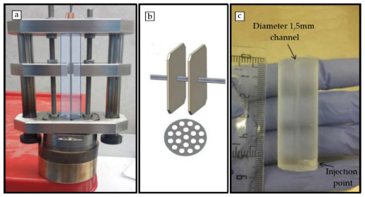
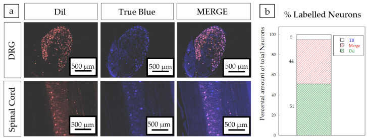
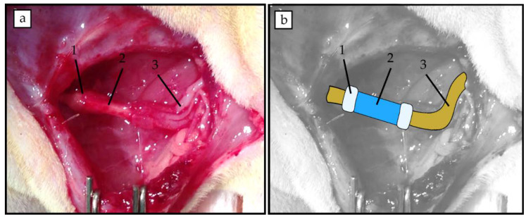
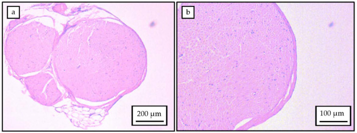
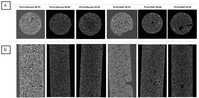
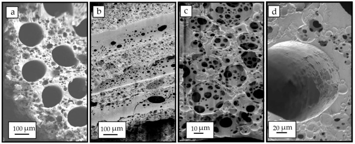
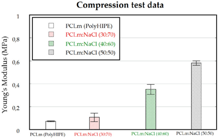
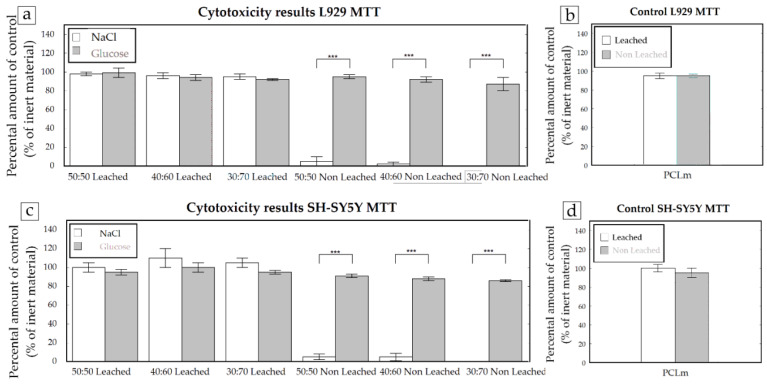
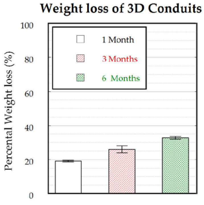
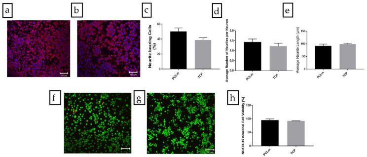
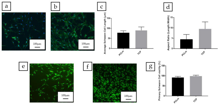
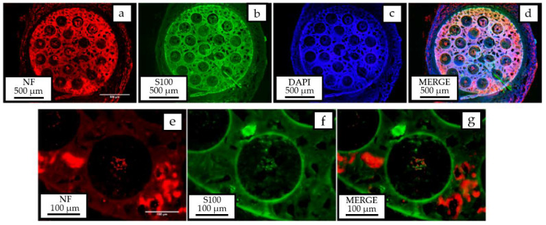
Peripheral nerves are basic communication structures guiding motor and sensory information from the central nervous system to receptor units. Severed peripheral nerve injuries represent a large clinical problem with relevant challenges to successful synthetic nerve repair scaffolds as substitutes to autologous nerve grafting. Numerous studies reported the use of hollow tubes made of synthetic polymers sutured between severed nerve stumps to promote nerve regeneration while providing protection for external factors, such as scar tissue formation and inflammation. Few approaches have described the potential use of a lumen structure comprised of microchannels or microfibers to provide axon growth avoiding misdirection and fostering proper healing. Here, we report the use of a 3D porous microchannel-based structure made of a photocurable methacrylated polycaprolactone, whose mechanical properties are comparable to native nerves. The neuro-regenerative properties of the polymer were assessed in vitro, prior to the implantation of the 3D porous structure, in a 6-mm rat sciatic nerve gap injury. The manufactured implants were biocompatible and able to be resorbed by the host's body at a suitable rate, allowing the complete healing of the nerve. The innovative design of the highly porous structure with the axon guiding microchannels, along with the observation of myelinated axons and Schwann cells in the in vivo tests, led to a significant progress towards the standardized use of synthetic 3D multichannel-based structures in peripheral nerve surgery.
周围神经是引导运动和感觉信息从中枢神经系统至受体单元的基本通信结构。周围神经切断伤是一个重大临床问题,对于成功合成神经修复支架以替代自体神经移植而言是相关挑战。众多研究报道了使用由合成聚合物制成的中空管缝合于切断的神经残端之间,以促进神经再生,同时为诸如瘢痕组织形成和炎症等外部因素提供保护。很少有方法描述过由微通道或微纤维组成的管腔结构的潜在用途,以提供轴突生长,避免方向错误并促进适当愈合。在此,我们报道了使用一种由光固化甲基丙烯酸化聚己内酯制成的基于三维多孔微通道的结构,其机械性能与天然神经相当。在植入三维多孔结构之前,先在6毫米大鼠坐骨神经间隙损伤模型中对该聚合物的神经再生特性进行了体外评估。制造的植入物具有生物相容性,并且能够以合适的速率被宿主身体吸收,从而使神经完全愈合。具有轴突引导微通道的高度多孔结构的创新设计,以及体内试验中观察到的有髓轴突和施万细胞,朝着在外周神经手术中标准化使用基于合成三维多通道的结构取得了重大进展。