• 文献检索
  • 文档翻译
  • 深度研究
  • 学术资讯
  • Suppr Zotero 插件Zotero 插件
  • 邀请有礼
  • 套餐&价格
  • 历史记录
应用&插件
Suppr Zotero 插件Zotero 插件浏览器插件Mac 客户端Windows 客户端微信小程序
定价
高级版会员购买积分包购买API积分包
服务
文献检索文档翻译深度研究API 文档MCP 服务
关于我们
关于 Suppr公司介绍联系我们用户协议隐私条款
关注我们

Suppr 超能文献

核心技术专利:CN118964589B侵权必究
粤ICP备2023148730 号-1Suppr @ 2026

文献检索

告别复杂PubMed语法,用中文像聊天一样搜索,搜遍4000万医学文献。AI智能推荐,让科研检索更轻松。

立即免费搜索

文件翻译

保留排版,准确专业,支持PDF/Word/PPT等文件格式,支持 12+语言互译。

免费翻译文档

深度研究

AI帮你快速写综述,25分钟生成高质量综述,智能提取关键信息,辅助科研写作。

立即免费体验

扫频源光学相干断层扫描血管造影减轻了视网膜下液引起的遮蔽伪影。

Swept-source optical coherence tomography angiography alleviates shadowing artifacts caused by subretinal fluid.

机构信息

Eye Center, Faculty of Medicine, University of Freiburg, Killianstrasse 5D-79106, Freiburg, Germany.

Faculty of Medicine, University of Freiburg, Freiburg, Germany.

出版信息

Int Ophthalmol. 2020 Aug;40(8):2007-2016. doi: 10.1007/s10792-020-01376-7. Epub 2020 Apr 24.

DOI:10.1007/s10792-020-01376-7
PMID:32333339
原文链接:https://pmc.ncbi.nlm.nih.gov/articles/PMC7358920/
Abstract

PURPOSE

To characterize the choriocapillaris (CC) structure in relation to subretinal fluid (SRF) as a possible systematic error source using spectral domain (SD-OCTA) compared to swept-source optical coherence tomography angiography (SS-OCTA).

METHODS

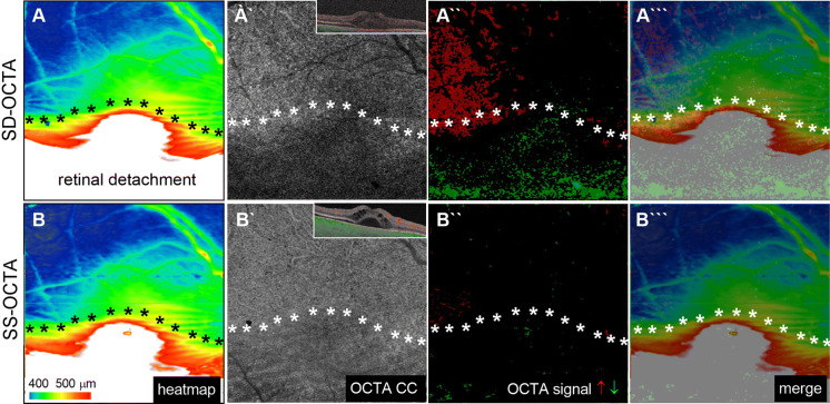
This is a prospective case-control study of 23 eyes. Ten patients with acute central serous chorioretinopathy (CSC), three patients with partial macular-off retinal detachment (RD) and ten healthy, age-matched controls were included. Abnormal CC decorrelation signals were quantitatively compared in CSC and controls by means of custom image processing. To investigate the influence of SRF on CC OCTA signal, the extent of SRF was quantified with a macular heatmap and compared with the corresponding OCTA signal of the CC.

RESULTS

SS-OCTA yielded a more homogeneous OCTA signal from the CC than SD-OCTA, offering less signal dispersion and variability in healthy and diseased eyes. Both devices demonstrated CC signal voids in CSC and RD, respectively. In CCS, the voids were predominantly located in the area with SRF. Compared to SD-OCTA, SS-OCTA delivered a more homogenous OCTA signal and reduced signal voids in the CC underneath SRF in both RD and CSC (CSC, 7.6% ± 6.3% vs, 19.7% ± 9.6%, p < 0.01). Despite this significant attenuation of signal voids, SS-OCTA continued to reveal signal voids below SRF and more pixels with reduced OCTA signals in CSC patients compared to controls (7.6% ± 6.3%, 0.1% ± 0.1%, p < 0.0001).

CONCLUSION

Understanding OCTA artifacts is critical to ensure accurate clinical evaluations. In this study, we describe the presence of SRF as an important shadow-causing artifact source for CC OCTA analysis which can be mitigated but not completely eliminated by employing SS-OCTA.

摘要

目的

利用谱域(SD-OCTA)与扫频源光学相干断层扫描血管造影术(SS-OCTA)相比,分析脉络膜毛细血管(CC)结构与视网膜下液(SRF)的关系,探讨其是否为系统性误差源。

方法

本研究为前瞻性病例对照研究,共纳入 23 只眼。纳入对象为 10 例急性中心性浆液性脉络膜视网膜病变(CSC)患者、3 例部分性黄斑区视网膜脱离(RD)患者和 10 例年龄匹配的健康对照者。采用定制的图像处理方法对 CSC 患者和对照组患者的异常 CC 去相关信号进行定量比较。为了研究 SRF 对 CC OCTA 信号的影响,通过黄斑热图量化 SRF 的范围,并与相应的 CC OCTA 信号进行比较。

结果

与 SD-OCTA 相比,SS-OCTA 提供了更均匀的 CC OCTA 信号,在健康眼和病变眼中信号的离散度和可变性更小。两种设备均显示 CSC 和 RD 中存在 CC 信号缺失。在 CSC 中,这些缺失主要位于 SRF 区域。与 SD-OCTA 相比,SS-OCTA 在 RD 和 CSC 中均能减少 SRF 下方 CC 的 OCTA 信号缺失(CSC:7.6%±6.3%比 19.7%±9.6%,p<0.01)。尽管信号缺失明显减少,但 SS-OCTA 在 CSC 患者中仍能显示 SRF 下方的信号缺失,以及更多的 OCTA 信号降低的像素(7.6%±6.3%比 0.1%±0.1%,p<0.0001)。

结论

了解 OCTA 伪影对于确保准确的临床评估至关重要。在本研究中,我们描述了 SRF 作为 CC OCTA 分析中重要的阴影伪影来源的存在,通过采用 SS-OCTA 可以减轻但不能完全消除该伪影。

https://cdn.ncbi.nlm.nih.gov/pmc/blobs/ae73/7358920/87520358ceb0/10792_2020_1376_Fig3_HTML.jpg
https://cdn.ncbi.nlm.nih.gov/pmc/blobs/ae73/7358920/b591ff5e97e1/10792_2020_1376_Fig1_HTML.jpg
https://cdn.ncbi.nlm.nih.gov/pmc/blobs/ae73/7358920/62ecdec098b6/10792_2020_1376_Fig2_HTML.jpg
https://cdn.ncbi.nlm.nih.gov/pmc/blobs/ae73/7358920/87520358ceb0/10792_2020_1376_Fig3_HTML.jpg
https://cdn.ncbi.nlm.nih.gov/pmc/blobs/ae73/7358920/b591ff5e97e1/10792_2020_1376_Fig1_HTML.jpg
https://cdn.ncbi.nlm.nih.gov/pmc/blobs/ae73/7358920/62ecdec098b6/10792_2020_1376_Fig2_HTML.jpg
https://cdn.ncbi.nlm.nih.gov/pmc/blobs/ae73/7358920/87520358ceb0/10792_2020_1376_Fig3_HTML.jpg

相似文献

1
Swept-source optical coherence tomography angiography alleviates shadowing artifacts caused by subretinal fluid.扫频源光学相干断层扫描血管造影减轻了视网膜下液引起的遮蔽伪影。
Int Ophthalmol. 2020 Aug;40(8):2007-2016. doi: 10.1007/s10792-020-01376-7. Epub 2020 Apr 24.
2
Longitudinal Analysis of the Choriocapillaris Using Optical Coherence Tomography Angiography Reveals Subretinal Fluid as a Substantial Confounder in Patients with Acute Central Serous Chorioretinopathy.使用光学相干断层扫描血管造影术对脉络膜毛细血管进行纵向分析发现,视网膜下液是急性中心性浆液性脉络膜视网膜病变患者的一个重要混杂因素。
Ophthalmol Ther. 2019 Dec;8(4):599-610. doi: 10.1007/s40123-019-00218-9. Epub 2019 Oct 25.
3
Clinical evaluation of neovascular and non-neovascular chronic central serous chorioretinopathy (CSC) diagnosed by swept source optical coherence tomography angiography (SS OCTA).经扫频源光学相干断层扫描血管造影(SS OCTA)诊断的新生血管性和非新生血管性慢性中心性浆液性脉络膜视网膜病变(CSC)的临床评估
Graefes Arch Clin Exp Ophthalmol. 2019 Aug;257(8):1581-1590. doi: 10.1007/s00417-019-04297-z. Epub 2019 Apr 29.
4
OCT Angiography of the Choriocapillaris in Central Serous Chorioretinopathy: A Quantitative Subgroup Analysis.中心性浆液性脉络膜视网膜病变中脉络膜毛细血管的光学相干断层扫描血管造影:一项定量亚组分析
Ophthalmol Ther. 2019 Mar;8(1):75-86. doi: 10.1007/s40123-018-0159-1. Epub 2019 Jan 7.
5
Early alterations in retinal microvasculature on swept-source optical coherence tomography angiography in acute central serous chorioretinopathy.急性中心性浆液性脉络膜视网膜病变的频域光相干断层扫描血管造影术对视网膜微血管早期改变的观察。
Sci Rep. 2021 Feb 4;11(1):3129. doi: 10.1038/s41598-021-82650-1.
6
Signal reduction in choriocapillaris and segmentation errors in spectral domain OCT angiography caused by soft drusen.软性玻璃膜疣导致的脉络膜毛细血管信号减弱及频域光学相干断层扫描血管造影中的分割误差。
Graefes Arch Clin Exp Ophthalmol. 2017 Dec;255(12):2347-2355. doi: 10.1007/s00417-017-3813-8. Epub 2017 Oct 5.
7
Early changes in choriocapillaris flow voids as an efficacy biomarker of photodynamic therapy in central serous chorioretinopathy.脉络膜毛细血管血流空泡的早期变化作为中心性浆液性脉络膜视网膜病变光动力疗法疗效的生物标志物。
Photodiagnosis Photodyn Ther. 2022 Jun;38:102862. doi: 10.1016/j.pdpdt.2022.102862. Epub 2022 Apr 11.
8
Artifact-Removed Quantitative Analysis of Choriocapillaris Flow Voids.去除血管内物质的脉络膜毛细血管血流空洞的定量分析。
Turk J Ophthalmol. 2023 Feb 24;53(1):37-43. doi: 10.4274/tjo.galenos.2022.23855.
9
Repeatability of choriocapillaris flow voids by optical coherence tomography angiography in central serous chorioretinopathy.光学相干断层扫描血管造影在中心性浆液性脉络膜视网膜病变中脉络膜毛细血管无灌注区的可重复性。
PLoS One. 2022 Dec 16;17(12):e0279243. doi: 10.1371/journal.pone.0279243. eCollection 2022.
10
Visualizing the Choriocapillaris Under Drusen: Comparing 1050-nm Swept-Source Versus 840-nm Spectral-Domain Optical Coherence Tomography Angiography.观察玻璃膜疣下的脉络膜毛细血管:1050纳米扫频源与840纳米谱域光学相干断层扫描血管造影术的比较
Invest Ophthalmol Vis Sci. 2016 Jul 1;57(9):OCT585-90. doi: 10.1167/iovs.15-18915.

引用本文的文献

1
Comparison of microvasculature between neovascular, non-neovascular chronic central serous chorioretinopathy cases and asymptomatic fellow eyes using swept-source optical coherence tomography angiography.使用扫频光学相干断层扫描血管造影术比较新生血管性、非新生血管性慢性中心性浆液性脉络膜视网膜病变病例与无症状对侧眼之间的微血管系统。
Int Ophthalmol. 2025 Jul 18;45(1):297. doi: 10.1007/s10792-025-03673-5.
2
Quantitative Changes in Vascular and Neural Fibers Induced by Subretinal Fluid Excluding the Peripapillary Region in Patients with Chronic Central Serous Chorioretinopathy.慢性中心性浆液性脉络膜视网膜病变患者视网膜下液(不包括视乳头周围区域)诱导的血管和神经纤维的定量变化
Diagnostics (Basel). 2025 Jan 14;15(2):174. doi: 10.3390/diagnostics15020174.
3

本文引用的文献

1
Characterisation of vascular changes in different stages of Stargardt disease using double swept-source optical coherence tomography angiography.使用双扫频源光学相干断层扫描血管造影术对Stargardt病不同阶段的血管变化进行特征分析。
BMJ Open Ophthalmol. 2019 Nov 2;4(1):e000318. doi: 10.1136/bmjophth-2019-000318. eCollection 2019.
2
Longitudinal Analysis of the Choriocapillaris Using Optical Coherence Tomography Angiography Reveals Subretinal Fluid as a Substantial Confounder in Patients with Acute Central Serous Chorioretinopathy.使用光学相干断层扫描血管造影术对脉络膜毛细血管进行纵向分析发现,视网膜下液是急性中心性浆液性脉络膜视网膜病变患者的一个重要混杂因素。
Ophthalmol Ther. 2019 Dec;8(4):599-610. doi: 10.1007/s40123-019-00218-9. Epub 2019 Oct 25.
3
Choroidal and Choriocapillaris OCT-A Analysis in Patients Affected by Active Central Serous Chorioretinopathy.活动性中心性浆液性脉络膜视网膜病变患者的脉络膜和脉络膜毛细血管光学相干断层扫描血管造影分析
Transl Vis Sci Technol. 2024 Dec 2;13(12):14. doi: 10.1167/tvst.13.12.14.
4
Optical Coherence Tomography Angiography-Navigated Laser Photocoagulation of Retinal Hemangioblastomas in Patients With von Hippel-Lindau Disease.光学相干断层扫描血管造影引导下激光光凝治疗 von Hippel-Lindau 病相关视网膜血管母细胞瘤。
Transl Vis Sci Technol. 2024 Jul 1;13(7):8. doi: 10.1167/tvst.13.7.8.
5
ZEISS PLEX Elite 9000 Widefield Optical Coherence Tomography Angiography as Screening Method for Early Detection of Retinal Hemangioblastomas in von Hippel-Lindau Disease.ZEISS PLEX Elite 9000 超广角光学相干断层扫描血管造影作为 von Hippel-Lindau 病视网膜血管母细胞瘤早期检测的筛查方法。
Transl Vis Sci Technol. 2024 Feb 1;13(2):8. doi: 10.1167/tvst.13.2.8.
6
Optical Coherence Tomography Angiography as a Diagnostic Tool for Diabetic Retinopathy.光学相干断层扫描血管造影术作为糖尿病视网膜病变的诊断工具
Diagnostics (Basel). 2024 Feb 2;14(3):326. doi: 10.3390/diagnostics14030326.
7
The OSCAR-MP Consensus Criteria for Quality Assessment of Retinal Optical Coherence Tomography Angiography.OSCAR-MP 视网膜光学相干断层扫描血管造影质量评估共识标准。
Neurol Neuroimmunol Neuroinflamm. 2023 Oct 9;10(6). doi: 10.1212/NXI.0000000000200169. Print 2023 Nov.
8
Central serous chorioretinopathy: updates in the pathogenesis, diagnosis and therapeutic strategies.中心性浆液性脉络膜视网膜病变:发病机制、诊断及治疗策略的新进展
Eye Vis (Lond). 2023 Jul 11;10(1):33. doi: 10.1186/s40662-023-00349-y.
9
Optical Coherence Tomography Angiography (OCT-A) in Uveitis: A Literature Review and a Reassessment of Its Real Role.葡萄膜炎中的光学相干断层扫描血管造影(OCT-A):文献综述及其实际作用的重新评估
Diagnostics (Basel). 2023 Feb 7;13(4):601. doi: 10.3390/diagnostics13040601.
10
Repeatability of choriocapillaris flow voids by optical coherence tomography angiography in central serous chorioretinopathy.光学相干断层扫描血管造影在中心性浆液性脉络膜视网膜病变中脉络膜毛细血管无灌注区的可重复性。
PLoS One. 2022 Dec 16;17(12):e0279243. doi: 10.1371/journal.pone.0279243. eCollection 2022.
Features of the choriocapillaris on four different optical coherence tomography angiography devices.四种不同光学相干断层扫描血管造影设备的脉络膜毛细血管特征。
Int Ophthalmol. 2020 Feb;40(2):325-333. doi: 10.1007/s10792-019-01182-w. Epub 2019 Sep 28.
4
Optical Coherence Tomography Angiography Compared with Indocyanine Green Angiography in Central Serous Chorioretinopathy.光学相干断层扫描血管造影与吲哚菁绿血管造影在中心性浆液性脉络膜视网膜病变中的比较。
Sci Rep. 2019 Apr 16;9(1):6149. doi: 10.1038/s41598-019-42623-x.
5
Choriocapillaris Flow Impairments in Association with Pachyvessel in Early Stages of Pachychoroid.脉络膜血流异常与厚脉络膜早期病变的厚血管相关。
Sci Rep. 2019 Apr 3;9(1):5565. doi: 10.1038/s41598-019-42052-w.
6
OCT Angiography of the Choriocapillaris in Central Serous Chorioretinopathy: A Quantitative Subgroup Analysis.中心性浆液性脉络膜视网膜病变中脉络膜毛细血管的光学相干断层扫描血管造影:一项定量亚组分析
Ophthalmol Ther. 2019 Mar;8(1):75-86. doi: 10.1007/s40123-018-0159-1. Epub 2019 Jan 7.
7
Comparing imaging capabilities of spectral domain and swept source optical coherence tomography angiography in healthy subjects and central serous retinopathy.比较健康受试者和中心性浆液性视网膜病变患者中光谱域光学相干断层扫描血管造影和扫频源光学相干断层扫描血管造影的成像能力。
Eye Vis (Lond). 2018 Aug 8;5:19. doi: 10.1186/s40662-018-0113-2. eCollection 2018.
8
PATTERNS OF CHORIOCAPILLARIS FLOW SIGNAL VOIDS IN CENTRAL SEROUS CHORIORETINOPATHY: An Optical Coherence Tomography Angiography Study.脉络膜毛细血管血流信号缺失在中心性浆液性脉络膜视网膜病变中的表现:一项光学相干断层扫描血管造影研究。
Retina. 2019 Nov;39(11):2178-2188. doi: 10.1097/IAE.0000000000002271.
9
Optical Coherence Tomography Angiography Offers New Insights into Choriocapillaris Perfusion.光学相干断层扫描血管造影术为脉络膜毛细血管灌注提供了新见解。
Ophthalmologica. 2018;239(2-3):74-84. doi: 10.1159/000485261. Epub 2018 Jan 19.
10
Diabetic Neuroglial Changes in the Superficial and Deep Nonperfused Areas on Optical Coherence Tomography Angiography.光学相干断层扫描血管造影术显示的糖尿病患者浅表和深部无灌注区域的神经胶质变化
Invest Ophthalmol Vis Sci. 2017 Nov 1;58(13):5870-5879. doi: 10.1167/iovs.17-22156.