• 文献检索
  • 文档翻译
  • 深度研究
  • 学术资讯
  • Suppr Zotero 插件Zotero 插件
  • 邀请有礼
  • 套餐&价格
  • 历史记录
应用&插件
Suppr Zotero 插件Zotero 插件浏览器插件Mac 客户端Windows 客户端微信小程序
定价
高级版会员购买积分包购买API积分包
服务
文献检索文档翻译深度研究API 文档MCP 服务
关于我们
关于 Suppr公司介绍联系我们用户协议隐私条款
关注我们

Suppr 超能文献

核心技术专利:CN118964589B侵权必究
粤ICP备2023148730 号-1Suppr @ 2026

文献检索

告别复杂PubMed语法,用中文像聊天一样搜索,搜遍4000万医学文献。AI智能推荐,让科研检索更轻松。

立即免费搜索

文件翻译

保留排版,准确专业,支持PDF/Word/PPT等文件格式,支持 12+语言互译。

免费翻译文档

深度研究

AI帮你快速写综述,25分钟生成高质量综述,智能提取关键信息,辅助科研写作。

立即免费体验

葡萄膜炎中的光学相干断层扫描血管造影(OCT-A):文献综述及其实际作用的重新评估

Optical Coherence Tomography Angiography (OCT-A) in Uveitis: A Literature Review and a Reassessment of Its Real Role.

作者信息

Herbort Carl P, Takeuchi Masaru, Papasavvas Ioannis, Tugal-Tutkun Ilknur, Hedayatfar Alireza, Usui Yoshihiko, Ozdal Pinar C, Urzua Cristhian A

机构信息

Retinal and Inflammatory Eye Diseases, Center for Ophthalmic Specialised Care (COS), Rue Charles-Monnard 6, 1003 Lausanne, Switzerland.

Department of Ophthalmology, National Defence Medical College, Tokorozawa 258-8513, Japan.

出版信息

Diagnostics (Basel). 2023 Feb 7;13(4):601. doi: 10.3390/diagnostics13040601.

DOI:10.3390/diagnostics13040601
PMID:36832089
原文链接:https://pmc.ncbi.nlm.nih.gov/articles/PMC9955936/
Abstract

BACKGROUND

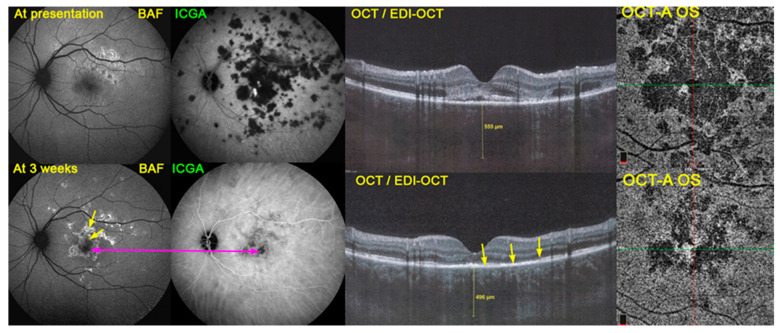
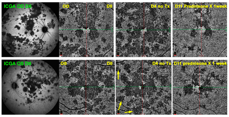
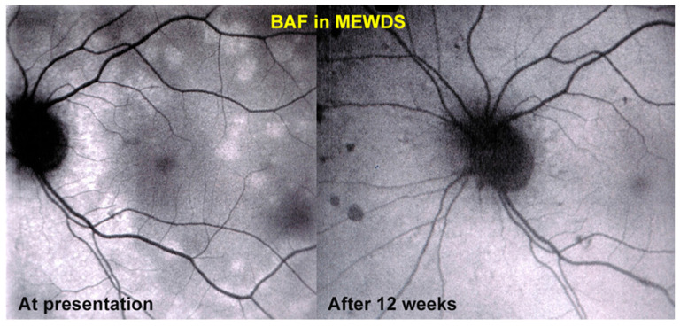
The global and precise follow-up of uveitis has become possible with the availability of dual fluorescein (FA) and indocyanine green angiography (ICGA) since the mid-1990s. Progressively, additional non-invasive imaging methods have emerged, bringing value-added precision to the imaging appraisal of uveitis, including, among others, optical coherence tomography (OCT), enhanced-depth imaging OCT (EDI-OCT) and blue light fundus autofluorescence (BAF). More recently, another complementary imaging method, OCT-angiography (OCT-A), further allowed retinal and choroidal circulation to be imaged without the need for dye injection.

PURPOSE

The purpose of this review was aimed at examining the evidence in published reports indicating whether OCT-A could possibly replace dye angiographic methods, as well as the real practical impact of OCT-A.

METHODS

A literature search in the PubMed database was performed using the terms OCT-angiography and uveitis, OCTA and uveitis and OCT-A and uveitis. Case reports were excluded. Articles were classified into technical reports, research reports and reviews. Articles in the two latter categories were analyzed in a more detailed, individual fashion. Special attention was paid to whether there were arguments in favor of an exclusive rather than complementary use of OCT-A. Furthermore, a synthesis of the main practical applications of OCT-A in the management of uveitis was attempted.

RESULTS

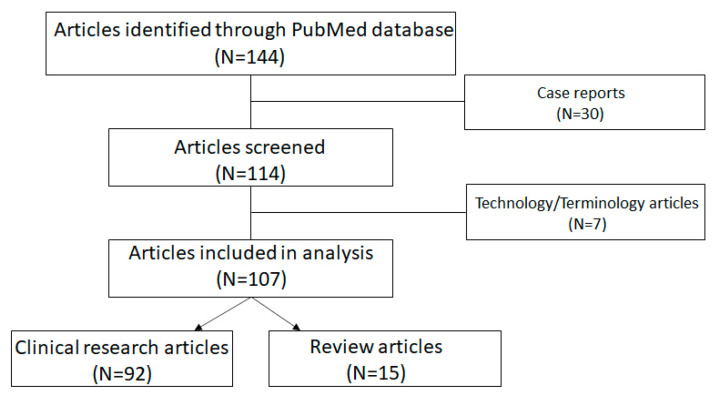
Between 2016 (the year of the first articles) and 2022, 144 articles containing the search terms were identified. After excluding case report articles, 114 articles were retained: 4 in 2016, 17 in 2017, 14 in 2018, 21 in 2019, 14 in 2020, 18 in 2021 and 26 in 2022. Seven articles contained technical information or consensus-based terminology. Ninety-two articles could be considered as clinical research articles. Of those, only two hinted in their conclusions that OCT-A could hypothetically replace dye methods. The terms mostly used to qualify the contribution of the articles in this group were "complementary to dye methods", "adjunct", "supplementing" and other similar terms. Fifteen articles were reviews, none of which hinted that OCT-A could replace dye methods. The situations where OCT-A represented a significant practical contribution to the practical appraisal of uveitis were identified.

CONCLUSION

To date, no evidence was found in the literature that OCT-A can replace the classic dye methods; however, it can complement them. Promoting the possibility that non-invasive OCT-A can substitute the invasive dye methods is deleterious, giving the elusive impression that dye methods are no longer inevitable for evaluating uveitis patients. Nevertheless, OCT-A is a precious tool in uveitis research.

摘要

背景

自20世纪90年代中期以来,随着双荧光素血管造影(FA)和吲哚菁绿血管造影(ICGA)技术的出现,对葡萄膜炎进行全面而精确的随访成为可能。逐渐地,更多的非侵入性成像方法应运而生,为葡萄膜炎的成像评估带来了更高的精准度,其中包括光学相干断层扫描(OCT)、增强深度成像OCT(EDI - OCT)和蓝光眼底自发荧光(BAF)。最近,另一种辅助成像方法——光学相干断层扫描血管造影(OCT - A),进一步实现了无需注射染料即可对视网膜和脉络膜循环进行成像。

目的

本综述旨在研究已发表报告中的证据,以确定OCT - A是否有可能取代染料血管造影方法,以及OCT - A的实际应用影响。

方法

在PubMed数据库中使用搜索词“OCT - angiography”和“uveitis”、“OCTA”和“uveitis”以及“OCT - A”和“uveitis”进行文献检索。排除病例报告。文章分为技术报告、研究报告和综述。对后两类文章进行更详细的单独分析。特别关注是否有支持OCT - A单独使用而非辅助使用的观点。此外,尝试总结OCT - A在葡萄膜炎管理中的主要实际应用。

结果

在2016年(第一篇相关文章发表年份)至2022年期间,共识别出144篇包含搜索词的文章。排除病例报告文章后,保留114篇:2016年4篇,2017年17篇,2018年14篇,2019年21篇,2020年14篇,2021年18篇,2022年26篇。7篇文章包含技术信息或基于共识的术语。92篇文章可视为临床研究文章。其中,只有两篇在结论中暗示OCT - A可能理论上取代染料方法。用于描述该组文章贡献的术语大多是“对染料方法的补充”、“辅助”、“补充”等类似术语。15篇文章为综述,均未暗示OCT - A可取代染料方法。确定了OCT - A对葡萄膜炎实际评估有重大实际贡献的情况。

结论

迄今为止,文献中未发现OCT - A可取代经典染料方法的证据;然而,它可以对其进行补充。宣扬非侵入性的OCT - A可替代侵入性染料方法的可能性是有害的,会给人一种错误印象,即评估葡萄膜炎患者时染料方法不再是必不可少的。尽管如此, OCT - A仍是葡萄膜炎研究中的宝贵工具。

https://cdn.ncbi.nlm.nih.gov/pmc/blobs/294e/9955936/f5626dc8eebe/diagnostics-13-00601-g003.jpg
https://cdn.ncbi.nlm.nih.gov/pmc/blobs/294e/9955936/02654788d928/diagnostics-13-00601-g001.jpg
https://cdn.ncbi.nlm.nih.gov/pmc/blobs/294e/9955936/92f007f391d1/diagnostics-13-00601-g002.jpg
https://cdn.ncbi.nlm.nih.gov/pmc/blobs/294e/9955936/fa3512a2ad76/diagnostics-13-00601-sch001.jpg
https://cdn.ncbi.nlm.nih.gov/pmc/blobs/294e/9955936/f5626dc8eebe/diagnostics-13-00601-g003.jpg

相似文献

1
Optical Coherence Tomography Angiography (OCT-A) in Uveitis: A Literature Review and a Reassessment of Its Real Role.葡萄膜炎中的光学相干断层扫描血管造影(OCT-A):文献综述及其实际作用的重新评估
Diagnostics (Basel). 2023 Feb 7;13(4):601. doi: 10.3390/diagnostics13040601.
2
Benefits and Limitations of OCT-A in the Diagnosis and Follow-Up of Posterior Intraocular Inflammation in Current Clinical Practice: A Valuable Tool or a Deceiver?光学相干断层扫描血管造影(OCT-A)在当前临床实践中诊断和随访眼后段炎症的益处与局限性:是有用工具还是误导?
Diagnostics (Basel). 2022 Sep 30;12(10):2384. doi: 10.3390/diagnostics12102384.
3
Need for Quantitative Measurement Methods for Posterior Uveitis: Comparison of Dual FA/ICGA Angiography, EDI-OCT Choroidal Thickness and SUN Vitreous Haze Evaluation in Stromal Choroiditis.后葡萄膜炎定量测量方法的必要性:基质性脉络膜炎中荧光素血管造影/吲哚菁绿血管造影双造影、增强深度成像光学相干断层扫描脉络膜厚度测量及SUN玻璃体混浊评估的比较
Klin Monbl Augenheilkd. 2018 Apr;235(4):424-435. doi: 10.1055/s-0043-124966. Epub 2018 Apr 18.
4
Clinicopathology of non-infectious choroiditis: evolution of its appraisal during the last 2-3 decades from "white dot syndromes" to precise classification.非感染性脉络膜炎的临床病理学:过去20至30年间其评估从“白点综合征”到精确分类的演变
J Ophthalmic Inflamm Infect. 2021 Nov 17;11(1):43. doi: 10.1186/s12348-021-00274-y.
5
Diagnosis, Mechanisms, and Differentiation of Inflammatory Diseases of the Outer Retina: Photoreceptoritis versus Choriocapillaritis; A Multimodal Imaging Perspective.外层视网膜炎症性疾病的诊断、机制及鉴别:光感受器炎与脉络膜毛细血管炎;多模态影像学视角
Diagnostics (Basel). 2022 Sep 9;12(9):2179. doi: 10.3390/diagnostics12092179.
6
Optical coherence tomography angiography (OCTA) as a new diagnostic tool in uveitis.光学相干断层扫描血管造影(OCTA)作为葡萄膜炎的一种新型诊断工具。
J Ophthalmic Inflamm Infect. 2019 May 28;9(1):10. doi: 10.1186/s12348-019-0176-9.
7
Classification of Non-Infectious and/or Immune Mediated Choroiditis: A Brief Overview of the Essentials.非感染性和/或免疫介导性脉络膜炎的分类:要点概述
Diagnostics (Basel). 2021 May 24;11(6):939. doi: 10.3390/diagnostics11060939.
8
Diagnosis and Treatment of Primary Inflammatory Choriocapillaropathies (PICCPs): A Comprehensive Overview.原发性炎症性脉络膜毛细血管病变(PICCPs)的诊断与治疗:全面综述。
Medicina (Kaunas). 2022 Jan 21;58(2):165. doi: 10.3390/medicina58020165.
9
Masqueraders of multiple evanescent white dot syndrome (MEWDS).多发性一过性白点综合征(MEWDS)的伪装者。
Int Ophthalmol. 2020 Mar;40(3):627-638. doi: 10.1007/s10792-019-01223-4. Epub 2019 Dec 2.
10
Review of the Current Literature and Our Experience on the Value of OCT-angiography in White Dot Syndromes.关于光学相干断层扫描血管造影术在白点综合征中的价值的当前文献综述及我们的经验
Ocul Immunol Inflamm. 2022 Feb 17;30(2):364-378. doi: 10.1080/09273948.2020.1837185. Epub 2021 Feb 22.

引用本文的文献

1
Inadequacies in uveitis: misnomers, incongruencies, persistence of obsolete terminologies & inappropriate guidelines, treatment inadequacies, and misinterpretations.葡萄膜炎的不足之处:名称不当、不一致、过时术语的持续存在及不恰当的指南、治疗不足和误解。
J Ophthalmic Inflamm Infect. 2025 Mar 6;15(1):19. doi: 10.1186/s12348-025-00470-0.
2
An update of multimodal imaging in white dot syndrome.白点综合征的多模态成像最新进展。
Oman J Ophthalmol. 2024 Oct 24;17(3):325-333. doi: 10.4103/ojo.ojo_116_24. eCollection 2024 Sep-Dec.
3
Indocyanine green fluorescence video-angiography for flap perfusion assessment in head and neck reconstruction: a prospective study.

本文引用的文献

1
Benefits and Limitations of OCT-A in the Diagnosis and Follow-Up of Posterior Intraocular Inflammation in Current Clinical Practice: A Valuable Tool or a Deceiver?光学相干断层扫描血管造影(OCT-A)在当前临床实践中诊断和随访眼后段炎症的益处与局限性:是有用工具还是误导?
Diagnostics (Basel). 2022 Sep 30;12(10):2384. doi: 10.3390/diagnostics12102384.
2
Longitudinal Comparison of Constant Artifacts in Optical Coherence Tomography Angiography in Patients with Posterior Uveitis Compared to Healthy Subjects.与健康受试者相比,后葡萄膜炎患者光学相干断层扫描血管造影中恒定伪像的纵向比较。
J Clin Med. 2022 Sep 13;11(18):5376. doi: 10.3390/jcm11185376.
3
吲哚菁绿荧光视频血管造影术在头颈部重建中皮瓣灌注评估的前瞻性研究。
Eur Arch Otorhinolaryngol. 2025 Feb;282(2):961-970. doi: 10.1007/s00405-024-08959-5. Epub 2024 Sep 6.
4
Automated robot-assisted wide-field optical coherence tomography using structured light camera.使用结构光相机的自动机器人辅助宽视野光学相干断层扫描
Biomed Opt Express. 2023 Jul 26;14(8):4310-4325. doi: 10.1364/BOE.496710. eCollection 2023 Aug 1.
The Role of Widefield and Ultra Widefield Optical Coherence Tomography in the Diagnosis and Management of Vitreoretinal Diseases.
广角和超广角光学相干断层扫描在玻璃体视网膜疾病诊断与治疗中的作用
Diagnostics (Basel). 2022 Sep 17;12(9):2247. doi: 10.3390/diagnostics12092247.
4
Acute Posterior Multifocal Placoid Pigment Epitheliopathy (APMPPE): A Comprehensive Approach and Case Series: Systemic Corticosteroid Therapy Is Necessary in a Large Proportion of Cases.急性后极部多发性斑状色素上皮病变(APMPPE):全面的处理方法和病例系列:大部分病例需要全身皮质类固醇治疗。
Medicina (Kaunas). 2022 Aug 8;58(8):1070. doi: 10.3390/medicina58081070.
5
Use of optical coherence tomography angiography in the uveitis clinic.光学相干断层扫描血管造影术在葡萄膜炎临床中的应用。
Graefes Arch Clin Exp Ophthalmol. 2023 Jan;261(1):23-36. doi: 10.1007/s00417-022-05763-x. Epub 2022 Jul 16.
6
Experts Opinion: OCTA vs. FFA/ICG in Uveitis - Which Will Survive?专家观点:OCTA 与 FFA/ICG 在葡萄膜炎中的应用——谁将幸存?
Ocul Immunol Inflamm. 2023 Oct;31(8):1561-1568. doi: 10.1080/09273948.2022.2084421. Epub 2022 Jul 7.
7
Anterior Segment Optical Coherence Tomography (AS-OCT) Visualization of Anterior Vitritis.眼前节光学相干断层扫描(AS-OCT)对眼前段炎症的可视化观察。
Ocul Immunol Inflamm. 2023 Jul;31(5):1101-1102. doi: 10.1080/09273948.2022.2079535. Epub 2022 Jun 16.
8
Comparison of Indocyanine Green Angiography and Swept-Source Wide-Field Optical Coherence Tomography Angiography in Posterior Uveitis.吲哚菁绿血管造影术与扫频源超广角光学相干断层扫描血管造影术在后部葡萄膜炎中的比较
Front Med (Lausanne). 2022 May 2;9:853315. doi: 10.3389/fmed.2022.853315. eCollection 2022.
9
Diagnosis and Treatment of Primary Inflammatory Choriocapillaropathies (PICCPs): A Comprehensive Overview.原发性炎症性脉络膜毛细血管病变(PICCPs)的诊断与治疗:全面综述。
Medicina (Kaunas). 2022 Jan 21;58(2):165. doi: 10.3390/medicina58020165.
10
Consensus-based recommendations for optical coherence tomography angiography reporting in uveitis.葡萄膜炎光学相干断层扫描血管造影报告的基于共识的建议。
Br J Ophthalmol. 2023 Jul;107(7):959-965. doi: 10.1136/bjophthalmol-2021-320021. Epub 2022 Feb 8.