• 文献检索
  • 文档翻译
  • 深度研究
  • 学术资讯
  • Suppr Zotero 插件Zotero 插件
  • 邀请有礼
  • 套餐&价格
  • 历史记录
应用&插件
Suppr Zotero 插件Zotero 插件浏览器插件Mac 客户端Windows 客户端微信小程序
定价
高级版会员购买积分包购买API积分包
服务
文献检索文档翻译深度研究API 文档MCP 服务
关于我们
关于 Suppr公司介绍联系我们用户协议隐私条款
关注我们

Suppr 超能文献

核心技术专利:CN118964589B侵权必究
粤ICP备2023148730 号-1Suppr @ 2026

文献检索

告别复杂PubMed语法,用中文像聊天一样搜索,搜遍4000万医学文献。AI智能推荐,让科研检索更轻松。

立即免费搜索

文件翻译

保留排版,准确专业,支持PDF/Word/PPT等文件格式,支持 12+语言互译。

免费翻译文档

深度研究

AI帮你快速写综述,25分钟生成高质量综述,智能提取关键信息,辅助科研写作。

立即免费体验

骨/骨髓微环境对前列腺癌细胞和 CD59 表达的影响。

Effects of the Bone/Bone Marrow Microenvironments on Prostate Cancer Cells and CD59 Expression.

机构信息

Department of Urology, Guizhou Provincial People's Hospital, Medical College of Guizhou University, Guiyang, Guizhou, China.

Department of Urology, Zhujiang Hospital, Southern Medical University, Guangzhou, Guangdong, China.

出版信息

Biomed Res Int. 2020 Apr 6;2020:2753414. doi: 10.1155/2020/2753414. eCollection 2020.

DOI:10.1155/2020/2753414
PMID:32337233
原文链接:https://pmc.ncbi.nlm.nih.gov/articles/PMC7165328/
Abstract

OBJECTIVE

To evaluate the effects of human bone marrow mesenchymal stem cells (hBMSCs) and osteoblasts (hFOB1.19) on PC3 prostate cancer cells.

METHODS

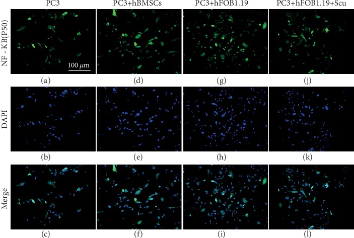
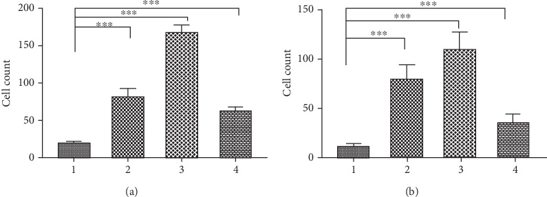
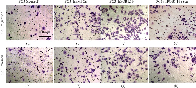
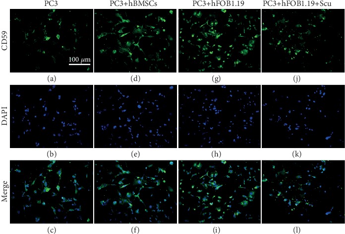
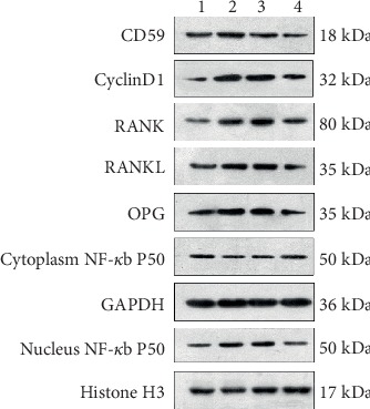
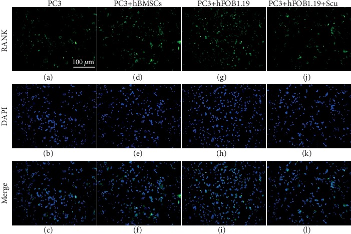
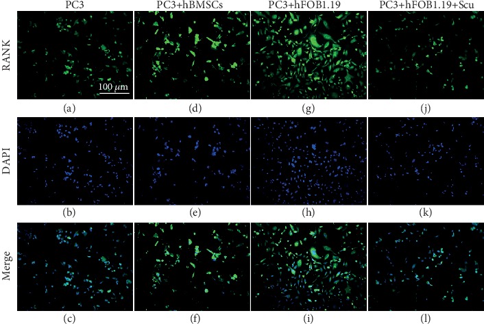
To simulate the interaction between the bone/bone marrow microenvironments and prostate cancer cells, we established cocultures of PC3 cells with hBMSC or hFOB1.19 cells and evaluated their effects on the proliferation, cell cycle distribution, cell migration, and invasion of PC3 cells. Quantitative reverse transcription polymerase chain reaction was used to detect mRNA expression in PC3 cells. The expression of receptor activator of nuclear factor- (NF-) B (RANK), RANK ligand (RANKL), osteoprotegerin (OPG), CD59, NF-B (p50 subunit), and cyclin D1 in PC3 cells was analyzed by immunofluorescence and western blotting.

RESULTS

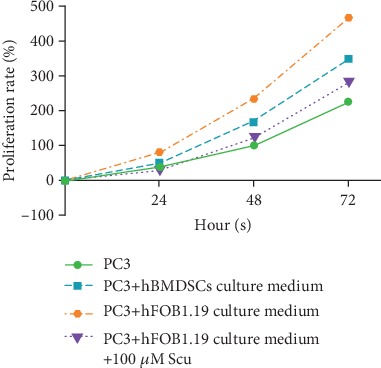
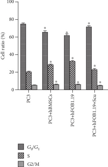
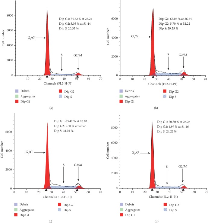
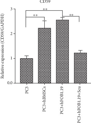
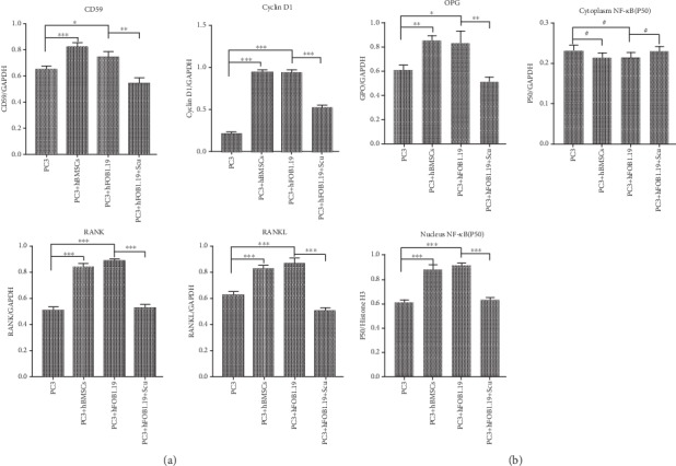
hBMSCs and hFOB1.19 cells enhanced the proliferation, migration, and invasion of PC3 cells; increased the proportion of PC3 cells in the S and G/M phases of the cell cycle; and upregulated RANK, RANKL, OPG, CD59, cyclin D1, and NF-B (p50 subunit) expression by PC3 cells. The RANKL inhibitor, scutellarin, inhibited these effects in PC3-hFOB1.19 cocultures.

CONCLUSION

hBMSCs and hFOB1.19 cells modulate the phenotype of PC3 prostate cancer cells and the expression of CD59 by activating the RANK/RANKL/OPG signaling pathway.

摘要

目的

评估人骨髓间充质干细胞(hBMSCs)和成骨细胞(hFOB1.19)对 PC3 前列腺癌细胞的影响。

方法

为了模拟骨/骨髓微环境与前列腺癌细胞之间的相互作用,我们建立了 PC3 细胞与 hBMSC 或 hFOB1.19 细胞共培养的模型,并评估了它们对 PC3 细胞增殖、细胞周期分布、细胞迁移和侵袭的影响。采用实时定量聚合酶链反应检测 PC3 细胞中 mRNA 的表达。采用免疫荧光和蛋白质印迹法分析 PC3 细胞中核因子-κB(NF-κB)(p50 亚基)、核因子受体激活剂(RANK)、RANK 配体(RANKL)、骨保护素(OPG)、CD59、细胞周期蛋白 D1 的表达。

结果

hBMSCs 和 hFOB1.19 细胞增强了 PC3 细胞的增殖、迁移和侵袭能力;增加了 PC3 细胞在细胞周期 S 和 G/M 期的比例;并上调了 PC3 细胞中 RANK、RANKL、OPG、CD59、细胞周期蛋白 D1 和 NF-κB(p50 亚基)的表达。RANKL 抑制剂 scutellarin 抑制了 PC3-hFOB1.19 共培养物中的这些作用。

结论

hBMSCs 和 hFOB1.19 细胞通过激活 RANK/RANKL/OPG 信号通路调节 PC3 前列腺癌细胞的表型和 CD59 的表达。

https://cdn.ncbi.nlm.nih.gov/pmc/blobs/bde8/7165328/e6f353b9514e/BMRI2020-2753414.012.jpg
https://cdn.ncbi.nlm.nih.gov/pmc/blobs/bde8/7165328/e7faead79bd1/BMRI2020-2753414.001.jpg
https://cdn.ncbi.nlm.nih.gov/pmc/blobs/bde8/7165328/e7fa79fd34a7/BMRI2020-2753414.002.jpg
https://cdn.ncbi.nlm.nih.gov/pmc/blobs/bde8/7165328/ba42e027ca0d/BMRI2020-2753414.003.jpg
https://cdn.ncbi.nlm.nih.gov/pmc/blobs/bde8/7165328/b7ba9bfca601/BMRI2020-2753414.004.jpg
https://cdn.ncbi.nlm.nih.gov/pmc/blobs/bde8/7165328/4d40c410db4c/BMRI2020-2753414.005.jpg
https://cdn.ncbi.nlm.nih.gov/pmc/blobs/bde8/7165328/1bd3652a9747/BMRI2020-2753414.006.jpg
https://cdn.ncbi.nlm.nih.gov/pmc/blobs/bde8/7165328/a535f8b703fb/BMRI2020-2753414.007.jpg
https://cdn.ncbi.nlm.nih.gov/pmc/blobs/bde8/7165328/b6561024da5e/BMRI2020-2753414.008.jpg
https://cdn.ncbi.nlm.nih.gov/pmc/blobs/bde8/7165328/d14213416240/BMRI2020-2753414.009.jpg
https://cdn.ncbi.nlm.nih.gov/pmc/blobs/bde8/7165328/af86ac069bdd/BMRI2020-2753414.010.jpg
https://cdn.ncbi.nlm.nih.gov/pmc/blobs/bde8/7165328/453b12a3fbf0/BMRI2020-2753414.011.jpg
https://cdn.ncbi.nlm.nih.gov/pmc/blobs/bde8/7165328/e6f353b9514e/BMRI2020-2753414.012.jpg

相似文献

1
Effects of the Bone/Bone Marrow Microenvironments on Prostate Cancer Cells and CD59 Expression.骨/骨髓微环境对前列腺癌细胞和 CD59 表达的影响。
Biomed Res Int. 2020 Apr 6;2020:2753414. doi: 10.1155/2020/2753414. eCollection 2020.
2
Induction of retinol-binding protein 4 and placenta-specific 8 expression in human prostate cancer cells remaining in bone following osteolytic tumor growth inhibition by osteoprotegerin.骨保护素抑制溶骨性肿瘤生长后,人前列腺癌细胞在骨中残留诱导视黄醇结合蛋白 4 和胎盘特异性蛋白 8 的表达。
Int J Oncol. 2013 Aug;43(2):365-74. doi: 10.3892/ijo.2013.1954. Epub 2013 May 24.
3
DU145 human prostate cancer cells express functional receptor activator of NFkappaB: new insights in the prostate cancer bone metastasis process.DU145人前列腺癌细胞表达核因子κB受体激活剂:前列腺癌骨转移过程中的新见解。
Bone. 2007 Apr;40(4):981-90. doi: 10.1016/j.bone.2006.11.006. Epub 2006 Dec 28.
4
RANKL acts directly on RANK-expressing prostate tumor cells and mediates migration and expression of tumor metastasis genes.核因子κB受体活化因子配体(RANKL)直接作用于表达核因子κB受体活化因子(RANK)的前列腺肿瘤细胞,并介导肿瘤转移基因的迁移和表达。
Prostate. 2008 Jan 1;68(1):92-104. doi: 10.1002/pros.20678.
5
Targeting IL-6 and RANKL signaling inhibits prostate cancer growth in bone.靶向白细胞介素-6和核因子κB受体活化因子配体信号通路可抑制前列腺癌在骨中的生长。
Clin Exp Metastasis. 2014 Dec;31(8):921-33. doi: 10.1007/s10585-014-9680-3. Epub 2014 Sep 16.
6
Caprylic acid (C8:0) promotes bone metastasis of prostate cancer by dysregulated adipo-osteogenic balance in bone marrow.辛酸(C8:0)通过失调的脂肪成骨平衡在骨髓中促进前列腺癌的骨转移。
Cancer Sci. 2020 Oct;111(10):3600-3612. doi: 10.1111/cas.14606. Epub 2020 Aug 28.
7
Activation of NF-kappa B signaling promotes growth of prostate cancer cells in bone.NF-κB 信号的激活促进了前列腺癌细胞在骨中的生长。
PLoS One. 2013;8(4):e60983. doi: 10.1371/journal.pone.0060983. Epub 2013 Apr 5.
8
CCN3 promotes prostate cancer bone metastasis by modulating the tumor-bone microenvironment through RANKL-dependent pathway.CCN3 通过 RANKL 依赖性途径调节肿瘤-骨微环境促进前列腺癌骨转移。
Carcinogenesis. 2013 Jul;34(7):1669-79. doi: 10.1093/carcin/bgt103. Epub 2013 Mar 27.
9
Effect of PI3K/Akt Signaling Pathway on the Process of Prostate Cancer Metastasis to Bone.PI3K/Akt信号通路对前列腺癌骨转移过程的影响
Cell Biochem Biophys. 2015 May;72(1):171-7. doi: 10.1007/s12013-014-0433-3.
10
The inhibition of RANKL/RANK signaling by osteoprotegerin suppresses bone invasion by oral squamous cell carcinoma cells.骨保护素通过抑制 RANKL/RANK 信号通路抑制口腔鳞状细胞癌细胞的骨侵袭。
Carcinogenesis. 2011 Nov;32(11):1634-40. doi: 10.1093/carcin/bgr198. Epub 2011 Sep 1.

引用本文的文献

1
Impact of Resolvin-E1 and Maresin-1 on Bone Marrow Stem Cell Osteogenesis under Inflammatory Stress.解析素 E1 和maresin-1 在炎症应激下对骨髓间充质干细胞成骨的影响。
Cells. 2024 May 28;13(11):932. doi: 10.3390/cells13110932.

本文引用的文献

1
iTRAQ-based Comparative Serum Proteomic Analysis of Prostate Cancer Patients with or without Bone Metastasis.基于iTRAQ的前列腺癌伴或不伴骨转移患者血清蛋白质组对比分析
J Cancer. 2019 Jul 10;10(18):4165-4177. doi: 10.7150/jca.33497. eCollection 2019.
2
Propofol attenuates osteoclastogenesis by lowering RANKL/OPG ratio in mouse osteoblasts.异丙酚通过降低小鼠成骨细胞中 RANKL/OPG 比值来抑制破骨细胞生成。
Int J Med Sci. 2018 May 14;15(7):723-729. doi: 10.7150/ijms.22713. eCollection 2018.
3
Metastases in Prostate Cancer.前列腺癌转移。
Cold Spring Harb Perspect Med. 2019 Mar 1;9(3):a033688. doi: 10.1101/cshperspect.a033688.
4
RANKL/RANK/OPG cytokine receptor system: mRNA expression pattern in BPH, primary and metastatic prostate cancer disease.RANKL/RANK/OPG 细胞因子受体系统:BPH、原发性和转移性前列腺癌疾病中的 mRNA 表达模式。
World J Urol. 2018 Feb;36(2):187-192. doi: 10.1007/s00345-017-2145-y. Epub 2017 Dec 4.
5
The RANK/RANKL/OPG system in tumorigenesis and metastasis of cancer stem cell: potential targets for anticancer therapy.RANK/RANKL/OPG系统在癌症干细胞肿瘤发生和转移中的作用:抗癌治疗的潜在靶点
Onco Targets Ther. 2017 Jul 27;10:3801-3810. doi: 10.2147/OTT.S135867. eCollection 2017.
6
Bone Metastases: An Overview.骨转移:概述
Oncol Rev. 2017 May 9;11(1):321. doi: 10.4081/oncol.2017.321. eCollection 2017 Mar 3.
7
Caffeic acid phenethyl ester protects against glucocorticoid-induced osteoporosis in vivo: Impact on oxidative stress and RANKL/OPG signals.咖啡酸苯乙酯在体内可预防糖皮质激素诱导的骨质疏松症:对氧化应激和RANKL/OPG信号的影响。
Toxicol Appl Pharmacol. 2017 Jun 1;324:26-35. doi: 10.1016/j.taap.2017.03.021. Epub 2017 Mar 29.
8
Cyclin D1, cancer progression, and opportunities in cancer treatment.细胞周期蛋白D1、癌症进展与癌症治疗机遇
J Mol Med (Berl). 2016 Dec;94(12):1313-1326. doi: 10.1007/s00109-016-1475-3. Epub 2016 Oct 2.
9
Molecular and Functional Imaging of Bone Metastases in Breast and Prostate Cancers: An Overview.乳腺癌和前列腺癌骨转移的分子和功能影像学:概述。
Clin Nucl Med. 2016 Jan;41(1):e44-50. doi: 10.1097/RLU.0000000000000993.
10
RANKL/OPG; Critical role in bone physiology.核因子κB受体活化因子配体/骨保护素;在骨生理学中起关键作用。
Rev Endocr Metab Disord. 2015 Jun;16(2):131-9. doi: 10.1007/s11154-014-9308-6.