Department of Biomedical Engineering, Duke University, Durham, NC, USA.
Department of Radiology, Duke University Medical Center, Durham, NC, USA.
Sci Rep. 2020 Apr 30;10(1):7385. doi: 10.1038/s41598-020-64361-1.
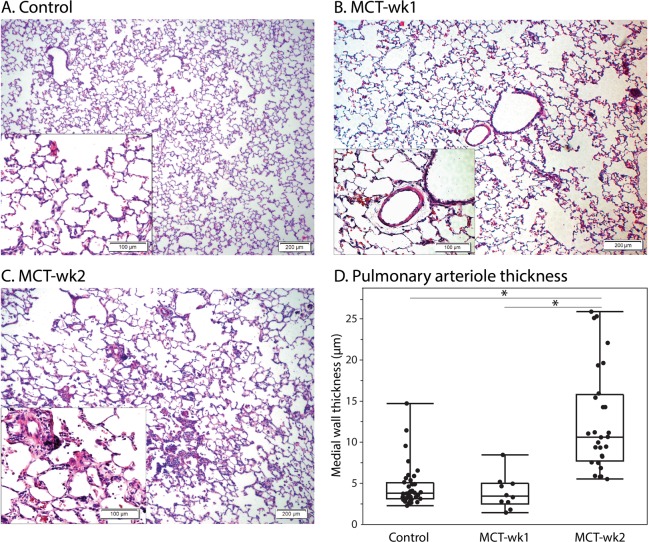
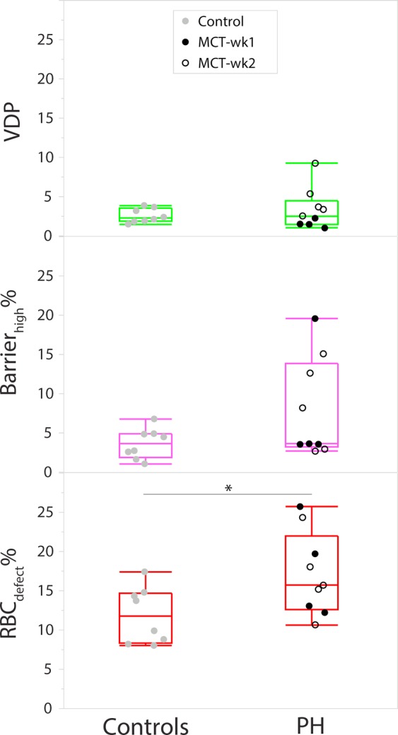
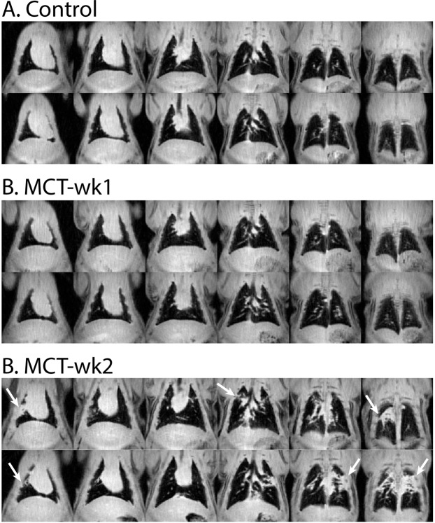
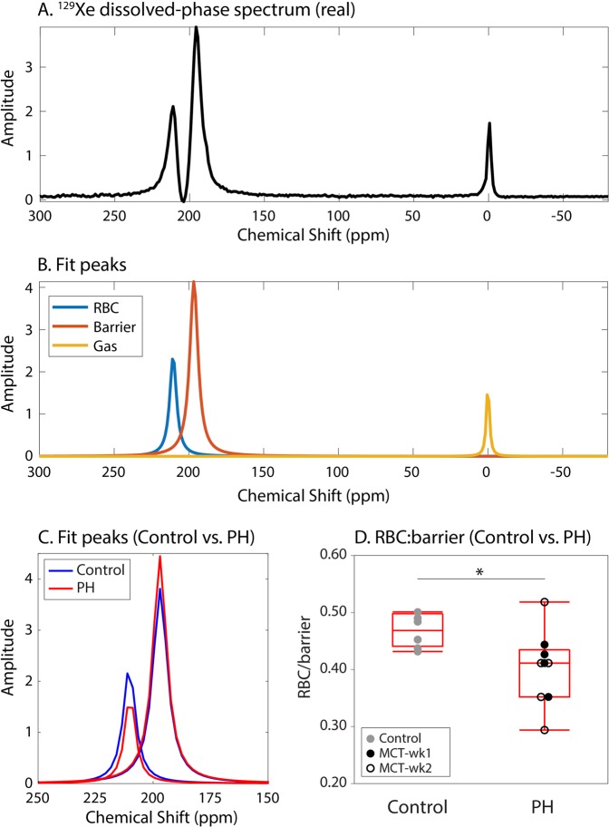
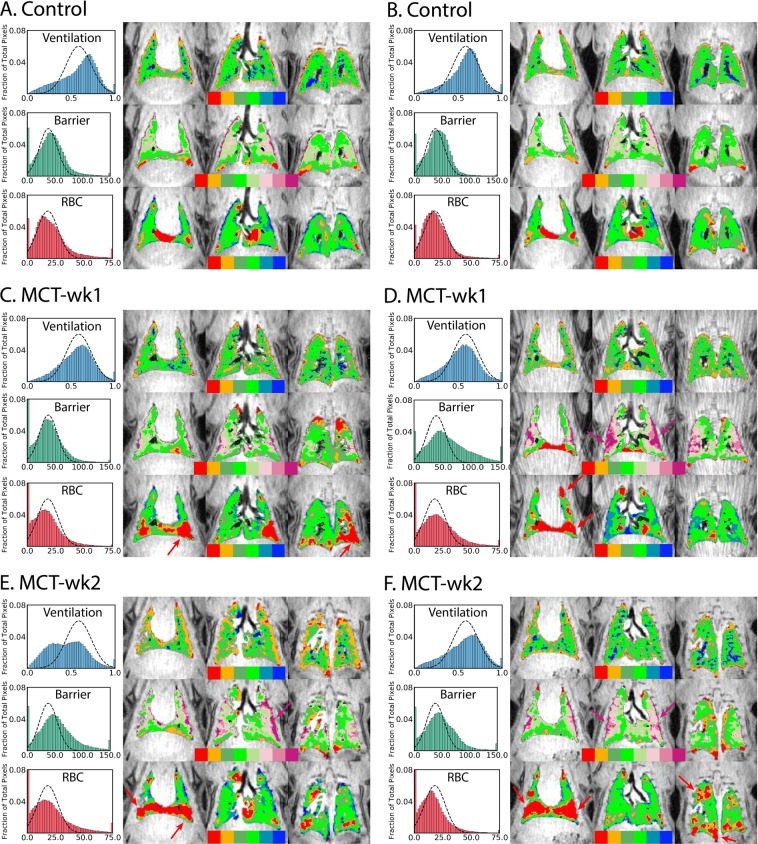
Hyperpolarized Xe magnetic resonance imaging (MRI) is capable of regional mapping of pulmonary gas-exchange and has found application in a wide range of pulmonary disorders in humans and animal model analogs. This study is the first application of Xe MRI to the monocrotaline rat model of pulmonary hypertension. Such models of preclinical pulmonary hypertension, a disease of the pulmonary vasculature that results in right heart failure and death, are usually assessed with invasive procedures such as right heart catheterization and histopathology. The work here adapted from protocols from clinical Xe MRI to enable preclinical imaging of rat models of pulmonary hypertension on a Bruker 7 T scanner. Xe spectroscopy and gas-exchange imaging showed reduced Xe uptake by red blood cells early in the progression of the disease, and at a later time point was accompanied by increased uptake by barrier tissues, edema, and ventilation defects-all of which are salient characteristics of the monocrotaline model. Imaging results were validated by H&E histology, which showed evidence of remodeling of arterioles. This proof-of-concept study has demonstrated that hyperpolarized Xe MRI has strong potential to be used to non-invasively monitor the progression of pulmonary hypertension in preclinical models and potentially to also assess response to therapy.
氙气磁共振成像(MRI)可对肺部气体交换进行区域性绘图,已在人类和动物模型类似物的多种肺部疾病中得到应用。本研究首次将 Xe MRI 应用于野百合碱诱导的肺动脉高压大鼠模型。此类临床前肺动脉高压模型是一种肺部血管疾病,可导致右心衰竭和死亡,通常采用右心导管术和组织病理学等有创程序进行评估。本研究根据临床 Xe MRI 方案进行了改编,使在布鲁克 7T 扫描仪上对肺动脉高压大鼠模型进行临床前成像成为可能。Xe 光谱和气体交换成像显示,在疾病进展早期,红细胞对 Xe 的摄取减少,而在稍后的时间点,屏障组织、水肿和通气缺陷对 Xe 的摄取增加,所有这些都是野百合碱模型的显著特征。成像结果通过 H&E 组织学得到了验证,该组织学显示出小动脉重构的证据。这项概念验证研究表明,极化氙气 MRI 具有很大的潜力可用于非侵入性地监测临床前模型中肺动脉高压的进展,并有可能评估对治疗的反应。