• 文献检索
  • 文档翻译
  • 深度研究
  • 学术资讯
  • Suppr Zotero 插件Zotero 插件
  • 邀请有礼
  • 套餐&价格
  • 历史记录
应用&插件
Suppr Zotero 插件Zotero 插件浏览器插件Mac 客户端Windows 客户端微信小程序
定价
高级版会员购买积分包购买API积分包
服务
文献检索文档翻译深度研究API 文档MCP 服务
关于我们
关于 Suppr公司介绍联系我们用户协议隐私条款
关注我们

Suppr 超能文献

核心技术专利:CN118964589B侵权必究
粤ICP备2023148730 号-1Suppr @ 2026

文献检索

告别复杂PubMed语法,用中文像聊天一样搜索,搜遍4000万医学文献。AI智能推荐,让科研检索更轻松。

立即免费搜索

文件翻译

保留排版,准确专业,支持PDF/Word/PPT等文件格式,支持 12+语言互译。

免费翻译文档

深度研究

AI帮你快速写综述,25分钟生成高质量综述,智能提取关键信息,辅助科研写作。

立即免费体验

血管内流动刺激内皮细胞中的 PKD2(多囊蛋白-2)通道,从而降低血压。

Intravascular flow stimulates PKD2 (polycystin-2) channels in endothelial cells to reduce blood pressure.

机构信息

Department of Physiology University of Tennessee Health Science Center Memphis, Memphis, United States.

出版信息

Elife. 2020 May 4;9:e56655. doi: 10.7554/eLife.56655.

DOI:10.7554/eLife.56655
PMID:32364494
原文链接:https://pmc.ncbi.nlm.nih.gov/articles/PMC7228764/
Abstract

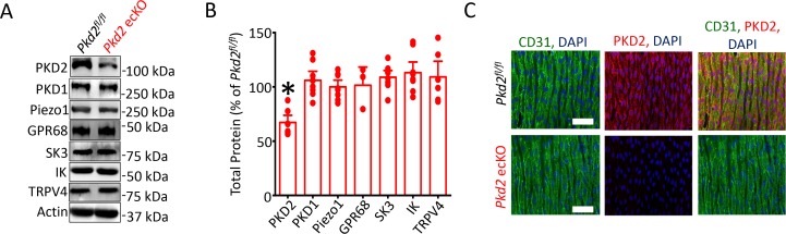
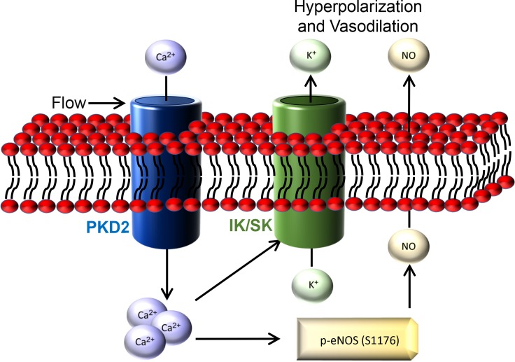
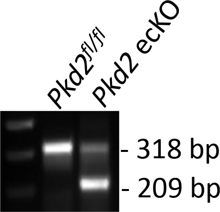
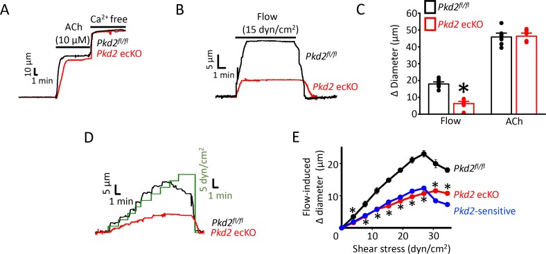
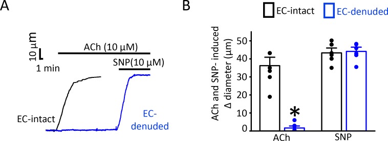
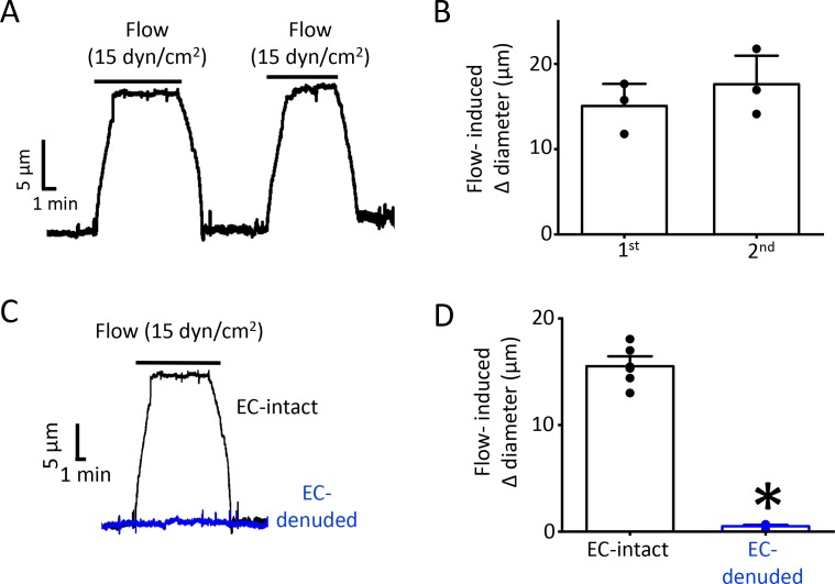
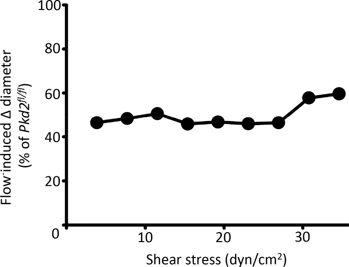
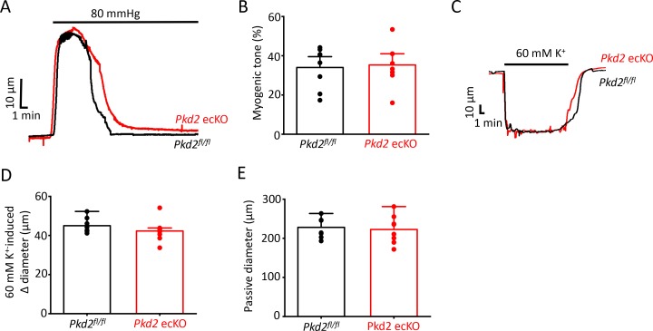
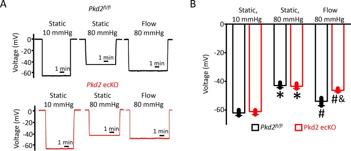
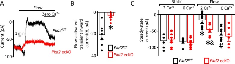
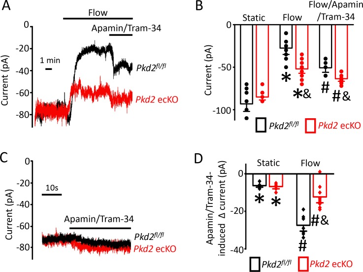
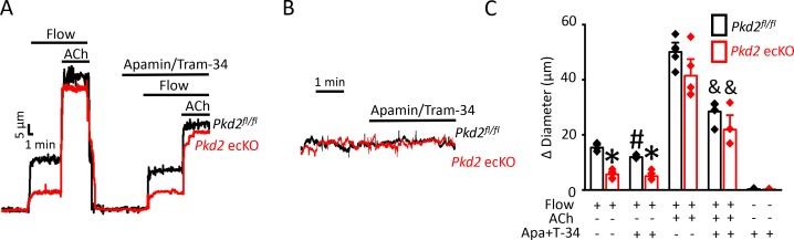
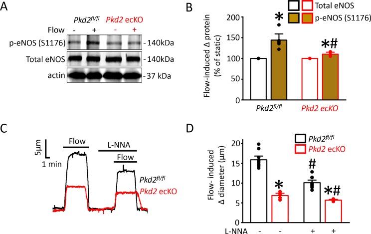
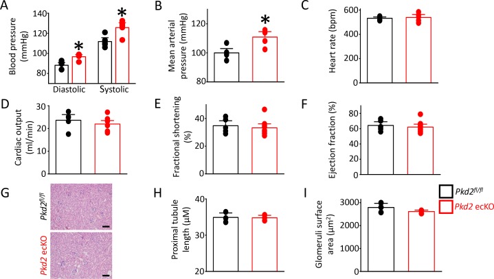
PKD2 (polycystin-2, TRPP1), a TRP polycystin channel, is expressed in endothelial cells (ECs), but its physiological functions in this cell type are unclear. Here, we generated inducible, EC-specific knockout mice to examine vascular functions of PKD2. Data show that a broad range of intravascular flow rates stimulate EC PKD2 channels, producing vasodilation. Flow-mediated PKD2 channel activation leads to calcium influx that activates SK/IK channels and eNOS serine 1176 phosphorylation in ECs. These signaling mechanisms produce arterial hyperpolarization and vasodilation. In contrast, EC PKD2 channels do not contribute to acetylcholine-induced vasodilation, suggesting stimulus-specific function. EC-specific PKD2 knockout elevated blood pressure in mice without altering cardiac function or kidney anatomy. These data demonstrate that flow stimulates PKD2 channels in ECs, leading to SK/IK channel and eNOS activation, hyperpolarization, vasodilation and a reduction in systemic blood pressure. Thus, PKD2 channels are a major component of functional flow sensing in the vasculature.

摘要

PKD2(多囊蛋白 2,TRPP1)是一种 TRP 多囊蛋白通道,在血管内皮细胞(ECs)中表达,但在这种细胞类型中的生理功能尚不清楚。在这里,我们生成了诱导型、内皮细胞特异性 PKD2 敲除小鼠,以研究 PKD2 在血管中的功能。数据表明,广泛的血管内血流速率刺激 EC PKD2 通道,引起血管舒张。血流介导的 PKD2 通道激活导致钙内流,激活 SK/IK 通道和 ECs 中 eNOS 丝氨酸 1176 磷酸化。这些信号转导机制产生动脉超极化和血管舒张。相比之下,EC PKD2 通道不参与乙酰胆碱诱导的血管舒张,表明其具有刺激特异性功能。内皮细胞特异性 PKD2 敲除导致小鼠血压升高,而不改变心脏功能或肾脏解剖结构。这些数据表明,血流刺激 ECs 中的 PKD2 通道,导致 SK/IK 通道和 eNOS 激活、超极化、血管舒张以及全身血压降低。因此,PKD2 通道是血管中功能性血流感应的主要组成部分。

https://cdn.ncbi.nlm.nih.gov/pmc/blobs/a653/7228764/6b1e82000e55/elife-56655-fig9.jpg
https://cdn.ncbi.nlm.nih.gov/pmc/blobs/a653/7228764/91d8d7e84751/elife-56655-fig1.jpg
https://cdn.ncbi.nlm.nih.gov/pmc/blobs/a653/7228764/1b1c5e8a1664/elife-56655-fig1-figsupp1.jpg
https://cdn.ncbi.nlm.nih.gov/pmc/blobs/a653/7228764/d01810a08607/elife-56655-fig2.jpg
https://cdn.ncbi.nlm.nih.gov/pmc/blobs/a653/7228764/77b5d807713c/elife-56655-fig2-figsupp1.jpg
https://cdn.ncbi.nlm.nih.gov/pmc/blobs/a653/7228764/25df2332e087/elife-56655-fig2-figsupp2.jpg
https://cdn.ncbi.nlm.nih.gov/pmc/blobs/a653/7228764/c24129a94022/elife-56655-fig2-figsupp3.jpg
https://cdn.ncbi.nlm.nih.gov/pmc/blobs/a653/7228764/c92f83731c97/elife-56655-fig2-figsupp4.jpg
https://cdn.ncbi.nlm.nih.gov/pmc/blobs/a653/7228764/16599f8bc58c/elife-56655-fig3.jpg
https://cdn.ncbi.nlm.nih.gov/pmc/blobs/a653/7228764/bfa2a5f6d718/elife-56655-fig4.jpg
https://cdn.ncbi.nlm.nih.gov/pmc/blobs/a653/7228764/3856e1c7b584/elife-56655-fig5.jpg
https://cdn.ncbi.nlm.nih.gov/pmc/blobs/a653/7228764/747499799c4f/elife-56655-fig6.jpg
https://cdn.ncbi.nlm.nih.gov/pmc/blobs/a653/7228764/60d1ced5e20b/elife-56655-fig7.jpg
https://cdn.ncbi.nlm.nih.gov/pmc/blobs/a653/7228764/0e28b57f6c4f/elife-56655-fig8.jpg
https://cdn.ncbi.nlm.nih.gov/pmc/blobs/a653/7228764/5a4915b7880b/elife-56655-fig8-figsupp1.jpg
https://cdn.ncbi.nlm.nih.gov/pmc/blobs/a653/7228764/6b1e82000e55/elife-56655-fig9.jpg

相似文献

1
Intravascular flow stimulates PKD2 (polycystin-2) channels in endothelial cells to reduce blood pressure.血管内流动刺激内皮细胞中的 PKD2(多囊蛋白-2)通道,从而降低血压。
Elife. 2020 May 4;9:e56655. doi: 10.7554/eLife.56655.
2
A plasma membrane-localized polycystin-1/polycystin-2 complex in endothelial cells elicits vasodilation.内皮细胞中一种定位于质膜的多囊蛋白-1/多囊蛋白-2复合物可引发血管舒张。
Elife. 2022 Mar 1;11:e74765. doi: 10.7554/eLife.74765.
3
Endothelial SK3 channel-associated Ca2+ microdomains modulate blood pressure.内皮细胞SK3通道相关的Ca2+微区调节血压。
Am J Physiol Heart Circ Physiol. 2016 May 1;310(9):H1151-63. doi: 10.1152/ajpheart.00787.2015. Epub 2016 Mar 4.
4
Ciliary polycystin-2 is a mechanosensitive calcium channel involved in nitric oxide signaling cascades.纤毛多囊蛋白-2是一种参与一氧化氮信号级联反应的机械敏感性钙通道。
Circ Res. 2009 Apr 10;104(7):860-9. doi: 10.1161/CIRCRESAHA.108.192765. Epub 2009 Mar 5.
5
Lisinopril alters contribution of nitric oxide and K(Ca) channels to vasodilatation in small mesenteric arteries of spontaneously hypertensive rats.赖诺普利改变一氧化氮和钾钙通道对自发性高血压大鼠肠系膜小动脉血管舒张的作用。
Physiol Res. 2015;64(1):39-49. doi: 10.33549/physiolres.932780. Epub 2014 Sep 5.
6
Small- and intermediate-conductance Ca2+-activated K+ channels directly control agonist-evoked nitric oxide synthesis in human vascular endothelial cells.小电导和中电导Ca2+激活的K+通道直接控制人血管内皮细胞中激动剂诱发的一氧化氮合成。
Am J Physiol Cell Physiol. 2007 Jul;293(1):C458-67. doi: 10.1152/ajpcell.00036.2007. Epub 2007 Apr 25.
7
Arterial smooth muscle cell PKD2 (TRPP1) channels regulate systemic blood pressure.动脉平滑肌细胞 PKD2(TRPP1)通道调节全身血压。
Elife. 2018 Dec 4;7:e42628. doi: 10.7554/eLife.42628.
8
Type 2 diabetes: increased expression and contribution of IKCa channels to vasodilation in small mesenteric arteries of ZDF rats.2 型糖尿病:ZDF 大鼠肠系膜小动脉中 IKCa 通道表达增加及其对血管舒张的贡献增加。
Am J Physiol Heart Circ Physiol. 2014 Oct 15;307(8):H1093-102. doi: 10.1152/ajpheart.00240.2013. Epub 2014 Aug 15.
9
Ca2+-activated K+ channels of small and intermediate conductance control eNOS activation through NAD(P)H oxidase.小电导和中电导钙激活钾通道通过 NAD(P)H 氧化酶控制 eNOS 激活。
Free Radic Biol Med. 2012 Mar 1;52(5):860-70. doi: 10.1016/j.freeradbiomed.2011.11.036. Epub 2011 Dec 23.
10
Inward rectifier potassium (Kir2.1) channels as end-stage boosters of endothelium-dependent vasodilators.内向整流钾通道(Kir2.1)作为内皮依赖性血管舒张剂的终末期增强剂。
J Physiol. 2016 Jun 15;594(12):3271-85. doi: 10.1113/JP271652. Epub 2016 Mar 4.

引用本文的文献

1
Metabolic Reprogramming in Autosomal Dominant Polycystic Kidney Disease: Role in Cystogenesis and Novel Therapeutic Approaches.常染色体显性多囊肾病中的代谢重编程:在囊肿形成中的作用及新的治疗方法
Biomedicines. 2025 Jun 30;13(7):1596. doi: 10.3390/biomedicines13071596.
2
Microvascular aberrations found in human polycystic kidneys are an early feature in a Pkd1 mutant mouse model.在人类多囊肾中发现的微血管异常是Pkd1突变小鼠模型中的一个早期特征。
Dis Model Mech. 2025 Apr 1;18(4). doi: 10.1242/dmm.052024. Epub 2025 Apr 28.
3
Physiologic mechanisms underlying polycystic kidney disease.

本文引用的文献

1
Polycystins as components of large multiprotein complexes of polycystin interactors.多囊蛋白作为多囊蛋白相互作用的大型多蛋白复合物的组成部分。
Cell Signal. 2020 Aug;72:109640. doi: 10.1016/j.cellsig.2020.109640. Epub 2020 Apr 17.
2
The ion channel function of polycystin-1 in the polycystin-1/polycystin-2 complex.多囊蛋白-1 多聚体中多囊蛋白-1 的离子通道功能。
EMBO Rep. 2019 Nov 5;20(11):e48336. doi: 10.15252/embr.201948336. Epub 2019 Aug 22.
3
Shear stress-induced endothelial adrenomedullin signaling regulates vascular tone and blood pressure.
多囊肾病的生理机制。
Physiol Rev. 2025 Jul 1;105(3):1553-1607. doi: 10.1152/physrev.00018.2024. Epub 2025 Feb 12.
4
The mutual interaction of TRPC5 channel with polycystin proteins.瞬时受体电位通道蛋白5(TRPC5)与多囊蛋白的相互作用
Korean J Physiol Pharmacol. 2025 Jan 1;29(1):93-108. doi: 10.4196/kjpp.24.265. Epub 2024 Nov 11.
5
Mechanosensory entities and functionality of endothelial cells.内皮细胞的机械感觉实体与功能
Front Cell Dev Biol. 2024 Oct 23;12:1446452. doi: 10.3389/fcell.2024.1446452. eCollection 2024.
6
PKD2: An Important Membrane Protein in Organ Development.PKD2:器官发育中的重要膜蛋白。
Cells. 2024 Oct 17;13(20):1722. doi: 10.3390/cells13201722.
7
Cardiac Localized Polycystin-2 in the Natriuretic Peptide Signaling Pathway and Hypertension.利钠肽信号通路中的心脏局部多囊蛋白-2与高血压
J Am Soc Nephrol. 2025 Jan 1;36(1):34-47. doi: 10.1681/ASN.0000000000000490. Epub 2024 Sep 20.
8
Mechanisms of endothelial flow sensing.内皮细胞流动感应的机制。
Nat Cardiovasc Res. 2023 Jun;2(6):517-529. doi: 10.1038/s44161-023-00276-0. Epub 2023 Jun 12.
9
Spontaneous Coronary Artery Dissection in Patients with Autosomal Dominant Polycystic Kidney Disease: A Systematic Review of the Literature.常染色体显性多囊肾病患者的自发性冠状动脉夹层:文献系统综述
J Pers Med. 2024 Jun 29;14(7):702. doi: 10.3390/jpm14070702.
10
Role of transient receptor potential channels in the regulation of vascular tone.瞬时受体电位通道在血管张力调节中的作用。
Drug Discov Today. 2024 Jul;29(7):104051. doi: 10.1016/j.drudis.2024.104051. Epub 2024 Jun 3.
切应力诱导的内皮素肾上腺髓质素信号调节血管张力和血压。
J Clin Invest. 2019 Jun 17;129(7):2775-2791. doi: 10.1172/JCI123825.
4
Arterial smooth muscle cell PKD2 (TRPP1) channels regulate systemic blood pressure.动脉平滑肌细胞 PKD2(TRPP1)通道调节全身血压。
Elife. 2018 Dec 4;7:e42628. doi: 10.7554/eLife.42628.
5
Endothelial NOS: perspective and recent developments.内皮型一氧化氮合酶:观点和最新进展。
Br J Pharmacol. 2019 Jan;176(2):189-196. doi: 10.1111/bph.14522. Epub 2018 Dec 9.
6
Structure of the human PKD1-PKD2 complex.人 PKD1-PKD2 复合物的结构。
Science. 2018 Sep 7;361(6406). doi: 10.1126/science.aat9819. Epub 2018 Aug 9.
7
GPR68 Senses Flow and Is Essential for Vascular Physiology.GPR68 感知流动,是血管生理学所必需的。
Cell. 2018 Apr 19;173(3):762-775.e16. doi: 10.1016/j.cell.2018.03.076.
8
Piezo1 channels sense whole body physical activity to reset cardiovascular homeostasis and enhance performance.Piezo1通道感知全身身体活动,以重置心血管稳态并提高运动表现。
Nat Commun. 2017 Aug 24;8(1):350. doi: 10.1038/s41467-017-00429-3.
9
Endothelial cation channel PIEZO1 controls blood pressure by mediating flow-induced ATP release.内皮阳离子通道PIEZO1通过介导血流诱导的ATP释放来控制血压。
J Clin Invest. 2016 Dec 1;126(12):4527-4536. doi: 10.1172/JCI87343. Epub 2016 Oct 31.
10
The Structure of the Polycystic Kidney Disease Channel PKD2 in Lipid Nanodiscs.脂质纳米盘多囊肾病通道PKD2的结构
Cell. 2016 Oct 20;167(3):763-773.e11. doi: 10.1016/j.cell.2016.09.048.