Department of Cardiology, Beijing Anzhen Hospital, Capital Medical University, Beijing Institute of Heart Lung and Blood Vessel Disease, Beijing Key Laboratory of Precision Medicine of Coronary Atherosclerotic Disease, Clinical Center for Coronary Heart Disease, Capital Medical University, Beijing, China.
Department of Cardiology, Nanlou Division, Chinese PLA General Hospital at Beijing; National Clinical Research Center for Geriatric Diseases, China.
Oxid Med Cell Longev. 2020 Apr 24;2020:5298483. doi: 10.1155/2020/5298483. eCollection 2020.
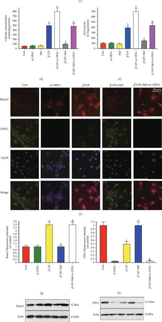
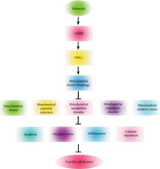
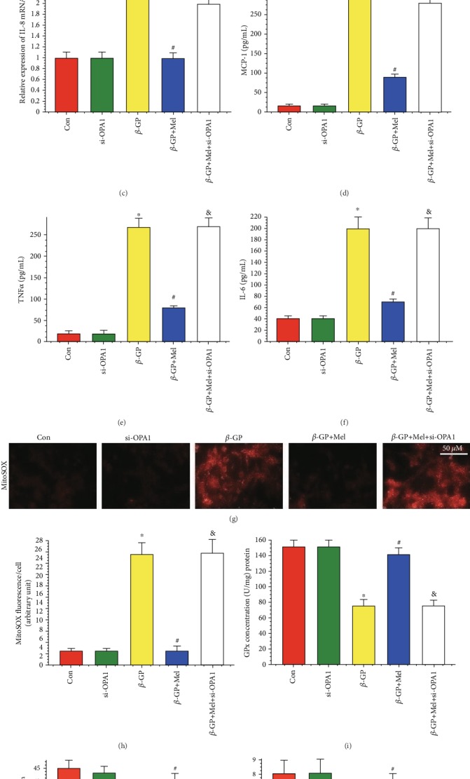
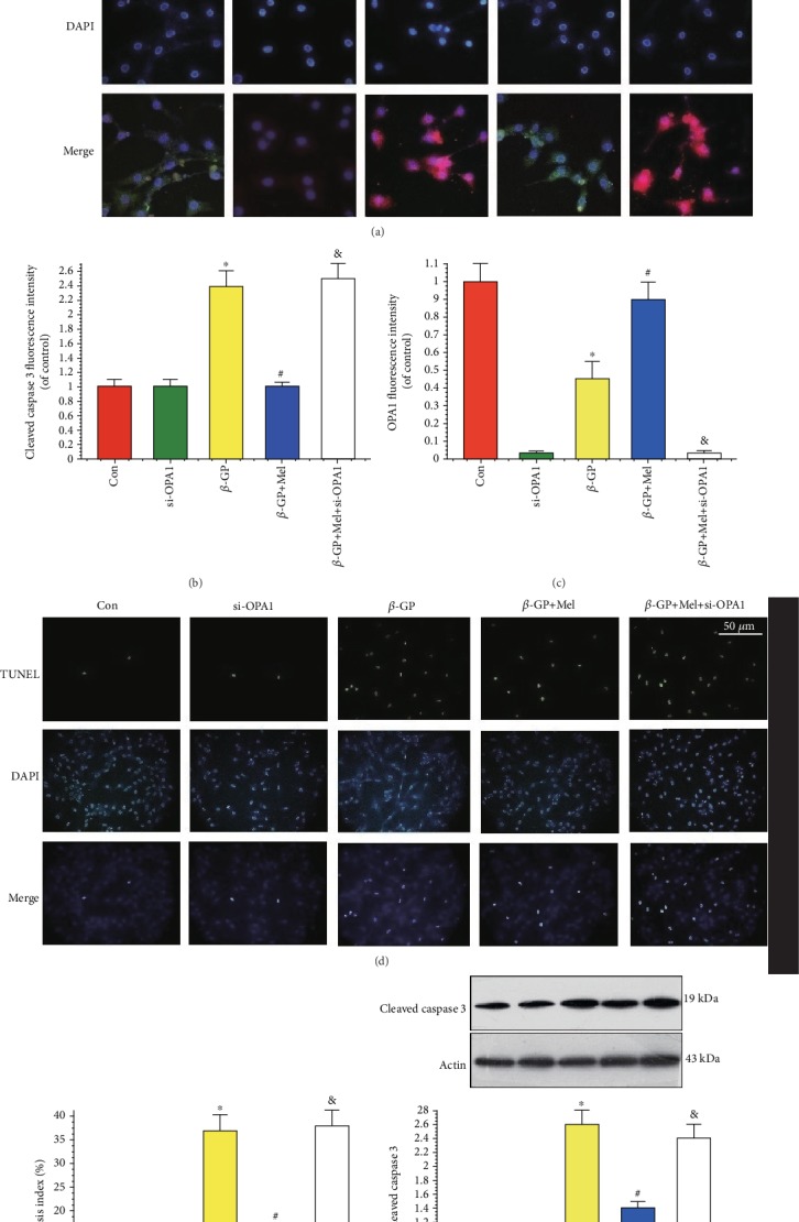
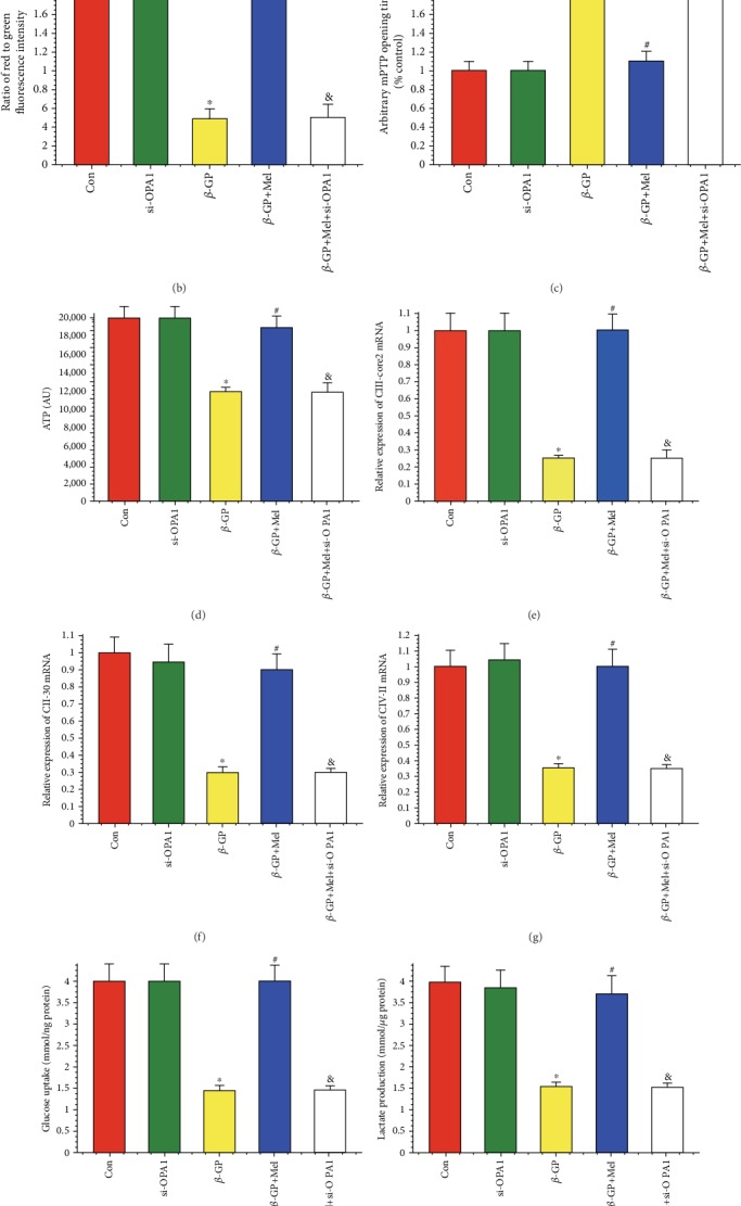
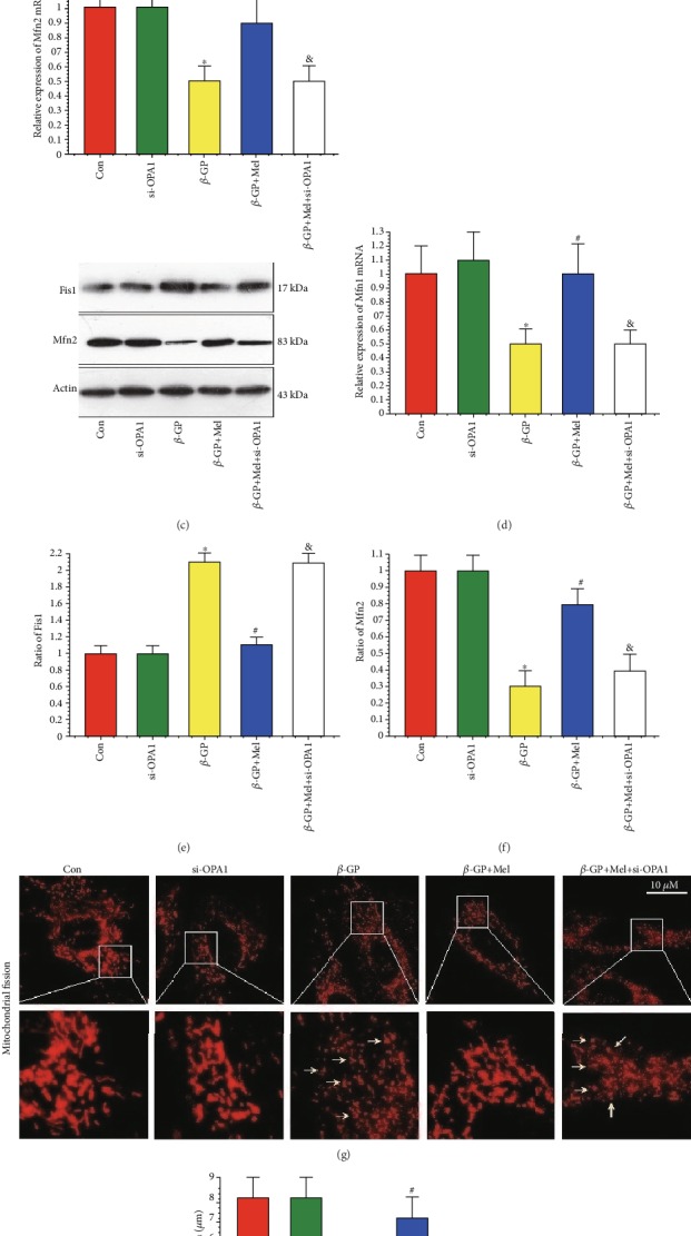
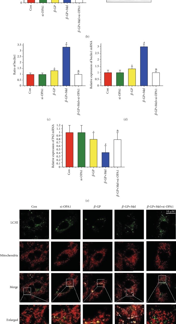
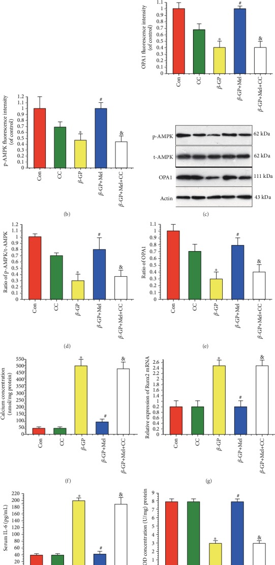
Mitochondrial fusion/mitophagy plays a role in cardiovascular calcification. Melatonin has been shown to protect against cardiovascular disease. This study sought to explore whether melatonin attenuates vascular calcification by regulating mitochondrial fusion/mitophagy via the AMP-activated protein kinase/optic atrophy 1 (AMPK/OPA1) signaling pathway. The effects of melatonin on vascular calcification were investigated in vascular smooth muscle cells (VSMCs). Calcium deposits were visualized by Alizarin Red S staining, while calcium content and alkaline phosphatase (ALP) activity were used to evaluate osteogenic differentiation. Western blots were used to measure expression of runt-related transcription factor 2 (Runx2), mitofusin 2 (Mfn2), mito-light chain 3 (mito-LC3) II, and cleaved caspase 3. Melatonin markedly reduced calcium deposition and ALP activity. Runx2 and cleaved caspase 3 were downregulated in response to melatonin, whereas Mfn2 and mito-LC3II were enhanced and accompanied by decreased mitochondrial superoxide levels. Melatonin also maintained mitochondrial function and promoted mitochondrial fusion/mitophagy via the OPA1 pathway. However, OPA1 deletion abolished the protective effects of melatonin on VSMC calcification. Melatonin treatment significantly increased p-AMPK and OPA1 protein expression, whereas treatment with compound C ablated the observed benefits of melatonin treatment. Collectively, our results demonstrate that melatonin protects VSMCs against calcification by promoting mitochondrial fusion/mitophagy via the AMPK/OPA1 pathway.
线粒体融合/自噬在心血管钙化中发挥作用。褪黑素已被证明可预防心血管疾病。本研究旨在探讨褪黑素是否通过调节 AMP 激活的蛋白激酶/视神经萎缩 1(AMPK/OPA1)信号通路的线粒体融合/自噬来减轻血管钙化。在血管平滑肌细胞(VSMCs)中研究了褪黑素对血管钙化的影响。通过茜素红 S 染色可视化钙沉积,而通过钙含量和碱性磷酸酶(ALP)活性来评估成骨分化。使用 Western blot 测定 runt 相关转录因子 2(Runx2)、融合蛋白 2(Mfn2)、线粒体轻链 3(mito-LC3)II 和裂解的半胱天冬酶 3 的表达。褪黑素明显减少钙沉积和 ALP 活性。褪黑素可下调 Runx2 和裂解的半胱天冬酶 3,而上调 Mfn2 和 mito-LC3II,并伴有线粒体超氧化物水平降低。褪黑素还通过 OPA1 途径维持线粒体功能并促进线粒体融合/自噬。然而,OPA1 缺失消除了褪黑素对 VSMC 钙化的保护作用。褪黑素处理显著增加了 p-AMPK 和 OPA1 蛋白表达,而用化合物 C 处理则消除了褪黑素处理的益处。总之,我们的结果表明,褪黑素通过 AMPK/OPA1 途径促进线粒体融合/自噬来保护 VSMCs 免受钙化。