Mulaosmanovic Emina, Lindblom Tobias U T, Bengtsson Marie, Windstam Sofia T, Mogren Lars, Marttila Salla, Stützel Hartmut, Alsanius Beatrix W
1Department of Biosystems and Technology, Microbial Horticulture Unit, Swedish University of Agricultural Sciences, PO Box 103, 23053 Alnarp, Sweden.
2Department of Crop Production Ecology, Plant Ecology Unit, Swedish University of Agricultural Sciences, PO Box 7043, 75007 Uppsala, Sweden.
Plant Methods. 2020 May 4;16:62. doi: 10.1186/s13007-020-00605-5. eCollection 2020.
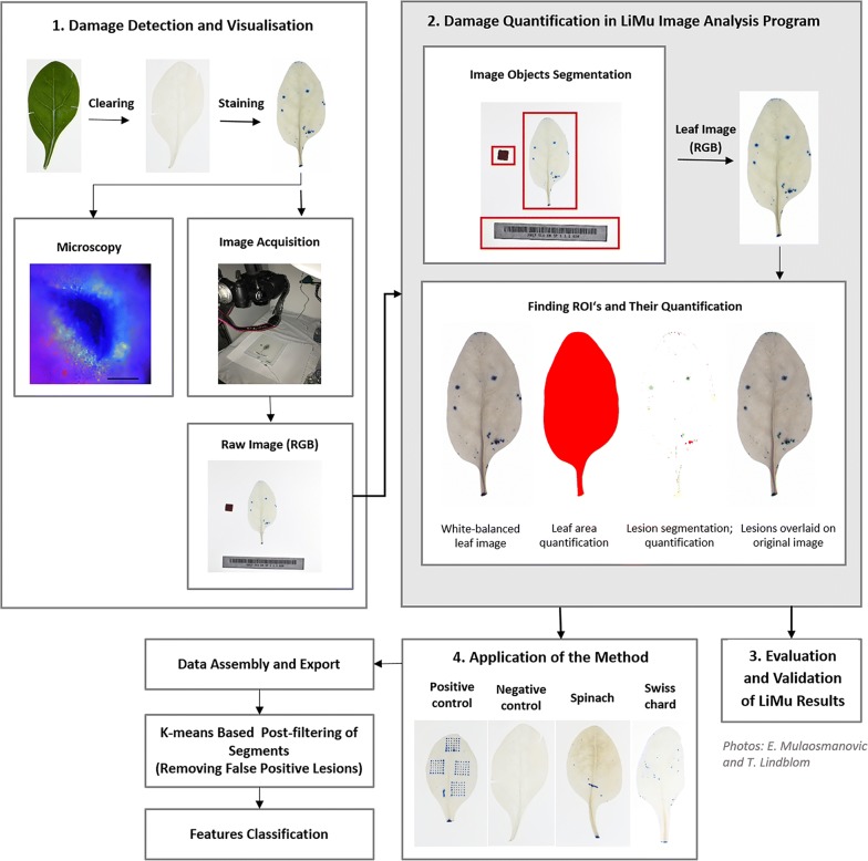
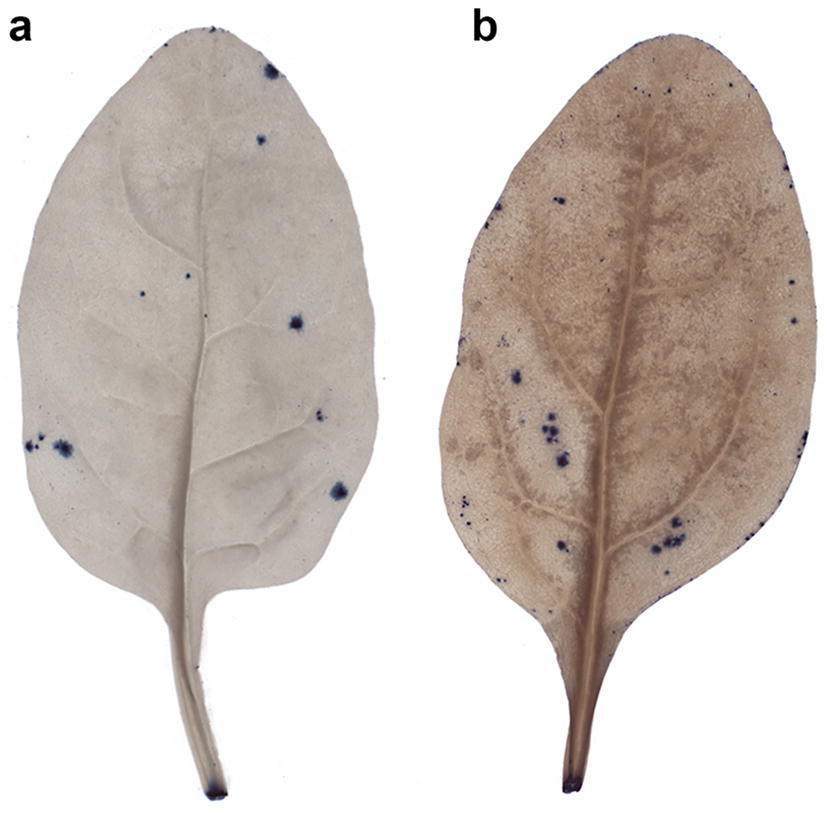
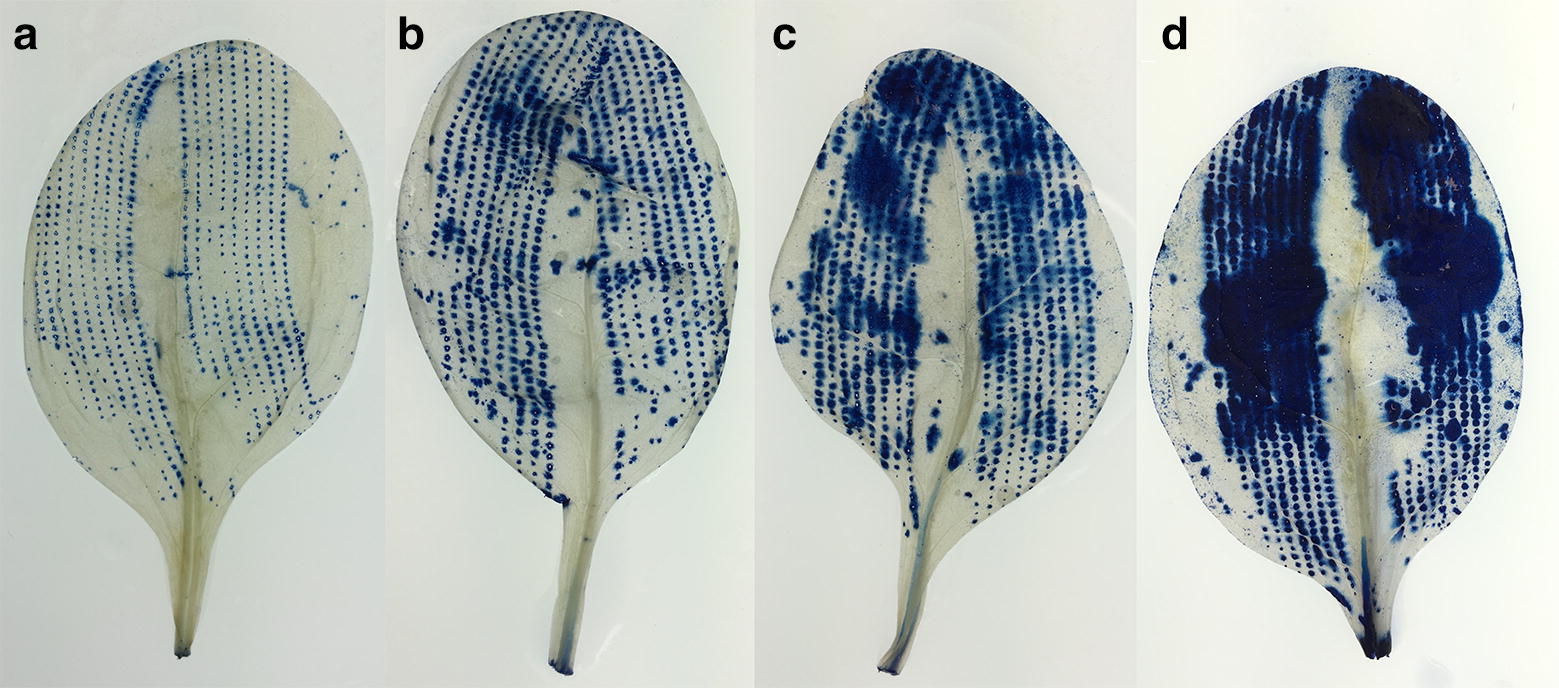
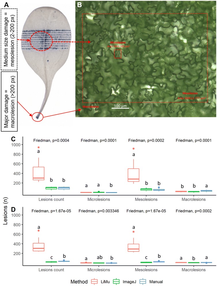
Field-grown leafy vegetables can be damaged by biotic and abiotic factors, or mechanically damaged by farming practices. Available methods to evaluate leaf tissue damage mainly rely on colour differentiation between healthy and damaged tissues. Alternatively, sophisticated equipment such as microscopy and hyperspectral cameras can be employed. Depending on the causal factor, colour change in the wounded area is not always induced and, by the time symptoms become visible, a plant can already be severely affected. To accurately detect and quantify damage on leaf scale, including microlesions, reliable differentiation between healthy and damaged tissue is essential. We stained whole leaves with trypan blue dye, which traverses compromised cell membranes but is not absorbed in viable cells, followed by automated quantification of damage on leaf scale.
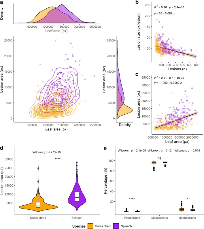
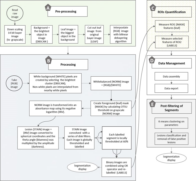
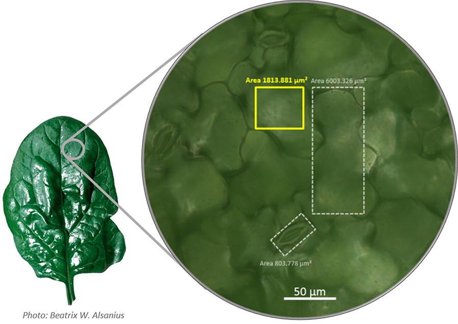
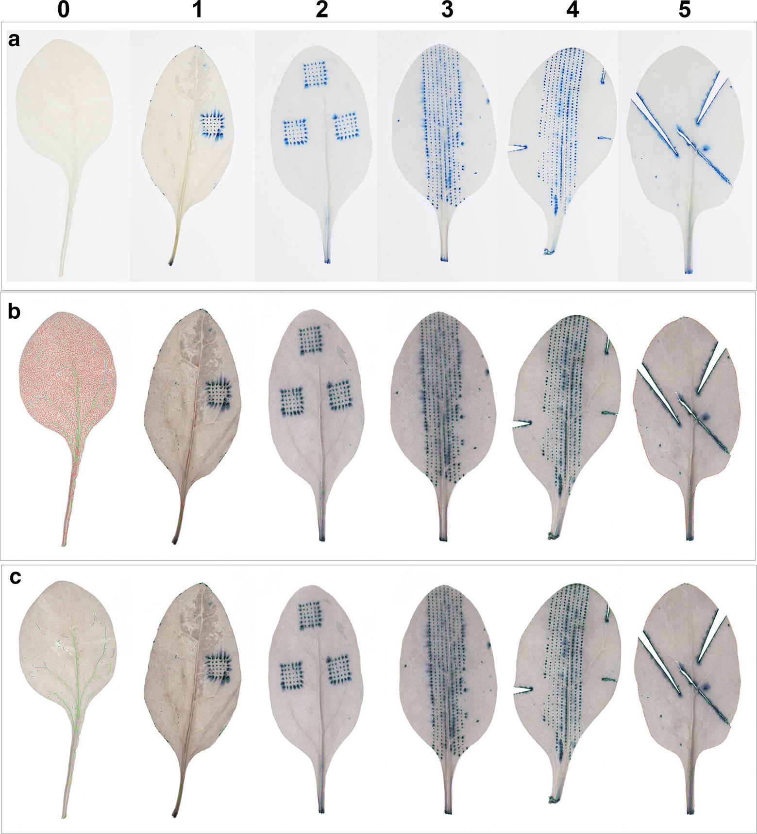
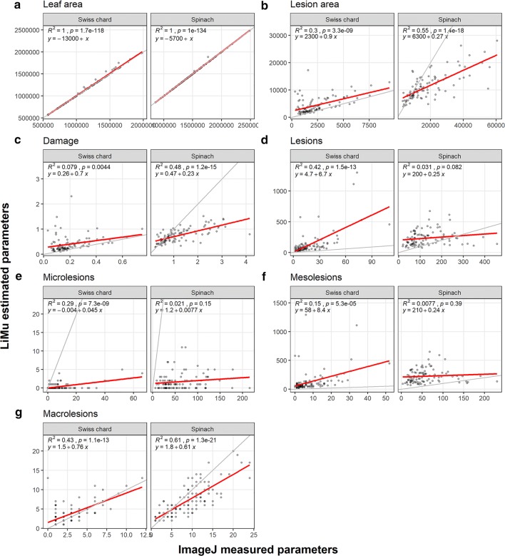
We present a robust, fast and sensitive method for leaf-scale visualisation, accurate automated extraction and measurement of damaged area on leaves of leafy vegetables. The image analysis pipeline we developed automatically identifies leaf area and individual stained (lesion) areas down to cell level. As proof of principle, we tested the methodology for damage detection and quantification on two field-grown leafy vegetable species, spinach and Swiss chard.
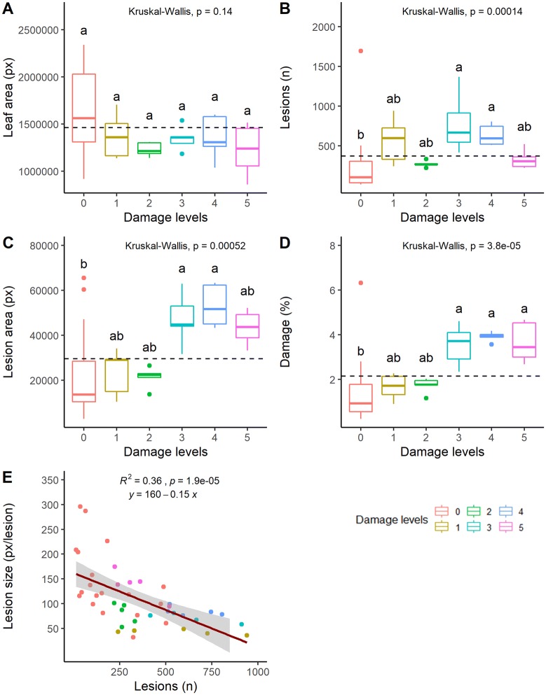
Our novel lesion quantification method can be used for detection of large (macro) or single-cell (micro) lesions on leaf scale, enabling quantification of lesions at any stage and without requiring symptoms to be in the visible spectrum. Quantifying the wounded area on leaf scale is necessary for generating prediction models for economic losses and produce shelf-life. In addition, risk assessments are based on accurate prediction of the relationship between leaf damage and infection rates by opportunistic pathogens and our method helps determine the severity of leaf damage at fine resolution.
田间种植的叶菜类蔬菜可能受到生物和非生物因素的损害,或因种植操作而受到机械损伤。现有的评估叶片组织损伤的方法主要依赖于健康组织和受损组织之间的颜色差异。另外,也可以使用显微镜和高光谱相机等精密设备。根据致病因素的不同,受伤区域并不总是会出现颜色变化,而且当症状可见时,植物可能已经受到严重影响。为了准确检测和量化叶片尺度上的损伤,包括微损伤,健康组织和受损组织之间的可靠区分至关重要。我们用台盼蓝染料对整片叶子进行染色,台盼蓝能穿过受损的细胞膜,但不会被活细胞吸收,然后自动量化叶片尺度上的损伤。
我们提出了一种强大、快速且灵敏的方法,用于叶菜类蔬菜叶片尺度上的可视化、准确的自动提取和受损面积测量。我们开发的图像分析流程能自动识别叶片面积以及直至细胞水平的单个染色(损伤)区域。作为原理验证,我们在两种田间种植的叶菜类蔬菜——菠菜和瑞士甜菜上测试了该损伤检测和量化方法。
我们新颖的损伤量化方法可用于检测叶片尺度上的大(宏观)损伤或单细胞(微观)损伤,能够在任何阶段对损伤进行量化,且无需症状处于可见光谱范围内。量化叶片尺度上的受伤面积对于生成经济损失和产品保质期的预测模型是必要的。此外,风险评估基于对叶片损伤与机会性病原体感染率之间关系的准确预测,而我们的方法有助于在高分辨率下确定叶片损伤的严重程度。