Mencucci Rita, Favuzza Eleonora, Marziali Elisa, Cennamo Michela, Mazzotta Cosimo, Lucenteforte Ersilia, Virgili Gianni, Rizzo Stanislao
1Eye Clinic, Department of Neuroscience, Psychology, Pharmacology and Child Health (NEUROFARBA), University of Florence, Eye Clinic, Largo Brambilla 3, 50134 Florence, Italy.
2Department of Medicine, Surgery and Neurosciences, Ophthalmology Unit, Siena University, Siena, Italy.
Eye Vis (Lond). 2020 May 6;7:25. doi: 10.1186/s40662-020-00191-6. eCollection 2020.
To compare the visual outcome and patients' satisfaction after ultrathin Descemet stripping automated endothelial keratoplasty (UT-DSAEK) and Descemet membrane endothelial keratoplasty (DMEK) performed on fellow eyes of the same patients.
In this retrospective study, the records of 18 pseudophakic patients affected by Fuchs endothelial dystrophy who underwent DMEK in one eye and UT-DSAEK in the fellow eye were reviewed. Best corrected visual acuity (BCVA), corneal pachymetry, keratometry, corneal aberrations, photopic and mesopic contrast sensitivity, and endothelial cell counts measured 12 months after surgery in either eye were analyzed and compared. The results of a satisfaction questionnaire were also reviewed.
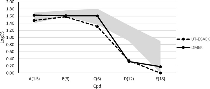
Twelve months after surgery, BCVA was not significantly different in UT-DSAEK and DMEK eyes (0.10 ± 0.04 and 0.07 ± 0.07 logMAR, respectively); at both 4- and 6 mm optical zones total and posterior corneal higher order aberrations (HOAs), posterior astigmatism and total coma were significantly lower after DMEK; BCVA in both groups was significantly correlated mainly with anterior corneal aberrations; contrast sensitivity was higher after DMEK especially in mesopic conditions and at medium spatial frequencies; the endothelial cell density was similar, although slightly higher in the UT-DSAEK group ( = 0.10). The satisfaction questionnaire showed that although patients were highly satisfied from both procedures, more than half of them preferred DMEK and reported a more comfortable and quicker postoperative recovery.
DMEK and UT-DSAEK showed no evidence of difference in terms of postoperative BCVA, although DMEK had a better performance in terms of contrast sensitivity, posterior corneal aberrations and overall patient satisfaction.
比较在同一患者的对侧眼上进行超薄Descemet膜剥除自动内皮角膜移植术(UT-DSAEK)和Descemet膜内皮角膜移植术(DMEK)后的视觉效果和患者满意度。
在这项回顾性研究中,回顾了18例患有Fuchs内皮营养不良的假晶状体患者的记录,这些患者一只眼睛接受了DMEK,对侧眼睛接受了UT-DSAEK。分析并比较了任何一只眼睛术后12个月时的最佳矫正视力(BCVA)、角膜测厚、角膜曲率测量、角膜像差、明视觉和暗视觉对比敏感度以及内皮细胞计数。还回顾了满意度调查问卷的结果。
术后12个月,UT-DSAEK组和DMEK组的BCVA无显著差异(分别为0.10±0.04和0.07±0.07 logMAR);在4毫米和6毫米光学区,DMEK术后总的和后角膜高阶像差(HOAs)、后散光和总彗差均显著降低;两组的BCVA主要与前角膜像差显著相关;DMEK术后对比敏感度更高,尤其是在暗视觉条件下和中等空间频率时;内皮细胞密度相似,尽管UT-DSAEK组略高(P = 0.10)。满意度调查问卷显示,尽管患者对两种手术都高度满意,但超过一半的患者更喜欢DMEK,并报告术后恢复更舒适、更快。
DMEK和UT-DSAEK在术后BCVA方面没有差异的证据,尽管DMEK在对比敏感度、后角膜像差和患者总体满意度方面表现更好。