,.
Invest Ophthalmol Vis Sci. 2020 May 11;61(5):8. doi: 10.1167/iovs.61.5.8.
The purpose of this study was to compare perfusion parameters of the parafovea with scans outside the parafovea to find an area most susceptible to changes secondary to diabetic retinopathy (DR).
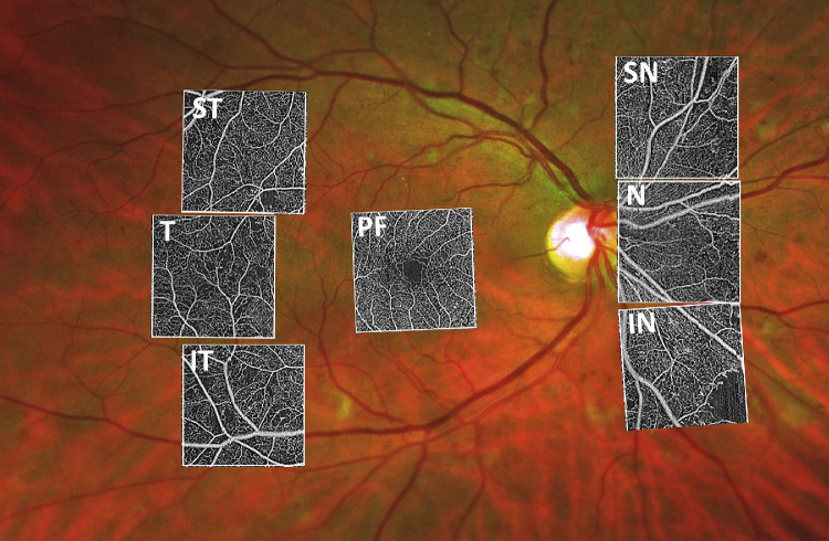
Patients with different DR severity levels as well as controls were included in this cross-sectional clinical trial. Seven standardized 3 × 3 mm areas were recorded with Swept Source Optical Coherence Tomography Angiography: one centered on the fovea, three were temporal to the fovea, and three nasally to the optic disc. The capillary perfusion density (PD) of the superficial capillary complex (SCC) and deep capillary complex (DCC) as well as the fractal dimension (FD) were generated. Statistical analyses were done with R software.
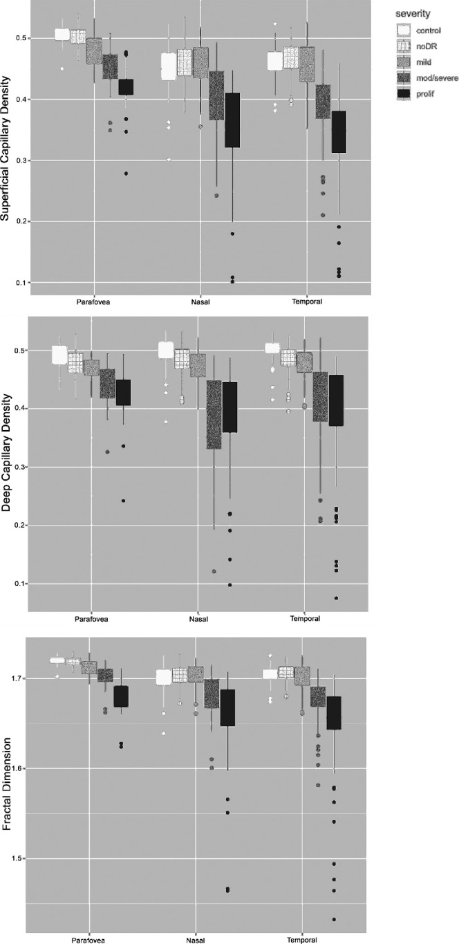
One hundred ninety-two eyes (33 controls, 51 no-DR, 41 mild DR, 37 moderate/severe DR, and 30 proliferative DR), of which 105 patients with diabetes and 25 healthy controls were included (59 ± 15 years; 62 women). Mean PD of the DCC was significantly less in patients without DR (parafovea = 0.48 ± 0.03; temporal = 0.48 ± 0.02; and nasal = 0.48 ± 0.03) compared to controls (parafovea = 0.49 ± 0.02; temporal = 0.50 ± 0.02; and nasal = 0.50 ± 0.03). With increasing DR severity, PD and FD of the SCC and DCC further decreased.
Capillary perfusion of the retina is affected early by diabetes. PD of the DCC was significantly reduced in patients with diabetes who did not have any clinical signs of DR. The capillary network outside the parafovea was more susceptible to capillary perfusion deficits compared to the capillaries close to the fovea.
clinicaltrial.gov, NCT03765112, https://clinicaltrials.gov/ct2/show/NCT03765112?term=NCT03765112&rank=1.
本研究旨在比较黄斑旁区与黄斑外区的灌注参数,以找到对糖尿病视网膜病变(DR)继发变化最敏感的区域。
本横断面临床试验纳入了不同 DR 严重程度的患者和对照者。使用扫频源光学相干断层扫描血管造影术记录了 7 个标准化的 3×3mm 区域:一个位于中心凹,三个位于中心凹颞侧,三个位于视盘鼻侧。生成了浅层毛细血管复合体(SCC)和深层毛细血管复合体(DCC)的毛细血管灌注密度(PD)和分形维数(FD)。统计分析使用 R 软件进行。
共纳入 192 只眼(33 只对照者,51 只无 DR 者,41 只轻度 DR 者,37 只中度/重度 DR 者和 30 只增殖性 DR 者),其中包括 105 例糖尿病患者和 25 名健康对照者(59±15 岁,62 名女性)。无 DR 患者的 DCC 平均 PD 明显低于对照组(黄斑旁 = 0.48±0.03;颞侧 = 0.48±0.02;鼻侧 = 0.48±0.03)(黄斑旁 = 0.49±0.02;颞侧 = 0.50±0.02;鼻侧 = 0.50±0.03)。随着 DR 严重程度的增加,SCC 和 DCC 的 PD 和 FD 进一步降低。
糖尿病早期即影响视网膜毛细血管灌注。无任何 DR 临床征象的糖尿病患者 DCC 的 PD 明显降低。与靠近黄斑的毛细血管相比,黄斑旁区的毛细血管网络更容易出现毛细血管灌注不足。
clinicaltrial.gov,NCT03765112,https://clinicaltrials.gov/ct2/show/NCT03765112?term=NCT03765112&rank=1.