Wofford Andrew, Bow Austin, Newby Steven, Brooks Seth, Rodriguez Rachel, Masi Tom, Stephenson Stacy, Gotcher Jack, Anderson David E, Campbell Josh, Dhar Madhu
Department of Biochemistry and Cellular and Molecular Biology, College of Arts and Sciences, University of Tennessee, Knoxville, TN 37916, USA.
Department of Large Animal Clinical Sciences, College of Veterinary Medicine, University of Tennessee, Knoxville, TN 37996, USA.
Stem Cells Int. 2020 Jan 13;2020:8142938. doi: 10.1155/2020/8142938. eCollection 2020.
Due to restorative concerns, bone regenerative therapies have garnered much attention in the field of human oral/maxillofacial surgery. Current treatments using autologous and allogenic bone grafts suffer from inherent challenges, hence the ideal bone replacement therapy is yet to be found. Establishing a model by which MSCs can be placed in a clinically acceptable bone defect to promote bone healing will prove valuable to oral/maxillofacial surgeons.
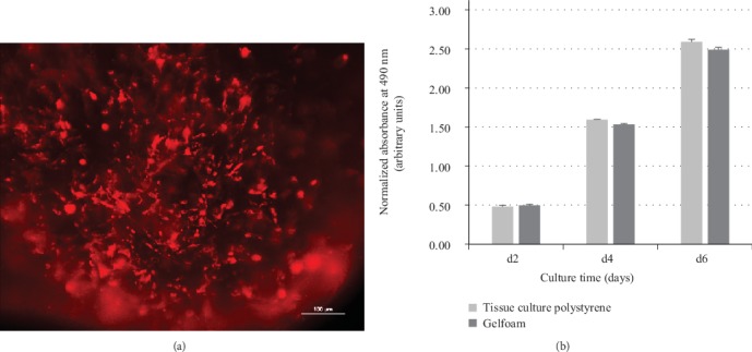
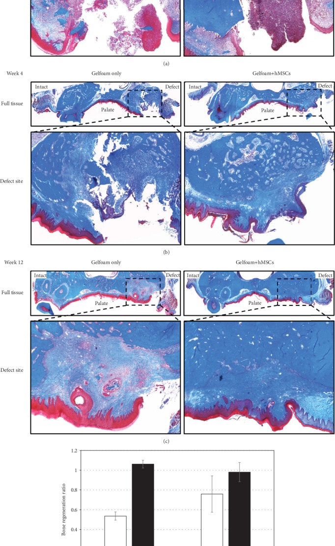
Human adipose tissue-derived MSCs were seeded onto Gelfoam® and their viability, proliferation, and osteogenic differentiation was evaluated . Subsequently, the construct was implanted in a rat maxillary alveolar bone defect to assess bone healing and regeneration.
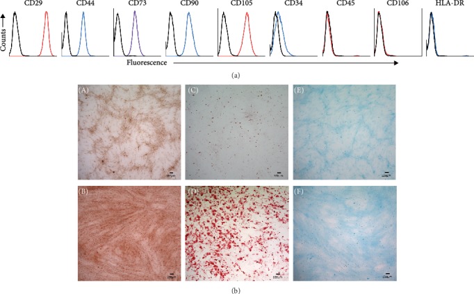
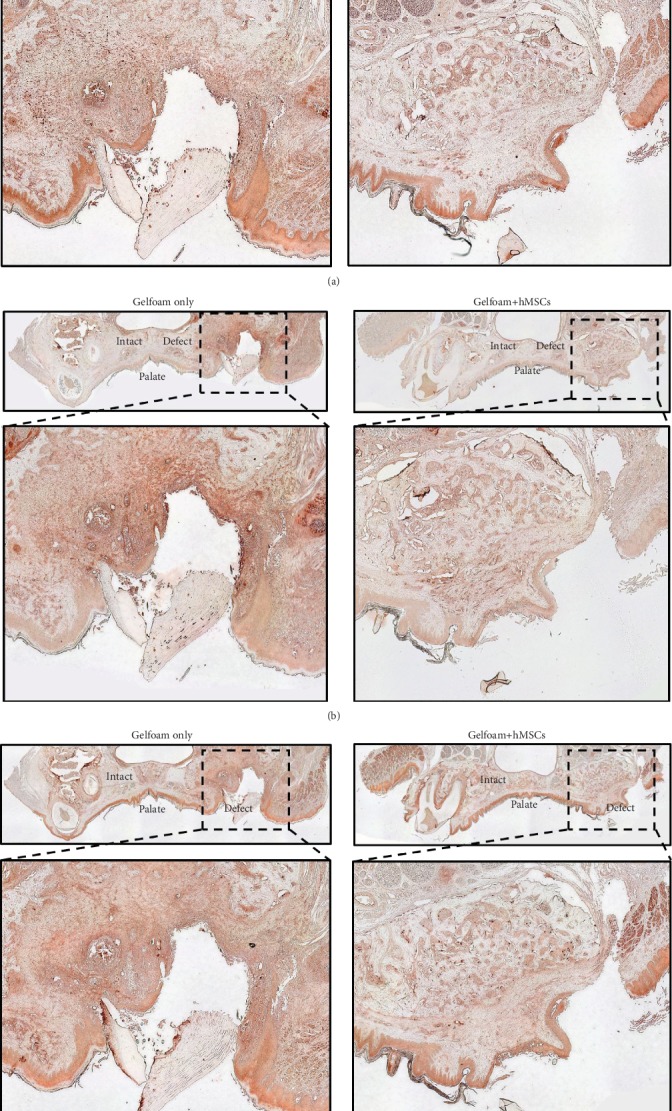
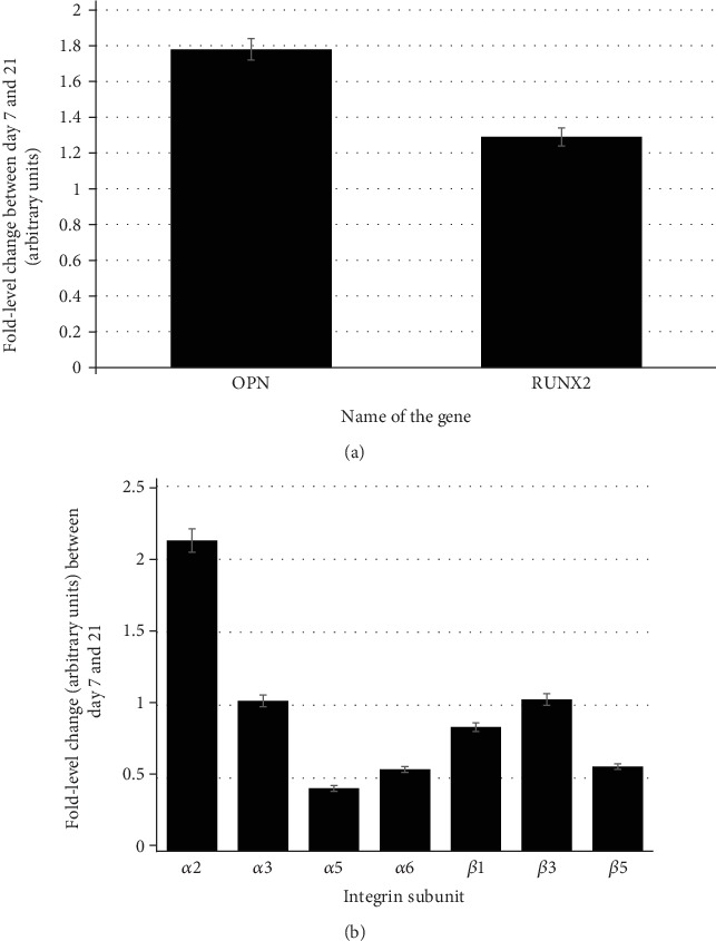
Human MSCs were adhered, proliferated, and uniformly distributed, and underwent osteogenic differentiation on Gelfoam®, comparable with the tissue culture surface. Data confirmed that Gelfoam® could be used as a scaffold for cell attachment and a delivery vehicle to implant MSCs . Histomorphometric analyses of bones harvested from rats treated with hMSCs showed statistically significant increase in collagen/early bone formation, with cells positive for osteogenic and angiogenic markers in the defect site. This pattern was visible as early as 4 weeks post treatment.
Xenogenically implanted human MSCs have the potential to heal an alveolar tooth defect in rats. Gelfoam®, a commonly used clinical biomaterial, can serve as a scaffold to deliver and maintain MSCs to the defect site. Translating this strategy to preclinical animal models provides hope for bone tissue engineering.
出于修复方面的考虑,骨再生疗法在人类口腔/颌面外科领域备受关注。目前使用自体和异体骨移植的治疗方法存在固有挑战,因此理想的骨替代疗法尚未找到。建立一个模型,使间充质干细胞(MSCs)能够置于临床上可接受的骨缺损处以促进骨愈合,这对口腔/颌面外科医生将具有重要价值。
将人脂肪组织来源的间充质干细胞接种到明胶海绵上,并评估其活力、增殖和骨向分化情况。随后,将构建物植入大鼠上颌牙槽骨缺损处,以评估骨愈合和再生情况。
人间充质干细胞在明胶海绵上黏附、增殖并均匀分布,且在明胶海绵上发生骨向分化,与组织培养表面相当。数据证实明胶海绵可作为细胞附着的支架和植入间充质干细胞的载体。对接受人源间充质干细胞治疗的大鼠所取骨骼进行组织形态计量学分析显示,胶原/早期骨形成有统计学意义的增加,缺损部位有成骨和血管生成标志物阳性的细胞。这种模式在治疗后4周就可见到。
异种植入的人源间充质干细胞有治愈大鼠牙槽骨缺损的潜力。明胶海绵,一种常用的临床生物材料,可作为将间充质干细胞递送至缺损部位并维持其存在的支架。将该策略转化至临床前动物模型为骨组织工程带来了希望。