Kühn Sonja, Bergqvist John, Gil Magdalena, Valenzuela Camila, Barrio Laura, Lebreton Stéphanie, Zurzolo Chiara, Enninga Jost
Institut Pasteur, Department of Cell Biology and Infection, Dynamics of Host-Pathogen Interactions Unit, 25 Rue du Dr. Roux, 75015 Paris, France; CNRS UMR3691, 25 Rue du Dr. Roux, 75015 Paris, France.
Institut Pasteur, Department of Cell Biology and Infection, Membrane Trafficking and Pathogenesis Unit, 28 Rue du Dr. Roux, 75015 Paris, France.
Cell Rep. 2020 May 12;31(6):107638. doi: 10.1016/j.celrep.2020.107638.
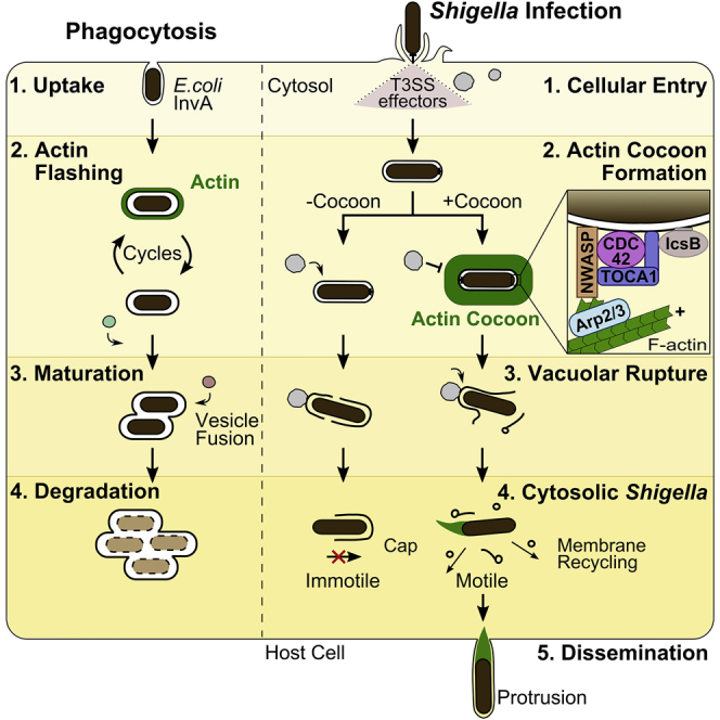
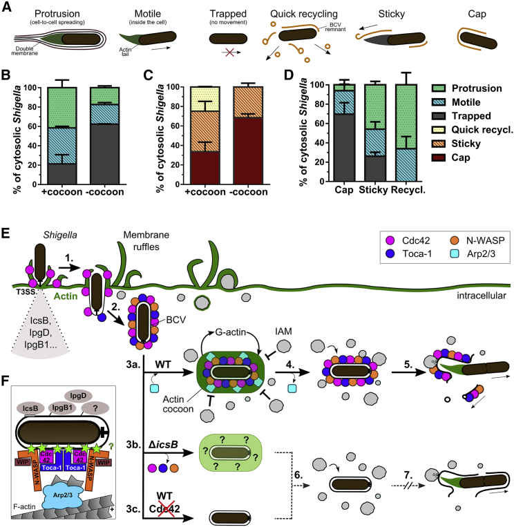
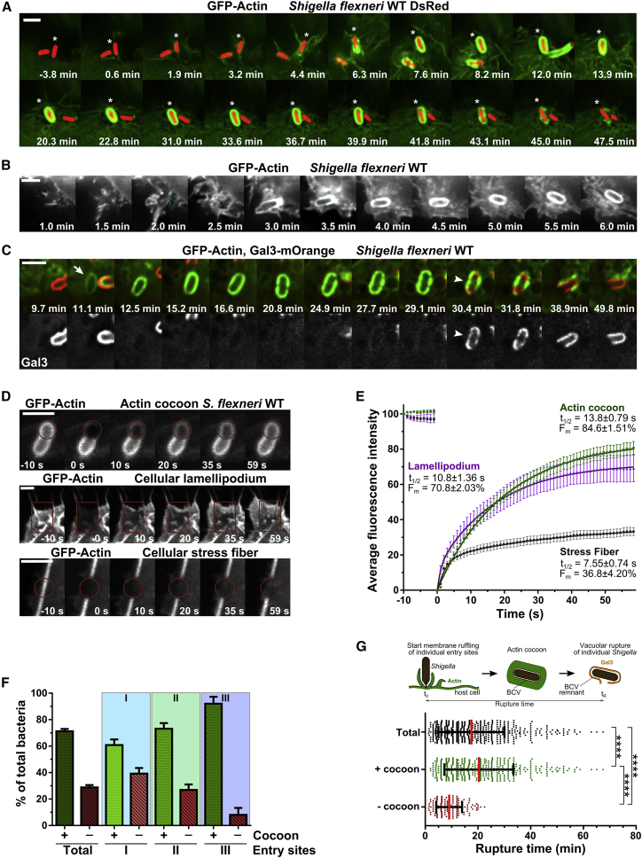
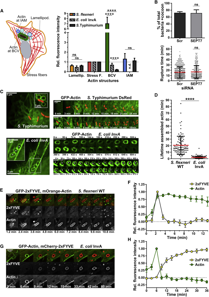
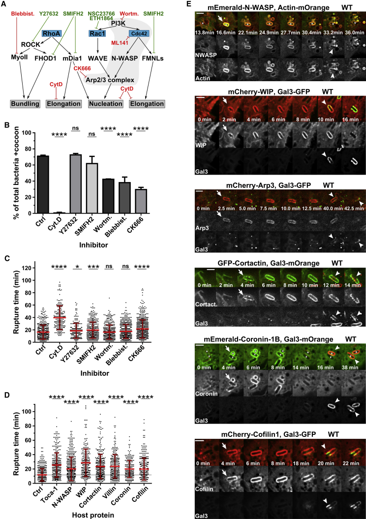
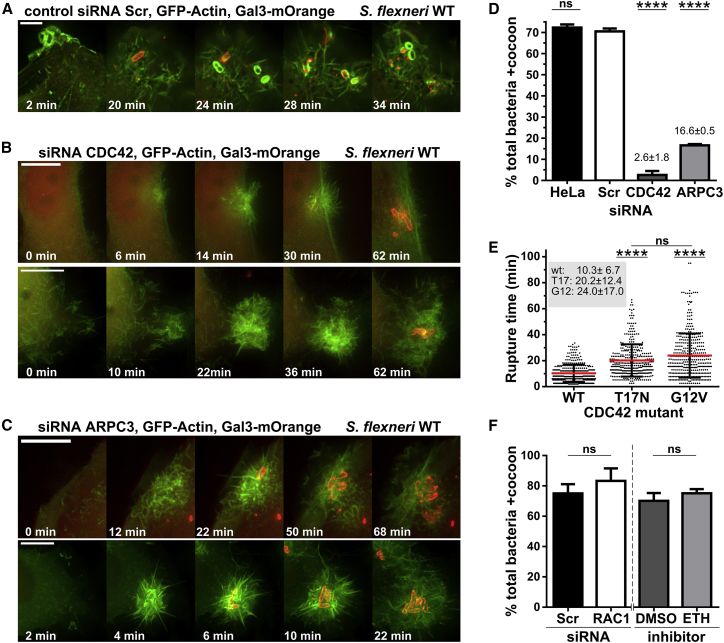
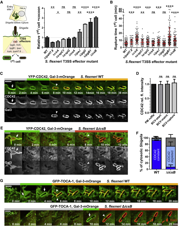
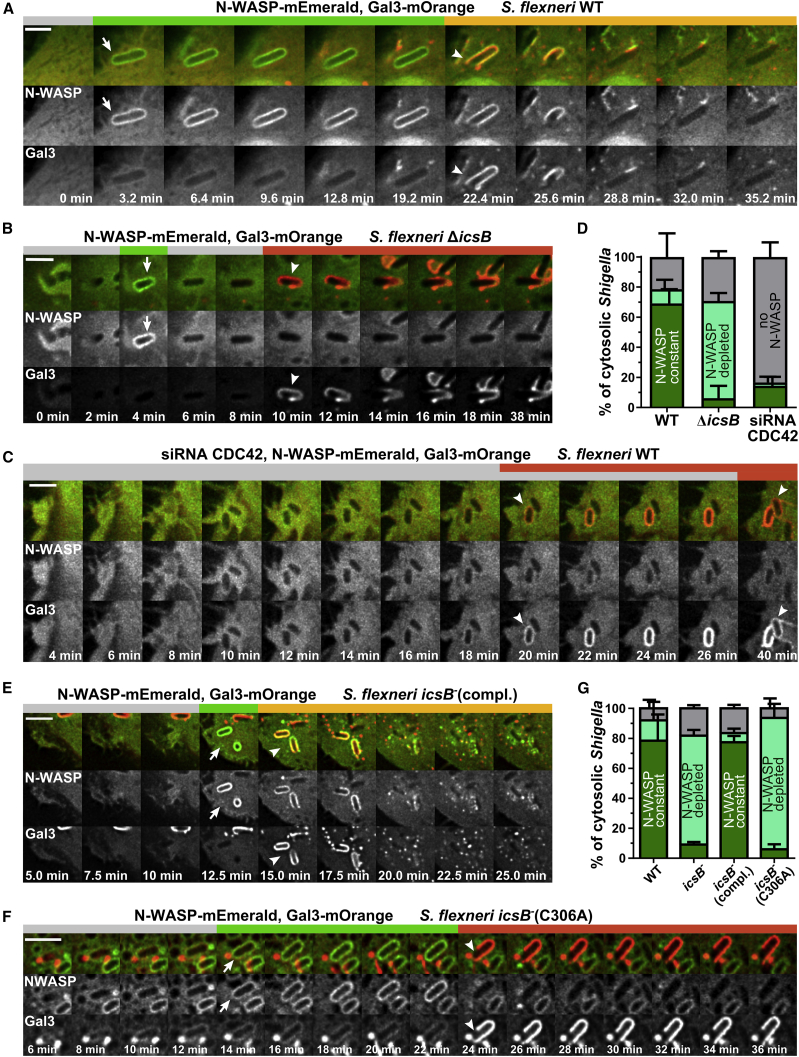
The enteroinvasive bacterium Shigella flexneri forces its uptake into non-phagocytic host cells through the translocation of T3SS effectors that subvert the actin cytoskeleton. Here, we report de novo actin polymerization after cellular entry around the bacterium-containing vacuole (BCV) leading to the formation of a dynamic actin cocoon. This cocoon is thicker than any described cellular actin structure and functions as a gatekeeper for the cytosolic access of the pathogen. Host CDC42, TOCA-1, N-WASP, WIP, the Arp2/3 complex, cortactin, coronin, and cofilin are recruited to the actin cocoon. They are subverted by T3SS effectors, such as IpgD, IpgB1, and IcsB. IcsB immobilizes components of the actin polymerization machinery at the BCV dependent on its fatty acyltransferase activity. This represents a unique microbial subversion strategy through localized entrapment of host actin regulators causing massive actin assembly. We propose that the cocoon promotes subsequent invasion steps for successful Shigella infection.
侵袭性细菌福氏志贺菌通过III型分泌系统(T3SS)效应蛋白的转运,迫使自身被非吞噬性宿主细胞摄取,这些效应蛋白会破坏肌动蛋白细胞骨架。在此,我们报告了细菌进入细胞后,围绕含菌液泡(BCV)发生的肌动蛋白从头聚合,导致形成动态的肌动蛋白茧。这个茧比任何已描述的细胞肌动蛋白结构都要厚,并且作为病原体胞质进入的守门人发挥作用。宿主的CDC42、TOCA-1、N-WASP、WIP、Arp2/3复合物、皮层肌动蛋白、冠蛋白和丝切蛋白被招募到肌动蛋白茧中。它们被T3SS效应蛋白(如IpgD、IpgB1和IcsB)破坏。IcsB根据其脂肪酰基转移酶活性,将肌动蛋白聚合机制的成分固定在BCV处。这代表了一种独特的微生物破坏策略,即通过局部捕获宿主肌动蛋白调节因子来导致大量肌动蛋白组装。我们提出,这个茧促进了福氏志贺菌成功感染的后续侵袭步骤。