Department of Orthopaedic Surgery, University of Wuerzburg, Koenig-Ludwig-Haus, Brettreichstr. 11, 97074, Wuerzburg, Germany.
Bernhard-Heine-Center for Locomotion Research, University of Wuerzburg, Wuerzburg, Germany.
BMC Musculoskelet Disord. 2020 May 13;21(1):297. doi: 10.1186/s12891-020-03340-z.
While multiple in vitro studies examined mesenchymal stromal cells (MSCs) derived from bone marrow or hyaline cartilage, there is little to no data about the presence of MSCs in the joint capsule or the ligamentum capitis femoris (LCF) of the hip joint. Therefore, this in vitro study examined the presence and differentiation potential of MSCs isolated from the bone marrow, arthritic hyaline cartilage, the LCF and full-thickness samples of the anterior joint capsule of the hip joint.
MSCs were isolated and multiplied in adherent monolayer cell cultures. Osteogenesis and adipogenesis were induced in monolayer cell cultures for 21 days using a differentiation medium containing specific growth factors, while chondrogenesis in the presence of TGF-ß1 was performed using pellet-culture for 27 days. Control cultures were maintained for comparison over the same duration of time. The differentiation process was analyzed using histological and immunohistochemical stainings as well as semiquantitative RT-PCR for measuring the mean expression levels of tissue-specific genes.
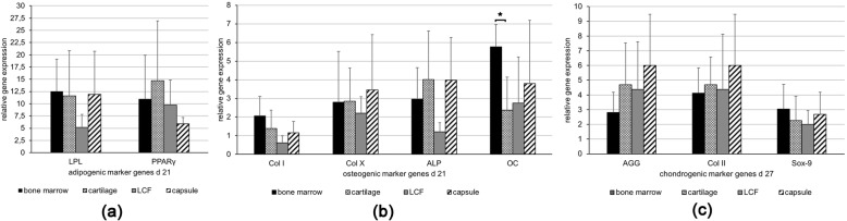
This in vitro research showed that the isolated cells from all four donor tissues grew plastic-adherent and showed similar adipogenic and osteogenic differentiation capacity as proven by the histological detection of lipid droplets or deposits of extracellular calcium and collagen type I. After 27 days of chondrogenesis proteoglycans accumulated in the differentiated MSC-pellets from all donor tissues. Immunohistochemical staining revealed vast amounts of collagen type II in all differentiated MSC-pellets, except for those from the LCF. Interestingly, all differentiated MSCs still showed a clear increase in mean expression of adipogenic, osteogenic and chondrogenic marker genes. In addition, the examination of an exemplary selected donor sample revealed that cells from all four donor tissues were clearly positive for the surface markers CD44, CD73, CD90 and CD105 by flow cytometric analysis.
This study proved the presence of MSC-like cells in all four examined donor tissues of the hip joint. No significant differences were observed during osteogenic or adipogenic differentiation depending on the source of MSCs used. Further research is necessary to fully determine the tripotent differentiation potential of cells isolated from the LCF and capsule tissue of the hip joint.
虽然有多项体外研究检查了骨髓或透明软骨来源的间充质基质细胞(MSCs),但关于髋关节关节囊或股骨头韧带(LCF)中 MSCs 的存在几乎没有数据。因此,这项体外研究检查了从骨髓、关节炎透明软骨、LCF 和髋关节前关节囊全层样本中分离的 MSCs 的存在和分化潜能。
MSCs 被分离并在贴壁单层细胞培养物中增殖。使用含有特定生长因子的分化培养基在单层细胞培养物中诱导成骨和成脂分化 21 天,而在 TGF-β1 存在的情况下使用微球培养物进行软骨分化 27 天。为了进行比较,对照培养物在相同的时间段内保持培养。使用组织学和免疫组织化学染色以及半定量 RT-PCR 分析分化过程,以测量组织特异性基因的平均表达水平。
这项体外研究表明,从所有四个供体组织分离的细胞呈塑料贴壁生长,并具有相似的成脂和成骨分化能力,这可以通过检测细胞内脂质滴或细胞外钙和 I 型胶原的沉积来证明。经过 27 天的软骨分化,来自所有供体组织的分化 MSC 微球中积累了蛋白聚糖。免疫组织化学染色显示,除了来自 LCF 的 MSC 微球外,所有分化的 MSC 微球中都有大量的 II 型胶原。有趣的是,所有分化的 MSC 仍表现出脂肪形成、成骨和软骨形成标记基因的明显增加。此外,对一个典型的供体样本的检查表明,来自四个供体组织的细胞在流式细胞术分析中均明显为 CD44、CD73、CD90 和 CD105 表面标记物阳性。
这项研究证明了髋关节四个检查供体组织中均存在 MSC 样细胞。在成骨或成脂分化过程中,没有观察到 MSCs 来源的明显差异。需要进一步的研究来完全确定从髋关节 LCF 和关节囊组织中分离的细胞的三向分化潜能。