Medical Biology Department, School of Medicine, İstanbul Medipol University, İstanbul, Turkey.
Regenerative and Restorative Medicine Research Center (REMER), Research Institute for Health Sciences and Technologies (SABITA), İstanbul Medipol University, İstanbul, Turkey.
PLoS One. 2020 May 14;15(5):e0228510. doi: 10.1371/journal.pone.0228510. eCollection 2020.
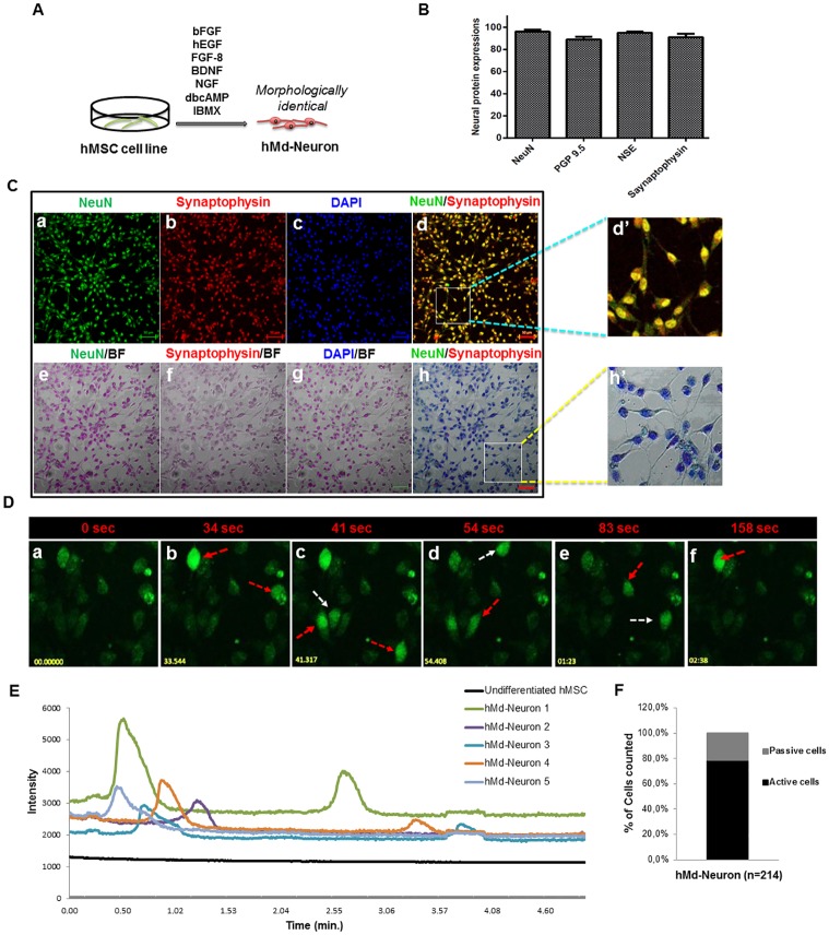
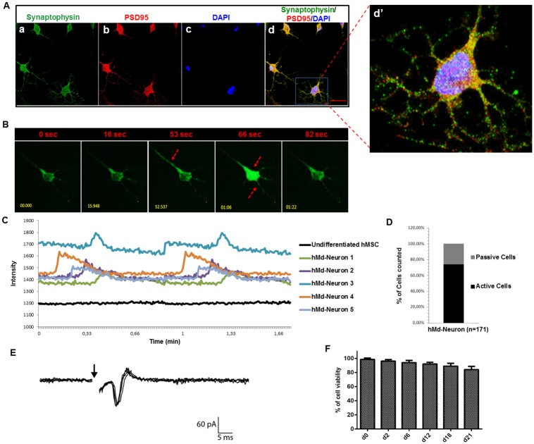
Mesenchymal stem cells have the ability to transdifferentiate into neurons and therefore one of the potential adult stem cell source for neuronal tissue regeneration applications and understanding neurodevelopmental processes. In many studies on human mesenchymal stem cell (hMSC) derived neurons, success in neuronal differentiation was limited to neuronal protein expressions which is not statisfactory in terms of neuronal activity. Established neuronal networks seen in culture have to be investigated in terms of synaptic signal transmission ability to develop a culture model for human neurons and further studying the mechanism of neuronal differentiation and neurological pathologies. Accordingly, in this study, we analysed the functionality of bone marrow hMSCs differentiated into neurons by a single step cytokine-based induction protocol. Neurons from both primary hMSCs and hMSC cell line displayed spontaneous activity (≥75%) as demonstrated by Ca++ imaging. Furthermore, when electrically stimulated, hMSC derived neurons (hMd-Neurons) matched the response of a typical neuron in the process of maturation. Our results reveal that a combination of neuronal inducers enhance differentiation capacity of bone marrow hMSCs into high yielding functional neurons with spontaneous activity and mature into electrophysiologically active state. Conceptually, we suggest these functional hMd-Neurons to be used as a tool for disease modelling of neuropathologies and neuronal differentiation studies.
间充质干细胞具有向神经元转分化的能力,因此是神经元组织再生应用和理解神经发育过程的潜在成体干细胞来源之一。在许多关于人骨髓间充质干细胞(hMSC)衍生神经元的研究中,神经元分化的成功仅限于神经元蛋白表达,这在神经元活性方面并不令人满意。在培养物中观察到的成熟神经元网络必须在突触信号传递能力方面进行研究,以开发用于人类神经元的培养模型,并进一步研究神经元分化和神经病理学的机制。因此,在这项研究中,我们通过基于细胞因子的一步诱导方案分析了分化为神经元的骨髓 hMSC 的功能。通过 Ca++成像显示,来自原代 hMSC 和 hMSC 细胞系的神经元均显示出自发性活动(≥75%)。此外,当电刺激时,hMSC 衍生的神经元(hMd-Neurons)在成熟过程中与典型神经元的反应相匹配。我们的结果表明,神经元诱导剂的组合增强了骨髓 hMSC 向具有自发性活动和成熟为电生理活性状态的高产量功能性神经元的分化能力。从概念上讲,我们建议将这些功能性 hMd-Neurons 用作神经病理学疾病模型和神经元分化研究的工具。