Royal (Dick) School of Veterinary Studies and The Roslin Institute, The University of Edinburgh, Midlothian, United Kingdom.
MRC Centre for Regenerative Medicine, The University of Edinburgh, Edinburgh BioQuarter, Edinburgh, United Kingdom.
J Vet Intern Med. 2020 Jul;34(4):1454-1463. doi: 10.1111/jvim.15792. Epub 2020 May 14.
Middle ear effusion is common in brachycephalic dogs with similarities to otitis media with effusion in children. Association with the cranial and eustachian tube morphology and bacterial infection is suspected in both species.
HYPOTHESIS/OBJECTIVES: To determine cytological and bacteriological features of middle ear effusions in dogs, provide information on histological features, and further assess the dog as a model of the human disease.
Sixteen live dogs, 3 postmortem cases of middle ear effusion, and 2 postmortem controls.
Prospective; clinical investigation using computed tomography, magnetic resonance imaging, video-otoscopy, myringotomy; cytological assessment of 30 and bacteriology of 28 effusions; histology and immunohistochemistry (CD3 for T-lymphocytes, Pax5 for B lymphocytes and MAC387 for macrophages) of 10 middle ear sections.
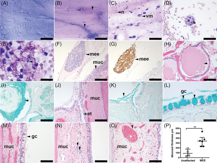
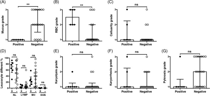
Effusions were associated with neurological deficits in 6/16 (38%) and concurrent atopic dermatitis and otitis externa in 9/16 (56%) of live cases. Neutrophils and macrophages predominated on cytology (median 60 [range 2%-95.5%] and 27 [2%-96.5%]) whether culture of effusions was positive or not. In histology sections, the mucosa was thickened in affected dogs but submucosal gland dilatation occurred in affected and unaffected dogs. There was no bacterial growth from 22/28 (79%) of effusions. Bacteria isolated from the other 6 (21%) were predominantly Staphylococcus pseudintermedius (4/6, 67%).
Clinical, morphological, and cytological findings in middle ear effusions of dogs and people suggest similar pathogeneses. Middle ear effusion of dogs could be a useful model of human otitis media with effusion. Such comparisons can improve understanding and management across species.
中耳积液在短头犬中很常见,与儿童分泌性中耳炎有相似之处。怀疑这两种物种的颅腔和咽鼓管形态以及细菌感染与之相关。
假设/目的:确定犬中耳积液的细胞学和细菌学特征,提供组织学特征信息,并进一步评估犬作为人类疾病模型的作用。
16 只活犬、3 例尸检中耳积液病例和 2 例尸检对照。
前瞻性;使用计算机断层扫描、磁共振成像、视频耳镜检查、鼓膜切开术进行临床研究;对 30 例积液进行细胞学评估,对 28 例积液进行细菌学评估;对 10 个中耳切片进行组织学和免疫组织化学(CD3 用于 T 淋巴细胞、Pax5 用于 B 淋巴细胞和 MAC387 用于巨噬细胞)分析。
在 16 例活犬中,6/16 (38%)伴有神经功能缺损,9/16 (56%)伴有特应性皮炎和外耳炎。无论积液培养是否为阳性,细胞学检查中以中性粒细胞和巨噬细胞为主(中位数分别为 60%[范围 2%-95.5%]和 27%[2%-96.5%])。受影响犬的黏膜增厚,但受影响和不受影响的犬均出现黏膜下腺扩张。28 份积液中有 22 份(79%)未检出细菌生长。从其他 6 份(21%)中分离出的细菌主要为中间葡萄球菌(4/6,67%)。
犬中耳积液的临床、形态学和细胞学发现提示相似的发病机制。犬中耳积液可能是人类分泌性中耳炎的有用模型。这些比较可以改善跨物种的理解和管理。