• 文献检索
  • 文档翻译
  • 深度研究
  • 学术资讯
  • Suppr Zotero 插件Zotero 插件
  • 邀请有礼
  • 套餐&价格
  • 历史记录
应用&插件
Suppr Zotero 插件Zotero 插件浏览器插件Mac 客户端Windows 客户端微信小程序
定价
高级版会员购买积分包购买API积分包
服务
文献检索文档翻译深度研究API 文档MCP 服务
关于我们
关于 Suppr公司介绍联系我们用户协议隐私条款
关注我们

Suppr 超能文献

核心技术专利:CN118964589B侵权必究
粤ICP备2023148730 号-1Suppr @ 2026

文献检索

告别复杂PubMed语法,用中文像聊天一样搜索,搜遍4000万医学文献。AI智能推荐,让科研检索更轻松。

立即免费搜索

文件翻译

保留排版,准确专业,支持PDF/Word/PPT等文件格式,支持 12+语言互译。

免费翻译文档

深度研究

AI帮你快速写综述,25分钟生成高质量综述,智能提取关键信息,辅助科研写作。

立即免费体验

基于扩散张量成像和支持向量机的肌萎缩侧索硬化症识别

Identification of Amyotrophic Lateral Sclerosis Based on Diffusion Tensor Imaging and Support Vector Machine.

作者信息

Chen Qiu-Feng, Zhang Xiao-Hong, Huang Nao-Xin, Chen Hua-Jun

机构信息

College of Computer and Information Sciences, Fujian Agriculture and Forestry University, Fuzhou, China.

Department of Radiology, Fujian Medical University Union Hospital, Fuzhou, China.

出版信息

Front Neurol. 2020 Apr 28;11:275. doi: 10.3389/fneur.2020.00275. eCollection 2020.

DOI:10.3389/fneur.2020.00275
PMID:32411072
原文链接:https://pmc.ncbi.nlm.nih.gov/articles/PMC7198809/
Abstract

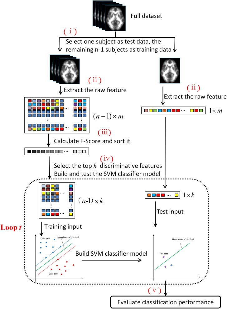
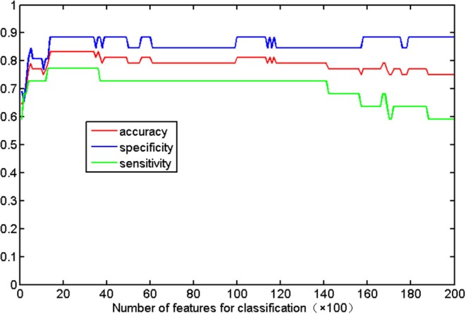
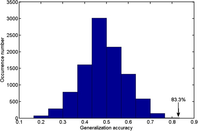
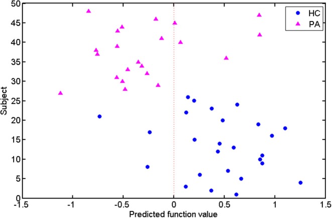
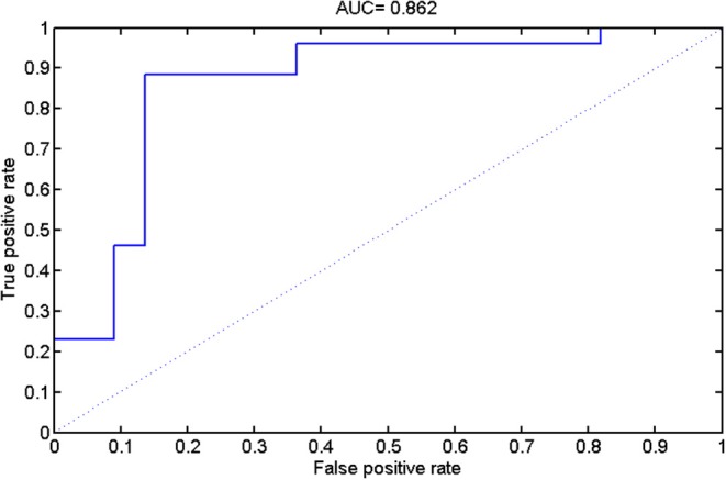
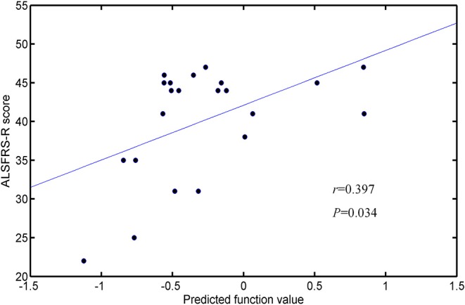
White matter (WM) impairments involving both motor and extra-motor areas have been well-documented in amyotrophic lateral sclerosis (ALS). This study tested the potential of diffusion measurements in WM for identifying ALS based on support vector machine (SVM). Voxel-wise fractional anisotropy (FA) values of diffusion tensor images (DTI) were extracted from 22 ALS patients and 26 healthy controls and served as discrimination features. The revised ALS Functional Rating Scale (ALSFRS-R) was employed to assess ALS severity. Feature ranking and selection were based on Fisher scores. A linear kernel SVM algorithm was applied to build the classification model, from which the classification performance was evaluated. To promote classifier generalization ability, a leave-one-out cross-validation (LOOCV) method was adopted. By using the 2,400~3,400 ranked features as optimal features, the highest classification accuracy of 83.33% (sensitivity = 77.27% and specificity = 88.46%, = 0.0001) was achieved, with an area under receiver operating characteristic curve of 0.862. The predicted function value was positively correlated with patient ALSFRS-R scores ( = 0.493, = 0.020). In the optimized SVM model, FA values from several regions mostly contributed to classification, primarily involving the corticospinal tract pathway, postcentral gyrus, and frontal and parietal areas. Our results suggest the feasibility of ALS diagnosis based on SVM analysis and diffusion measurements of WM. Additional investigations using a larger cohort is recommended in order to validate the results of this study.

摘要

在肌萎缩侧索硬化症(ALS)中,涉及运动和运动外区域的白质(WM)损伤已有充分记录。本研究基于支持向量机(SVM)测试了WM扩散测量用于识别ALS的潜力。从22例ALS患者和26名健康对照中提取扩散张量图像(DTI)的体素分数各向异性(FA)值,并将其用作判别特征。采用修订的ALS功能评定量表(ALSFRS-R)评估ALS严重程度。特征排序和选择基于Fisher分数。应用线性核SVM算法构建分类模型,并评估其分类性能。为提高分类器泛化能力,采用留一法交叉验证(LOOCV)方法。使用2400~3400个排序后的特征作为最佳特征,实现了83.33%的最高分类准确率(敏感性=77.27%,特异性=88.46%,P=0.0001),受试者工作特征曲线下面积为0.862。预测函数值与患者ALSFRS-R评分呈正相关(r=0.493,P=0.020)。在优化的SVM模型中,几个区域的FA值对分类贡献最大,主要涉及皮质脊髓束通路、中央后回以及额叶和顶叶区域。我们的结果表明基于SVM分析和WM扩散测量进行ALS诊断是可行的。建议使用更大的队列进行进一步研究以验证本研究结果。

https://cdn.ncbi.nlm.nih.gov/pmc/blobs/1e85/7198809/f22685b01f68/fneur-11-00275-g0007.jpg
https://cdn.ncbi.nlm.nih.gov/pmc/blobs/1e85/7198809/e41745f02e4f/fneur-11-00275-g0001.jpg
https://cdn.ncbi.nlm.nih.gov/pmc/blobs/1e85/7198809/fe78b58fd804/fneur-11-00275-g0002.jpg
https://cdn.ncbi.nlm.nih.gov/pmc/blobs/1e85/7198809/6e9b6c960a17/fneur-11-00275-g0003.jpg
https://cdn.ncbi.nlm.nih.gov/pmc/blobs/1e85/7198809/10fac18c1554/fneur-11-00275-g0004.jpg
https://cdn.ncbi.nlm.nih.gov/pmc/blobs/1e85/7198809/3137f1b6120f/fneur-11-00275-g0005.jpg
https://cdn.ncbi.nlm.nih.gov/pmc/blobs/1e85/7198809/1273f5e10b58/fneur-11-00275-g0006.jpg
https://cdn.ncbi.nlm.nih.gov/pmc/blobs/1e85/7198809/f22685b01f68/fneur-11-00275-g0007.jpg

相似文献

1
Identification of Amyotrophic Lateral Sclerosis Based on Diffusion Tensor Imaging and Support Vector Machine.基于扩散张量成像和支持向量机的肌萎缩侧索硬化症识别
Front Neurol. 2020 Apr 28;11:275. doi: 10.3389/fneur.2020.00275. eCollection 2020.
2
Brain white matter abnormalities and correlation with severity in amyotrophic lateral sclerosis: An atlas-based diffusion tensor imaging study.肌萎缩侧索硬化症患者脑白质异常及其与严重程度的相关性:一项基于图谱的弥散张量成像研究
J Neurol Sci. 2019 Oct 15;405:116438. doi: 10.1016/j.jns.2019.116438. Epub 2019 Aug 29.
3
Altered white matter microarchitecture in amyotrophic lateral sclerosis: A voxel-based meta-analysis of diffusion tensor imaging.肌萎缩侧索硬化症中的白质微结构改变:基于体素的弥散张量成像的荟萃分析。
Neuroimage Clin. 2018 Apr 4;19:122-129. doi: 10.1016/j.nicl.2018.04.005. eCollection 2018.
4
Interhemispheric connectivity in amyotrophic lateral sclerosis: A near-infrared spectroscopy and diffusion tensor imaging study.肌萎缩侧索硬化症的半球间连接性:一项近红外光谱和扩散张量成像研究。
Neuroimage Clin. 2016 Sep 29;12:666-672. doi: 10.1016/j.nicl.2016.09.020. eCollection 2016.
5
White matter microstructural impairments in amyotrophic lateral sclerosis: A mean apparent propagator MRI study.肌萎缩侧索硬化症的白质微观结构损伤:平均表观传播子 MRI 研究。
Neuroimage Clin. 2021;32:102863. doi: 10.1016/j.nicl.2021.102863. Epub 2021 Oct 23.
6
Segmental alterations of the corpus callosum in motor neuron disease: A DTI and texture analysis in 575 patients.运动神经元病胼胝体节段性改变:575 例患者的 DTI 和纹理分析。
Neuroimage Clin. 2022;35:103061. doi: 10.1016/j.nicl.2022.103061. Epub 2022 May 28.
7
Structural imaging differences and longitudinal changes in primary lateral sclerosis and amyotrophic lateral sclerosis.原发性侧索硬化症和肌萎缩性侧索硬化症的结构影像学差异和纵向变化。
Neuroimage Clin. 2012 Dec 24;2:151-60. doi: 10.1016/j.nicl.2012.12.003. eCollection 2012.
8
Role of diffusion tensor imaging or magnetic resonance spectroscopy in the diagnosis and disability assessment of amyotrophic lateral sclerosis.扩散张量成像或磁共振波谱在肌萎缩侧索硬化症诊断及功能障碍评估中的作用
J Neurol Sci. 2015 Jan 15;348(1-2):206-10. doi: 10.1016/j.jns.2014.12.004. Epub 2014 Dec 6.
9
Differential involvement of corticospinal tract (CST) fibers in UMN-predominant ALS patients with or without CST hyperintensity: A diffusion tensor tractography study.皮质脊髓束(CST)纤维在伴有或不伴有CST高信号的以UMN为主的肌萎缩侧索硬化症患者中的差异参与:一项扩散张量纤维束成像研究。
Neuroimage Clin. 2017 Feb 22;14:574-579. doi: 10.1016/j.nicl.2017.02.017. eCollection 2017.
10
A prospective harmonized multicenter DTI study of cerebral white matter degeneration in ALS.一项前瞻性的 ALS 脑白质退变的多中心协调弥散张量成像研究。
Neurology. 2020 Aug 25;95(8):e943-e952. doi: 10.1212/WNL.0000000000010235. Epub 2020 Jul 9.

引用本文的文献

1
Toward diffusion tensor imaging as a biomarker in neurodegenerative diseases: technical considerations to optimize recordings and data processing.迈向将扩散张量成像作为神经退行性疾病生物标志物:优化记录和数据处理的技术考量
Front Hum Neurosci. 2024 Apr 2;18:1378896. doi: 10.3389/fnhum.2024.1378896. eCollection 2024.
2
Digital Alternative Communication for Individuals with Amyotrophic Lateral Sclerosis: What We Have.肌萎缩侧索硬化症患者的数字替代沟通方式:我们所拥有的。
J Clin Med. 2023 Aug 11;12(16):5235. doi: 10.3390/jcm12165235.
3
Aberrant Multimodal Connectivity Pattern Involved in Default Mode Network and Limbic Network in Amyotrophic Lateral Sclerosis.

本文引用的文献

1
Inferior Longitudinal Fasciculus' Role in Visual Processing and Language Comprehension: A Combined MEG-DTI Study.下纵束在视觉处理和语言理解中的作用:一项MEG-DTI联合研究
Front Neurosci. 2019 Aug 23;13:875. doi: 10.3389/fnins.2019.00875. eCollection 2019.
2
Brain white matter abnormalities and correlation with severity in amyotrophic lateral sclerosis: An atlas-based diffusion tensor imaging study.肌萎缩侧索硬化症患者脑白质异常及其与严重程度的相关性:一项基于图谱的弥散张量成像研究
J Neurol Sci. 2019 Oct 15;405:116438. doi: 10.1016/j.jns.2019.116438. Epub 2019 Aug 29.
3
Precentral degeneration and cerebellar compensation in amyotrophic lateral sclerosis: A multimodal MRI analysis.
肌萎缩侧索硬化症中默认模式网络和边缘系统网络涉及的异常多模态连接模式
Brain Sci. 2023 May 15;13(5):803. doi: 10.3390/brainsci13050803.
4
Diffusion Tensor Imaging in Amyotrophic Lateral Sclerosis: Machine Learning for Biomarker Development.肌萎缩侧索硬化症的弥散张量成像:用于生物标志物开发的机器学习。
Int J Mol Sci. 2023 Jan 18;24(3):1911. doi: 10.3390/ijms24031911.
肌萎缩侧索硬化症中的中央前回变性和小脑代偿:多模态 MRI 分析。
Hum Brain Mapp. 2019 Aug 15;40(12):3464-3474. doi: 10.1002/hbm.24609. Epub 2019 Apr 24.
4
Survival prediction models in motor neuron disease.运动神经元病的生存预测模型。
Eur J Neurol. 2019 Sep;26(9):1143-1152. doi: 10.1111/ene.13957. Epub 2019 Apr 20.
5
Machine Learning in Amyotrophic Lateral Sclerosis: Achievements, Pitfalls, and Future Directions.肌萎缩侧索硬化症中的机器学习:成就、陷阱与未来方向。
Front Neurosci. 2019 Feb 28;13:135. doi: 10.3389/fnins.2019.00135. eCollection 2019.
6
From anatomy to function: the role of the somatosensory cortex in emotional regulation.从解剖学到功能:躯体感觉皮层在情绪调节中的作用。
Braz J Psychiatry. 2019 May-Jun;41(3):261-269. doi: 10.1590/1516-4446-2018-0183. Epub 2018 Dec 6.
7
Imaging Biomarkers for the Diagnosis and Prognosis of Neurodegenerative Diseases. The Example of Amyotrophic Lateral Sclerosis.用于神经退行性疾病诊断和预后的影像学生物标志物。以肌萎缩侧索硬化症为例。
Front Neurosci. 2018 Oct 25;12:784. doi: 10.3389/fnins.2018.00784. eCollection 2018.
8
Functional Anatomy of the Inferior Longitudinal Fasciculus: From Historical Reports to Current Hypotheses.下纵束的功能解剖:从历史报告到当前假说
Front Neuroanat. 2018 Sep 19;12:77. doi: 10.3389/fnana.2018.00077. eCollection 2018.
9
A Connectomic Atlas of the Human Cerebrum-Chapter 10: Tractographic Description of the Superior Longitudinal Fasciculus.人类大脑连接组图谱-第 10 章:上纵束的纤维束描绘。
Oper Neurosurg (Hagerstown). 2018 Dec 1;15(suppl_1):S407-S422. doi: 10.1093/ons/opy264.
10
Altered white matter microarchitecture in amyotrophic lateral sclerosis: A voxel-based meta-analysis of diffusion tensor imaging.肌萎缩侧索硬化症中的白质微结构改变:基于体素的弥散张量成像的荟萃分析。
Neuroimage Clin. 2018 Apr 4;19:122-129. doi: 10.1016/j.nicl.2018.04.005. eCollection 2018.