• 文献检索
  • 文档翻译
  • 深度研究
  • 学术资讯
  • Suppr Zotero 插件Zotero 插件
  • 邀请有礼
  • 套餐&价格
  • 历史记录
应用&插件
Suppr Zotero 插件Zotero 插件浏览器插件Mac 客户端Windows 客户端微信小程序
定价
高级版会员购买积分包购买API积分包
服务
文献检索文档翻译深度研究API 文档MCP 服务
关于我们
关于 Suppr公司介绍联系我们用户协议隐私条款
关注我们

Suppr 超能文献

核心技术专利:CN118964589B侵权必究
粤ICP备2023148730 号-1Suppr @ 2026

文献检索

告别复杂PubMed语法,用中文像聊天一样搜索,搜遍4000万医学文献。AI智能推荐,让科研检索更轻松。

立即免费搜索

文件翻译

保留排版,准确专业,支持PDF/Word/PPT等文件格式,支持 12+语言互译。

免费翻译文档

深度研究

AI帮你快速写综述,25分钟生成高质量综述,智能提取关键信息,辅助科研写作。

立即免费体验

运动神经元病胼胝体节段性改变:575 例患者的 DTI 和纹理分析。

Segmental alterations of the corpus callosum in motor neuron disease: A DTI and texture analysis in 575 patients.

机构信息

Department of Neurology, University of Ulm, Germany.

Department of Neurology, University of Ulm, Germany; German Center for Neurodegenerative Diseases (DZNE), Ulm, Germany.

出版信息

Neuroimage Clin. 2022;35:103061. doi: 10.1016/j.nicl.2022.103061. Epub 2022 May 28.

DOI:10.1016/j.nicl.2022.103061
PMID:35653913
原文链接:https://pmc.ncbi.nlm.nih.gov/articles/PMC9163839/
Abstract

INTRODUCTION

Within the core neuroimaging signature of amyotrophic lateral sclerosis (ALS), the corpus callosum (CC) is increasingly recognized as a consistent feature. The aim of this study was to investigate the sensitivity and specificity of the microstructural segmental CC morphology, assessed by diffusion tensor imaging (DTI) and high-resolution T1-weighted (T1w) imaging, in a large cohort of ALS patients including different clinical phenotypes.

METHODS

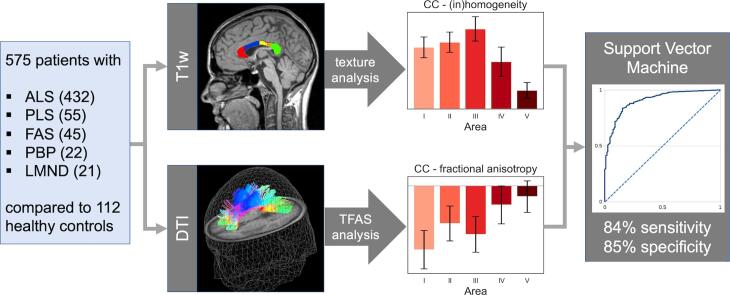
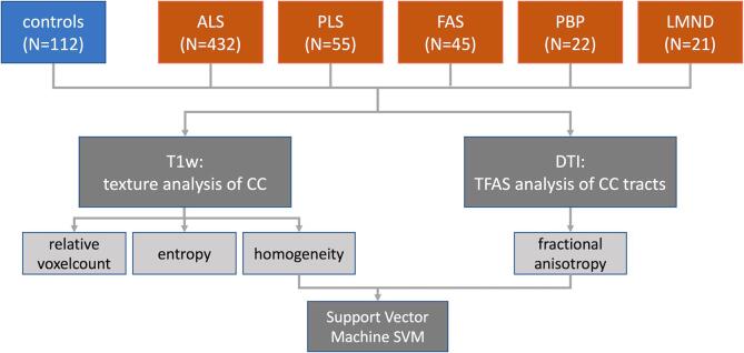
In a single-centre study, 575 patients with ALS (classical phenotype, N  = 432; restricted phenotypes primary lateral sclerosis (PLS) N = 55, flail arm syndrome (FAS) N = 45, progressive bulbar palsy (PBP) N = 22, lower motor neuron disease (LMND) N = 21) and 112 healthy controls underwent multiparametric MRI, i.e. volume-rendering T1w scans and DTI. Tract-based fractional anisotropy statistics (TFAS) was applied to callosal tracts of CC areas I-V, identified from DTI data (tract-of-interest (TOI) analysis), and texture analysis was applied to T1w data. In order to further specify the callosal alterations, a support vector machine (SVM) algorithm was used to discriminate between motor neuron disease patients and controls.

RESULTS

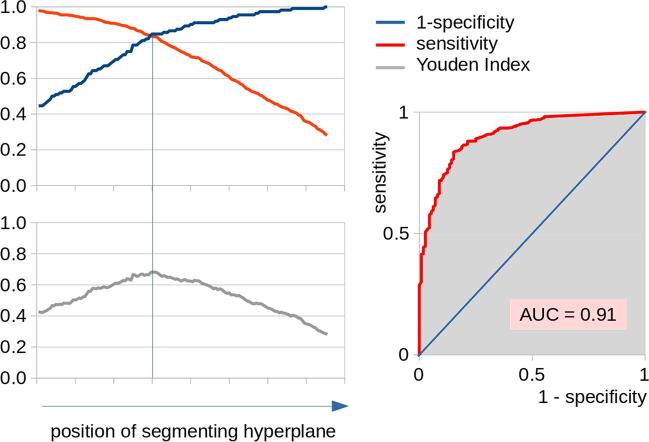
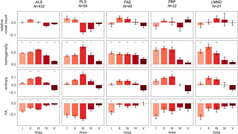
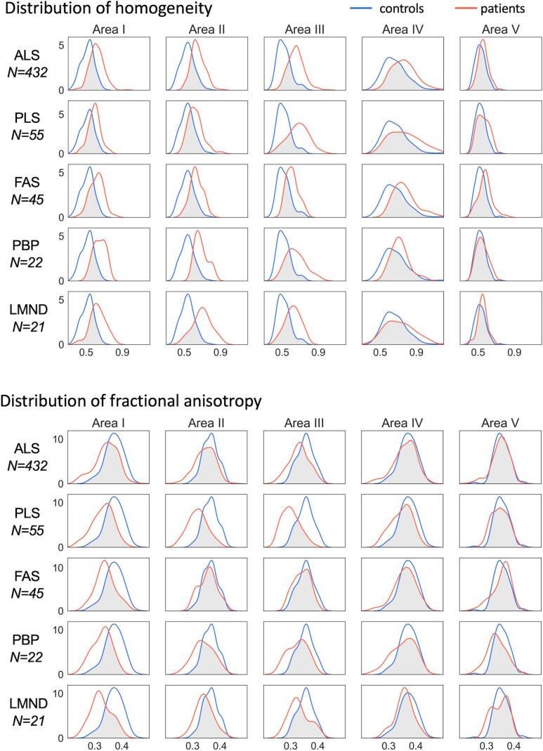
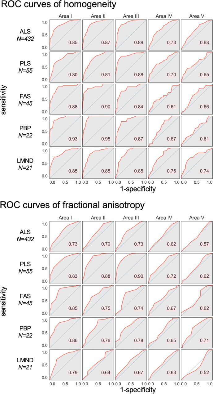
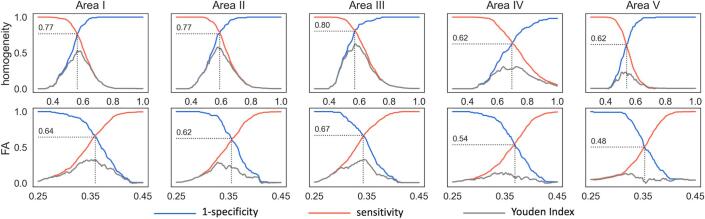
The analysis of white matter integrity revealed predominantly FA reductions for tracts of the callosal areas I, II, and III (with highest reductions in callosal area III) for all ALS patients and separately for each phenotype when compared to controls; texture analysis demonstrated significant alterations of the parameters entropy and homogeneity for ALS patients and all phenotypes for the CC areas I, II, and III (with again highest reductions in callosal area III) compared to controls. With SVM applied on multiparametric callosal parameters of area III, a separation of all ALS patients including phenotypes from controls with 72% sensitivity and 73% specificity was achieved. These results for callosal area III parameters could be improved by an SVM of six multiparametric callosal parameters of areas I, II, and III, achieving a separation of all ALS patients including phenotypes from controls with 84% sensitivity and 85% specificity.

DISCUSSION

The multiparametric MRI texture and DTI analysis demonstrated substantial alterations of the frontal and central CC with most significant alterations in callosal area III (motor segment) in ALS and separately in all investigated phenotypes (PLS, FAS, PBP, LMND) in comparison to controls, while no significant differences were observed between ALS and its phenotypes. The combination of the texture and the DTI parameters in an unbiased SVM-based approach might contribute as a neuroimaging marker for the assessment of the CC in ALS, including subtypes.

摘要

介绍

在肌萎缩侧索硬化症(ALS)的核心神经影像学特征中,胼胝体(CC)越来越被认为是一个一致的特征。本研究的目的是在包括不同临床表型的大量 ALS 患者中,通过弥散张量成像(DTI)和高分辨率 T1 加权(T1w)成像,研究 CC 微观结构节段形态的敏感性和特异性。

方法

在一项单中心研究中,575 名 ALS 患者(经典表型,N=432;限局性表型原发性侧索硬化症(PLS),N=55;臂丛张力障碍综合征(FAS),N=45;进行性球麻痹(PBP),N=22;下运动神经元病(LMND),N=21)和 112 名健康对照者接受了多参数 MRI 检查,包括容积再现 T1w 扫描和 DTI。应用基于束的各向异性分数统计量(TFAS)对 DTI 数据中的 CC 区域 I-V 进行感兴趣区(TOI)分析,以评估胼胝体束的各向异性分数。为了进一步明确胼胝体的改变,应用支持向量机(SVM)算法对运动神经元病患者和对照组进行判别分析。

结果

对脑白质完整性的分析显示,所有 ALS 患者和每个表型与对照组相比,CC 区域 I、II 和 III 的 FA 降低最为明显(CC 区域 III 的降低最明显);纹理分析显示,CC 区域 I、II 和 III 的熵和同质性参数在 ALS 患者和所有表型中均有显著改变(CC 区域 III 的降低最明显)。应用 SVM 对 CC 区域 III 的多参数进行分析,可实现对包括所有表型的 ALS 患者与对照组的区分,敏感性为 72%,特异性为 73%。通过对 CC 区域 I、II 和 III 的六个多参数进行 SVM 分析,可将包括所有表型的 ALS 患者与对照组区分开来,敏感性为 84%,特异性为 85%。

讨论

多参数 MRI 纹理和 DTI 分析显示,在 ALS 中,CC 的额极和中央部出现明显改变,以 CC 区域 III(运动节段)改变最明显,与对照组相比,在所有研究的表型(PLS、FAS、PBP、LMND)中也有明显改变,而 ALS 与其表型之间无显著差异。在基于 SVM 的无偏方法中,纹理和 DTI 参数的组合可能作为评估 ALS 包括亚型的 CC 的神经影像学标志物。

https://cdn.ncbi.nlm.nih.gov/pmc/blobs/57e0/9163839/cd8b9fb76c98/gr7.jpg
https://cdn.ncbi.nlm.nih.gov/pmc/blobs/57e0/9163839/b7efcc55cf5a/ga1.jpg
https://cdn.ncbi.nlm.nih.gov/pmc/blobs/57e0/9163839/a4d570e44a68/gr1.jpg
https://cdn.ncbi.nlm.nih.gov/pmc/blobs/57e0/9163839/7d197cad26ef/gr2.jpg
https://cdn.ncbi.nlm.nih.gov/pmc/blobs/57e0/9163839/f31b045b60e7/gr3.jpg
https://cdn.ncbi.nlm.nih.gov/pmc/blobs/57e0/9163839/00ade0f63cb7/gr4.jpg
https://cdn.ncbi.nlm.nih.gov/pmc/blobs/57e0/9163839/cac028e52f98/gr5.jpg
https://cdn.ncbi.nlm.nih.gov/pmc/blobs/57e0/9163839/43da749c3516/gr6.jpg
https://cdn.ncbi.nlm.nih.gov/pmc/blobs/57e0/9163839/cd8b9fb76c98/gr7.jpg

相似文献

1
Segmental alterations of the corpus callosum in motor neuron disease: A DTI and texture analysis in 575 patients.运动神经元病胼胝体节段性改变:575 例患者的 DTI 和纹理分析。
Neuroimage Clin. 2022;35:103061. doi: 10.1016/j.nicl.2022.103061. Epub 2022 May 28.
2
Focal alterations of the callosal area III in primary lateral sclerosis: An MRI planimetry and texture analysis.原发性侧索硬化症中胼胝体区 III 的局灶性改变:MRI 平面测量和纹理分析。
Neuroimage Clin. 2020;26:102223. doi: 10.1016/j.nicl.2020.102223. Epub 2020 Feb 21.
3
Segmental involvement of the corpus callosum in associated ALS: a tract of interest-based DTI study.肌萎缩侧索硬化症相关病例中胼胝体的节段性受累:一项基于感兴趣区的扩散张量成像研究
Ther Adv Chronic Dis. 2021 Mar 21;12:20406223211002969. doi: 10.1177/20406223211002969. eCollection 2021.
4
Segmental Alterations of the Corpus Callosum in Progressive Supranuclear Palsy: A Multiparametric Magnetic Resonance Imaging Study.进行性核上性麻痹中胼胝体的节段性改变:一项多参数磁共振成像研究
Front Aging Neurosci. 2021 Nov 19;13:720634. doi: 10.3389/fnagi.2021.720634. eCollection 2021.
5
Fast progressive lower motor neuron disease is an ALS variant: A two-centre tract of interest-based MRI data analysis.快速进展性下运动神经元病是肌萎缩侧索硬化症的一种变异型:基于感兴趣区的磁共振成像数据分析的两项研究。
Neuroimage Clin. 2017 Oct 14;17:145-152. doi: 10.1016/j.nicl.2017.10.008. eCollection 2018.
6
Identical patterns of cortico-efferent tract involvement in primary lateral sclerosis and amyotrophic lateral sclerosis: A tract of interest-based MRI study.原发性侧索硬化症和肌萎缩侧索硬化症皮质传出束受累的相同模式:基于感兴趣区的磁共振成像研究。
Neuroimage Clin. 2018 Mar 15;18:762-769. doi: 10.1016/j.nicl.2018.03.018. eCollection 2018.
7
The same cortico-efferent tract involvement in progressive bulbar palsy and in 'classical' ALS: A tract of interest-based MRI study.进行性球麻痹与“经典”肌萎缩侧索硬化症中相同的皮质传出束受累:一项基于感兴趣区域的 MRI 研究。
Neuroimage Clin. 2019;24:101979. doi: 10.1016/j.nicl.2019.101979. Epub 2019 Aug 9.
8
Cortico-efferent tract involvement in primary lateral sclerosis and amyotrophic lateral sclerosis: A two-centre tract of interest-based DTI analysis.皮质传出束在原发性侧索硬化症和肌萎缩侧索硬化症中的受累:基于两中心感兴趣区的 DTI 分析。
Neuroimage Clin. 2018;20:1062-1069. doi: 10.1016/j.nicl.2018.10.005. Epub 2018 Oct 9.
9
Structural and microstructural neuroimaging signature of C9orf72-associated ALS: A multiparametric MRI study.C9orf72 相关肌萎缩侧索硬化症的结构和微观结构神经影像学特征:多参数 MRI 研究。
Neuroimage Clin. 2023;39:103505. doi: 10.1016/j.nicl.2023.103505. Epub 2023 Sep 9.
10
Pathology of callosal damage in ALS: An , 7 T diffusion tensor MRI study.肌萎缩侧索硬化症胼胝体损伤的病理学:一项7T扩散张量磁共振成像研究
Neuroimage Clin. 2017 Apr 30;15:200-208. doi: 10.1016/j.nicl.2017.04.024. eCollection 2017.

引用本文的文献

1
Emerging biomarkers in amyotrophic lateral sclerosis: from pathogenesis to clinical applications.肌萎缩侧索硬化症中的新兴生物标志物:从发病机制到临床应用
Front Mol Biosci. 2025 Jun 30;12:1608853. doi: 10.3389/fmolb.2025.1608853. eCollection 2025.
2
Unveiling structural damage of the corpus callosum in amyotrophic lateral sclerosis through diffusion tensor imaging and spread direction perspectives.通过扩散张量成像和扩散方向视角揭示肌萎缩侧索硬化症中胼胝体的结构损伤
Ann Med. 2025 Dec;57(1):2490822. doi: 10.1080/07853890.2025.2490822. Epub 2025 Apr 15.
3
T1-relaxation times along the corticospinal tract as a diagnostic marker in patients with amyotrophic lateral sclerosis.
作为肌萎缩侧索硬化症患者诊断标志物的沿皮质脊髓束的T1弛豫时间
Front Neuroimaging. 2025 Feb 13;4:1549727. doi: 10.3389/fnimg.2025.1549727. eCollection 2025.
4
Regional Cerebral Atrophy Contributes to Personalized Survival Prediction in Amyotrophic Lateral Sclerosis: A Multicentre, Machine Learning, Deformation-Based Morphometry Study.脑区萎缩有助于肌萎缩侧索硬化症的个性化生存预测:一项多中心、机器学习、基于变形形态测量学的研究
Ann Neurol. 2025 Jun;97(6):1144-1157. doi: 10.1002/ana.27196. Epub 2025 Feb 22.
5
Altered interhemispheric connectivity in Huntington's Disease.亨廷顿病患者大脑两半球间连接的改变。
Neuroimage Clin. 2024;44:103670. doi: 10.1016/j.nicl.2024.103670. Epub 2024 Sep 12.
6
The burden of upper motor neuron involvement is correlated with the bilateral limb involvement interval in patients with amyotrophic lateral sclerosis: a retrospective observational study.肌萎缩侧索硬化症患者上运动神经元受累负担与双侧肢体受累间隔相关:一项回顾性观察研究。
Neural Regen Res. 2025 May 1;20(5):1505-1512. doi: 10.4103/NRR.NRR-D-23-01359. Epub 2024 Mar 1.
7
The integrity of the corticospinal tract and corpus callosum, and the risk of ALS: univariable and multivariable Mendelian randomization.皮质脊髓束和胼胝体的完整性与肌萎缩侧索硬化症的风险:单变量和多变量孟德尔随机化分析。
Sci Rep. 2024 Jul 26;14(1):17216. doi: 10.1038/s41598-024-68374-y.
8
Toward diffusion tensor imaging as a biomarker in neurodegenerative diseases: technical considerations to optimize recordings and data processing.迈向将扩散张量成像作为神经退行性疾病生物标志物:优化记录和数据处理的技术考量
Front Hum Neurosci. 2024 Apr 2;18:1378896. doi: 10.3389/fnhum.2024.1378896. eCollection 2024.
9
AI-assisted quantification of hypothalamic atrophy in amyotrophic lateral sclerosis by convolutional neural network-based automatic segmentation.基于卷积神经网络的自动分割的肌萎缩侧索硬化症下丘脑萎缩的 AI 辅助定量分析。
Sci Rep. 2023 Dec 6;13(1):21505. doi: 10.1038/s41598-023-48649-6.
10
Biomarkers in amyotrophic lateral sclerosis: current status and future prospects.肌萎缩侧索硬化症中的生物标志物:现状与未来展望。
Nat Rev Neurol. 2023 Dec;19(12):754-768. doi: 10.1038/s41582-023-00891-2. Epub 2023 Nov 10.