Department of Anatomy, Physiology and Pharmacology University of Saskatchewan, Saskatoon, Saskatchewan S7N 5E5, Canada.
Department of Psychiatry, University of Alberta, Edmonton, Alberta T6G 2B7, Canada.
Learn Mem. 2020 May 15;27(6):222-235. doi: 10.1101/lm.050245.119. Print 2020 Jun.
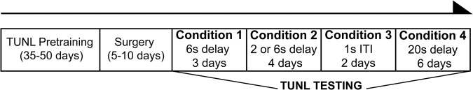
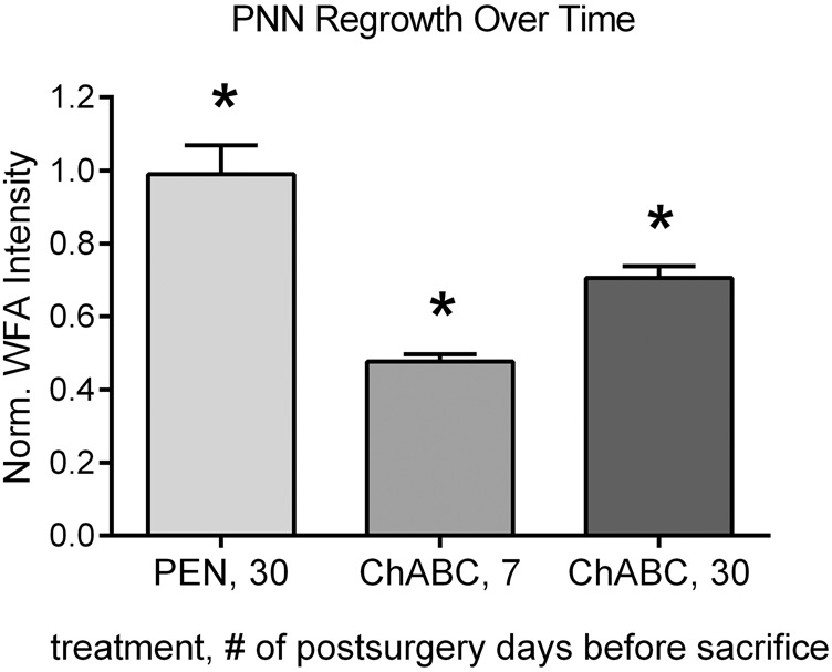
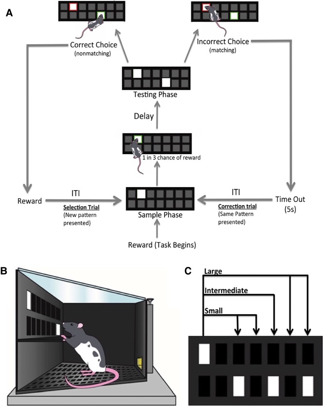
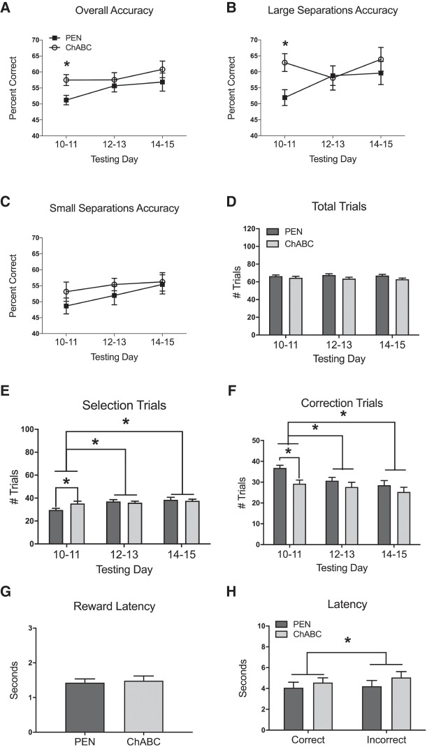
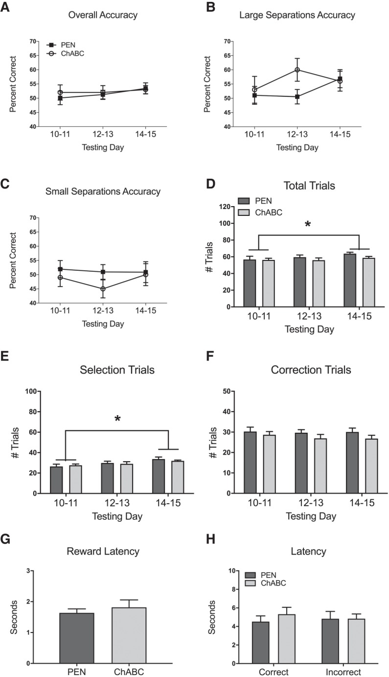
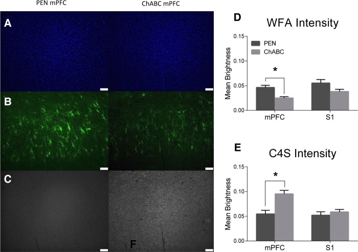
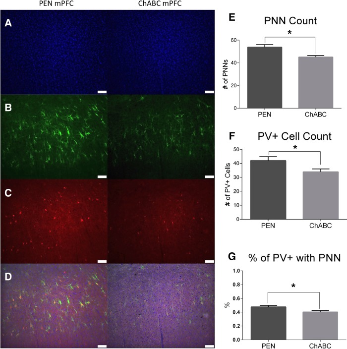
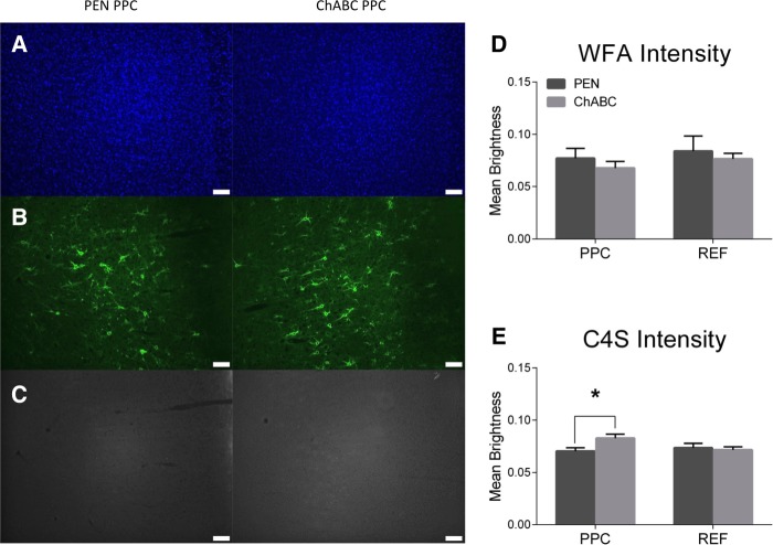
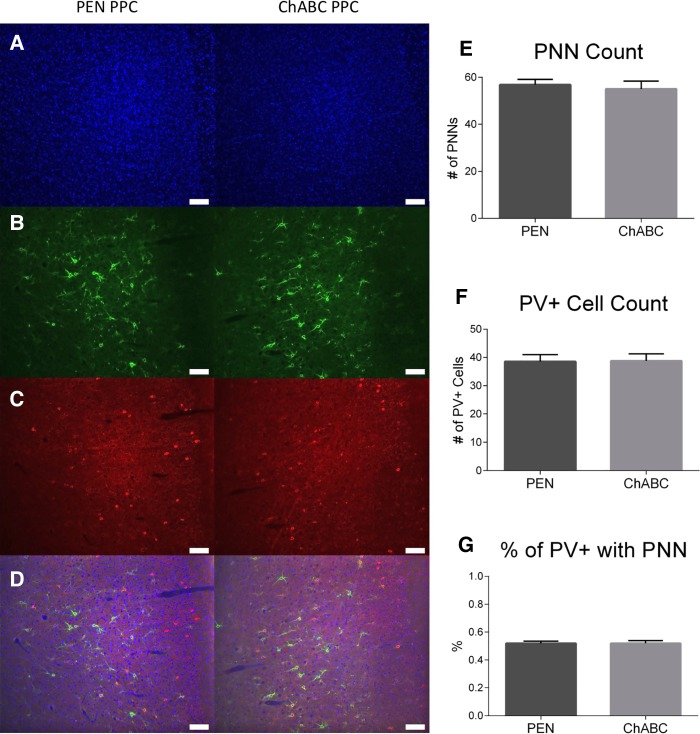
Perineuronal nets (PNNs) are specialized extracellular matrix structures that surround subsets of neurons throughout the central nervous system (CNS). They are made up of chondroitin sulfate proteoglycans (CSPGs), hyaluronan, tenascin-R, and many other link proteins that together make up their rigid and lattice-like structure. Modulation of PNNs can alter synaptic plasticity and thereby affect learning, memory, and cognition. In the present study, we degraded PNNs in the medial prefrontal (mPFC) and posterior parietal (PPC) cortices of Long-Evans rats using the enzyme chondroitinase ABC (ChABC), which cleaves apart CSPGs. We then measured the consequences of PNN degradation on spatial working memory (WM) with a trial-unique, non-matching-to location (TUNL) automated touchscreen task. All rats were trained with a standard 6 sec delay and 20 sec inter-trial interval (ITI) and then tested under four different conditions: a 6 sec delay, a variable 2 or 6 sec delay, a 2 sec delay with a 1 sec ITI (interference condition), and a 20 sec delay. Rats that received mPFC ChABC treatment initially performed TUNL with higher accuracy, more selection trials completed, and fewer correction trials completed compared to controls in the 20 sec delay condition but did not perform differently from controls in any other condition. Rats that received PPC ChABC treatment did not perform significantly differently from controls in any condition. Posthumous immunohistochemistry confirmed an increase in CSPG degradation products (C4S stain) in the mPFC and PPC following ChABC infusions while WFA staining intensity and parvalbumin positive neuron number were decreased following mPFC, but not PPC, ChABC infusions. These findings suggest that PNNs in the mPFC play a subtle role in spatial WM, but PNNs in the PPC do not. Furthermore, it appears that PNNs in the mPFC are involved in adapting to a challenging novel delay, but that they do not play an essential role in spatial WM function.
周围神经毡(PNNs)是中枢神经系统(CNS)中特定神经元周围的特殊细胞外基质结构。它们由软骨素硫酸盐蛋白聚糖(CSPGs)、透明质酸、腱糖蛋白-R 和许多其他连接蛋白组成,共同构成其刚性和晶格状结构。PNNs 的调节可以改变突触可塑性,从而影响学习、记忆和认知。在本研究中,我们使用酶软骨素酶 ABC(ChABC)降解长爪沙鼠内侧前额叶(mPFC)和后顶叶(PPC)皮质中的 PNNs,ChABC 可以分解 CSPGs。然后,我们使用独特试验、非位置匹配(TUNL)自动触摸屏任务测量 PNN 降解对空间工作记忆(WM)的影响。所有大鼠均在标准 6 秒延迟和 20 秒试验间间隔(ITI)下进行训练,然后在四种不同条件下进行测试:6 秒延迟、2 或 6 秒可变延迟、2 秒延迟加 1 秒 ITI(干扰条件)和 20 秒延迟。与对照组相比,接受 mPFC ChABC 处理的大鼠在 20 秒延迟条件下首次进行 TUNL 时表现出更高的准确性、更多的选择试验完成和更少的校正试验完成,但在其他任何条件下与对照组的表现没有差异。接受 PPC ChABC 处理的大鼠在任何条件下与对照组的表现均无显著差异。死后免疫组织化学证实 ChABC 输注后 mPFC 和 PPC 中的 CSPG 降解产物(C4S 染色)增加,而 WFA 染色强度和 parvalbumin 阳性神经元数量在 mPFC 但不在 PPC 输注后减少。这些发现表明 mPFC 中的 PNNs 在空间 WM 中起着微妙的作用,但 PPC 中的 PNNs 则不然。此外,似乎 mPFC 中的 PNNs 参与了适应具有挑战性的新延迟,但它们在空间 WM 功能中并非必不可少。