• 文献检索
  • 文档翻译
  • 深度研究
  • 学术资讯
  • Suppr Zotero 插件Zotero 插件
  • 邀请有礼
  • 套餐&价格
  • 历史记录
应用&插件
Suppr Zotero 插件Zotero 插件浏览器插件Mac 客户端Windows 客户端微信小程序
定价
高级版会员购买积分包购买API积分包
服务
文献检索文档翻译深度研究API 文档MCP 服务
关于我们
关于 Suppr公司介绍联系我们用户协议隐私条款
关注我们

Suppr 超能文献

核心技术专利:CN118964589B侵权必究
粤ICP备2023148730 号-1Suppr @ 2026

文献检索

告别复杂PubMed语法,用中文像聊天一样搜索,搜遍4000万医学文献。AI智能推荐,让科研检索更轻松。

立即免费搜索

文件翻译

保留排版,准确专业,支持PDF/Word/PPT等文件格式,支持 12+语言互译。

免费翻译文档

深度研究

AI帮你快速写综述,25分钟生成高质量综述,智能提取关键信息,辅助科研写作。

立即免费体验

卷积神经网络在超声成像中用于乳腺和甲状腺结节的诊断。

Convolutional Neural Network for Breast and Thyroid Nodules Diagnosis in Ultrasound Imaging.

机构信息

Department of Ultrasound Medicine, The Third Affiliated Hospital of Guangzhou Medical University, 63 Duobao Road, Guangzhou, 510000 Guangdong, China.

Laboratory of Ultrasound Medicine and Artificial Intelligence, Liwan Experimental Center, The Liwan Hospital of the Third Affiliated Hospital of Guangzhou Medical University, 35 Liwan Road, Guangzhou, 510000 Guangdong, China.

出版信息

Biomed Res Int. 2020 Jan 10;2020:1763803. doi: 10.1155/2020/1763803. eCollection 2020.

DOI:10.1155/2020/1763803
PMID:32420322
原文链接:https://pmc.ncbi.nlm.nih.gov/articles/PMC7199615/
Abstract

OBJECTIVE

The incidence of superficial organ diseases has increased rapidly in recent years. New methods such as computer-aided diagnosis (CAD) are widely used to improve diagnostic efficiency. Convolutional neural networks (CNNs) are one of the most popular methods, and further improvements of CNNs should be considered. This paper aims to develop a multiorgan CAD system based on CNNs for classifying both thyroid and breast nodules and investigate the impact of this system on the diagnostic efficiency of different preprocessing approaches.

METHODS

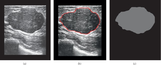
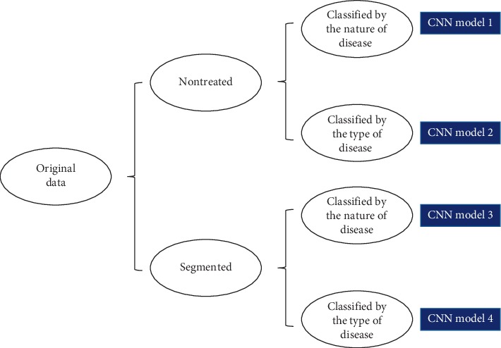
The training and validation sets comprised randomly selected thyroid and breast nodule images. The data were subgrouped into 4 models according to the different preprocessing methods (depending on segmentation and the classification method). A prospective data set was selected to verify the clinical value of the CNN model by comparison with ultrasound guidelines. Diagnostic efficiency was assessed based on receiver operating characteristic (ROC) curves.

RESULTS

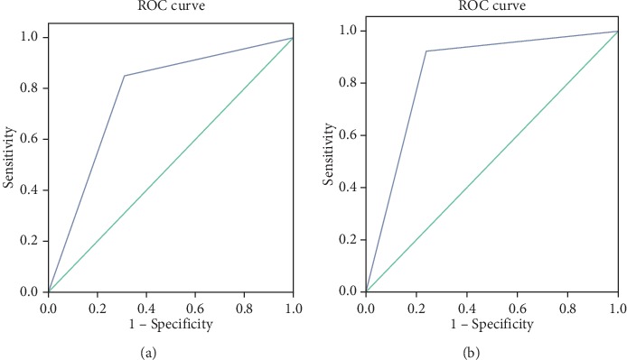
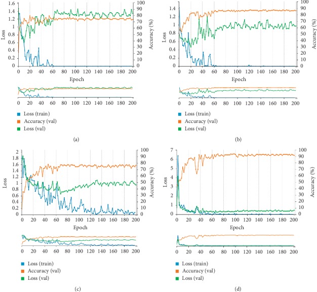
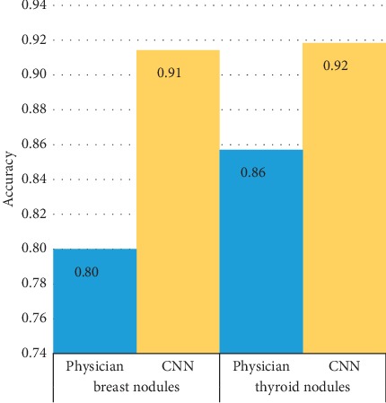
Among the 4 models, the CNN model using segmented images for classification achieved the best result. For the validation set, the sensitivity, specificity, positive predictive value (PPV), negative predictive value (NPV), accuracy, and area under the curve (AUC) of our CNN model were 84.9%, 69.0%, 62.5%, 88.2%, 75.0%, and 0.769, respectively. There was no statistically significant difference between the CNN model and the ultrasound guidelines. The combination of the two methods achieved superior diagnostic efficiency compared with their use individually.

CONCLUSIONS

The study demonstrates the probability, feasibility, and clinical value of CAD in the ultrasound diagnosis of multiple organs. The use of segmented images and classification by the nature of the disease are the main factors responsible for the improvement of the CNN model. Moreover, the combination of the CNN model and ultrasound guidelines results in better diagnostic performance, which will contribute to the improved diagnostic efficiency of CAD systems.

摘要

目的

近年来,浅层器官疾病的发病率迅速上升。计算机辅助诊断(CAD)等新方法被广泛用于提高诊断效率。卷积神经网络(CNN)是最受欢迎的方法之一,应考虑进一步改进 CNN。本文旨在开发一种基于 CNN 的多器官 CAD 系统,用于分类甲状腺和乳腺结节,并研究该系统对不同预处理方法诊断效率的影响。

方法

训练集和验证集包括随机选择的甲状腺和乳腺结节图像。根据不同的预处理方法(取决于分割和分类方法)将数据分为 4 个模型。选择前瞻性数据集,通过与超声指南进行比较来验证 CNN 模型的临床价值。诊断效率基于受试者工作特征(ROC)曲线进行评估。

结果

在 4 个模型中,使用分割图像进行分类的 CNN 模型取得了最佳结果。对于验证集,我们的 CNN 模型的灵敏度、特异性、阳性预测值(PPV)、阴性预测值(NPV)、准确性和曲线下面积(AUC)分别为 84.9%、69.0%、62.5%、88.2%、75.0%和 0.769。与超声指南相比,CNN 模型没有统计学上的显著差异。两种方法的结合比单独使用时具有更高的诊断效率。

结论

本研究证明了 CAD 在多器官超声诊断中的可能性、可行性和临床价值。使用分割图像和按疾病性质进行分类是提高 CNN 模型的主要因素。此外,CNN 模型与超声指南的结合可提高诊断性能,从而提高 CAD 系统的诊断效率。

https://cdn.ncbi.nlm.nih.gov/pmc/blobs/bea8/7199615/55f77e3823b3/BMRI2020-1763803.006.jpg
https://cdn.ncbi.nlm.nih.gov/pmc/blobs/bea8/7199615/d000e209cd64/BMRI2020-1763803.001.jpg
https://cdn.ncbi.nlm.nih.gov/pmc/blobs/bea8/7199615/eaf6a3f61ac1/BMRI2020-1763803.002.jpg
https://cdn.ncbi.nlm.nih.gov/pmc/blobs/bea8/7199615/1715f6153e01/BMRI2020-1763803.003.jpg
https://cdn.ncbi.nlm.nih.gov/pmc/blobs/bea8/7199615/44dfc7c09e4d/BMRI2020-1763803.004.jpg
https://cdn.ncbi.nlm.nih.gov/pmc/blobs/bea8/7199615/f5e2a3348a28/BMRI2020-1763803.005.jpg
https://cdn.ncbi.nlm.nih.gov/pmc/blobs/bea8/7199615/55f77e3823b3/BMRI2020-1763803.006.jpg

相似文献

1
Convolutional Neural Network for Breast and Thyroid Nodules Diagnosis in Ultrasound Imaging.卷积神经网络在超声成像中用于乳腺和甲状腺结节的诊断。
Biomed Res Int. 2020 Jan 10;2020:1763803. doi: 10.1155/2020/1763803. eCollection 2020.
2
Cascade convolutional neural networks for automatic detection of thyroid nodules in ultrasound images.级联卷积神经网络在超声图像中自动检测甲状腺结节
Med Phys. 2017 May;44(5):1678-1691. doi: 10.1002/mp.12134. Epub 2017 Apr 17.
3
Ultrasound Computer-Aided Diagnosis (CAD) Based on the Thyroid Imaging Reporting and Data System (TI-RADS) to Distinguish Benign from Malignant Thyroid Nodules and the Diagnostic Performance of Radiologists with Different Diagnostic Experience.基于甲状腺影像报告和数据系统 (TI-RADS) 的超声计算机辅助诊断 (CAD) 对甲状腺良恶性结节的鉴别诊断及不同诊断经验的放射科医生的诊断效能
Med Sci Monit. 2020 Jan 2;26:e918452. doi: 10.12659/MSM.918452.
4
Deep convolutional neural network for the diagnosis of thyroid nodules on ultrasound.基于深度卷积神经网络的甲状腺超声结节辅助诊断
Head Neck. 2019 Apr;41(4):885-891. doi: 10.1002/hed.25415. Epub 2019 Feb 4.
5
Ultrasound image-based thyroid nodule automatic segmentation using convolutional neural networks.基于超声图像的甲状腺结节自动分割卷积神经网络。
Int J Comput Assist Radiol Surg. 2017 Nov;12(11):1895-1910. doi: 10.1007/s11548-017-1649-7. Epub 2017 Jul 31.
6
Diagnosis of thyroid micronodules on ultrasound using a deep convolutional neural network.超声检查中应用深度卷积神经网络诊断甲状腺微小结节。
Sci Rep. 2023 May 4;13(1):7231. doi: 10.1038/s41598-023-34459-3.
7
A comparative study of pre-trained convolutional neural networks for semantic segmentation of breast tumors in ultrasound.用于超声乳腺肿瘤语义分割的预训练卷积神经网络的比较研究
Comput Biol Med. 2020 Nov;126:104036. doi: 10.1016/j.compbiomed.2020.104036. Epub 2020 Oct 8.
8
Evaluation of a deep learning-based computer-aided diagnosis system for distinguishing benign from malignant thyroid nodules in ultrasound images.基于深度学习的超声图像甲状腺良恶性结节鉴别计算机辅助诊断系统的评估
Med Phys. 2020 Sep;47(9):3952-3960. doi: 10.1002/mp.14301. Epub 2020 Jun 25.
9
Computer-aided diagnosis for classifying benign versus malignant thyroid nodules based on ultrasound images: A comparison with radiologist-based assessments.基于超声图像的甲状腺良恶性结节分类的计算机辅助诊断:与放射科医生评估的比较。
Med Phys. 2016 Jan;43(1):554. doi: 10.1118/1.4939060.
10
A comparison between deep learning convolutional neural networks and radiologists in the differentiation of benign and malignant thyroid nodules on CT images.深度学习卷积神经网络与放射科医生在CT图像上鉴别甲状腺良恶性结节的比较。
Endokrynol Pol. 2021;72(3):217-225. doi: 10.5603/EP.a2021.0015. Epub 2021 Feb 23.

引用本文的文献

1
Application research of artificial intelligence software in the analysis of thyroid nodule ultrasound image characteristics.人工智能软件在甲状腺结节超声图像特征分析中的应用研究
PLoS One. 2025 Jun 2;20(6):e0323343. doi: 10.1371/journal.pone.0323343. eCollection 2025.
2
Thyroid nodule classification in ultrasound imaging using deep transfer learning.基于深度迁移学习的超声成像甲状腺结节分类
BMC Cancer. 2025 Mar 25;25(1):544. doi: 10.1186/s12885-025-13917-3.
3
ThyroNet-X4 genesis: an advanced deep learning model for auxiliary diagnosis of thyroid nodules' malignancy.

本文引用的文献

1
Association between benign thyroid disease and breast cancer: a single center experience.良性甲状腺疾病与乳腺癌的相关性:单中心经验。
BMC Endocr Disord. 2019 Oct 17;19(1):104. doi: 10.1186/s12902-019-0426-8.
2
The role of two tumor foci for predicting central lymph node metastasis in papillary thyroid carcinoma: A meta-analysis.两个肿瘤灶在预测甲状腺乳头状癌中央淋巴结转移中的作用:一项荟萃分析。
Int J Surg. 2018 Apr;52:166-170. doi: 10.1016/j.ijsu.2018.02.029. Epub 2018 Feb 23.
3
Identifying Medical Diagnoses and Treatable Diseases by Image-Based Deep Learning.
甲状腺网络-X4起源:一种用于辅助诊断甲状腺结节恶性肿瘤的先进深度学习模型。
Sci Rep. 2025 Feb 4;15(1):4214. doi: 10.1038/s41598-025-86819-w.
4
From Bench-to-Bedside: How Artificial Intelligence is Changing Thyroid Nodule Diagnostics, a Systematic Review.从基础到临床:人工智能如何改变甲状腺结节诊断——系统评价。
J Clin Endocrinol Metab. 2024 Jun 17;109(7):1684-1693. doi: 10.1210/clinem/dgae277.
5
Bone age assessment based on three-dimensional ultrasound and artificial intelligence compared with paediatrician-read radiographic bone age: protocol for a prospective, diagnostic accuracy study.基于三维超声和人工智能的骨龄评估与儿科医生阅片评估骨龄的比较:一项前瞻性、诊断准确性研究方案。
BMJ Open. 2024 Feb 24;14(2):e079969. doi: 10.1136/bmjopen-2023-079969.
6
The Application of Artificial Intelligence in Thyroid Nodules: A Systematic Review Based on Bibliometric Analysis.人工智能在甲状腺结节中的应用:基于文献计量分析的系统评价。
Endocr Metab Immune Disord Drug Targets. 2024;24(11):1280-1290. doi: 10.2174/0118715303264254231117113456.
7
A Framework for Detecting Thyroid Cancer from Ultrasound and Histopathological Images Using Deep Learning, Meta-Heuristics, and MCDM Algorithms.一种使用深度学习、元启发式算法和多准则决策算法从超声和组织病理学图像中检测甲状腺癌的框架。
J Imaging. 2023 Aug 27;9(9):173. doi: 10.3390/jimaging9090173.
8
A Deep Learning Framework for the Characterization of Thyroid Nodules from Ultrasound Images Using Improved Inception Network and Multi-Level Transfer Learning.一种使用改进的Inception网络和多级迁移学习从超声图像中表征甲状腺结节的深度学习框架。
Diagnostics (Basel). 2023 Jul 24;13(14):2463. doi: 10.3390/diagnostics13142463.
9
Deep Learning in Different Ultrasound Methods for Breast Cancer, from Diagnosis to Prognosis: Current Trends, Challenges, and an Analysis.从诊断到预后:不同超声方法在乳腺癌中的深度学习——当前趋势、挑战及分析
Cancers (Basel). 2023 Jun 10;15(12):3139. doi: 10.3390/cancers15123139.
10
Ultrasound image-based deep learning to differentiate tubal-ovarian abscess from ovarian endometriosis cyst.基于超声图像的深度学习用于鉴别输卵管卵巢脓肿与卵巢子宫内膜异位囊肿。
Front Physiol. 2023 Feb 7;14:1101810. doi: 10.3389/fphys.2023.1101810. eCollection 2023.
基于图像的深度学习识别医学诊断和可治疗疾病。
Cell. 2018 Feb 22;172(5):1122-1131.e9. doi: 10.1016/j.cell.2018.02.010.
4
Dedicated computer-aided detection software for automated 3D breast ultrasound; an efficient tool for the radiologist in supplemental screening of women with dense breasts.用于自动化三维乳腺超声的专用计算机辅助检测软件;对于致密型乳腺女性补充筛查的放射科医生来说,是一种有效的工具。
Eur Radiol. 2018 Jul;28(7):2996-3006. doi: 10.1007/s00330-017-5280-3. Epub 2018 Feb 7.
5
Computer-aided system for diagnosing thyroid nodules on ultrasound: A comparison with radiologist-based clinical assessments.用于超声诊断甲状腺结节的计算机辅助系统:与基于放射科医生的临床评估的比较。
Head Neck. 2018 Apr;40(4):778-783. doi: 10.1002/hed.25049. Epub 2017 Dec 29.
6
Thyroid Nodule Classification in Ultrasound Images by Fine-Tuning Deep Convolutional Neural Network.通过微调深度卷积神经网络对超声图像中的甲状腺结节进行分类
J Digit Imaging. 2017 Aug;30(4):477-486. doi: 10.1007/s10278-017-9997-y.
7
Computer-aided diagnosis of malignant or benign thyroid nodes based on ultrasound images.基于超声图像的甲状腺良恶性结节的计算机辅助诊断
Eur Arch Otorhinolaryngol. 2017 Jul;274(7):2891-2897. doi: 10.1007/s00405-017-4562-3. Epub 2017 Apr 7.
8
ACR Thyroid Imaging, Reporting and Data System (TI-RADS): White Paper of the ACR TI-RADS Committee.美国放射学会甲状腺影像报告和数据系统(TI-RADS):美国放射学会TI-RADS委员会白皮书
J Am Coll Radiol. 2017 May;14(5):587-595. doi: 10.1016/j.jacr.2017.01.046. Epub 2017 Apr 2.
9
Ultrasound Standard Plane Detection Using a Composite Neural Network Framework.基于复合神经网络框架的超声标准切面检测。
IEEE Trans Cybern. 2017 Jun;47(6):1576-1586. doi: 10.1109/TCYB.2017.2685080. Epub 2017 Mar 30.
10
Cascade convolutional neural networks for automatic detection of thyroid nodules in ultrasound images.级联卷积神经网络在超声图像中自动检测甲状腺结节
Med Phys. 2017 May;44(5):1678-1691. doi: 10.1002/mp.12134. Epub 2017 Apr 17.