Department of Biomedical Engineering, Tulane University, New Orleans, Louisiana 70118, United States.
Langmuir. 2020 Jun 23;36(24):6626-6634. doi: 10.1021/acs.langmuir.0c00320. Epub 2020 Jun 11.
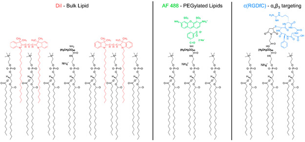
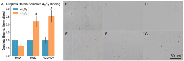
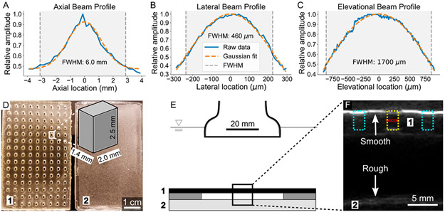
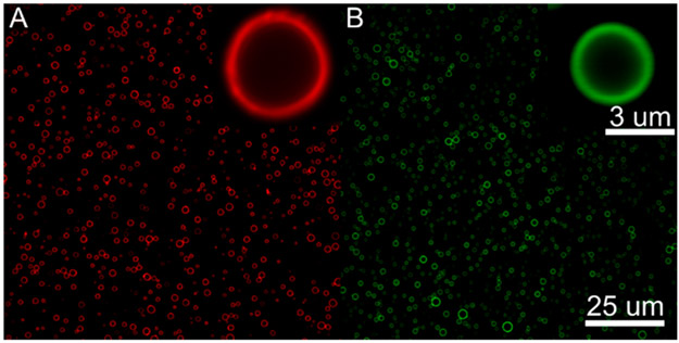
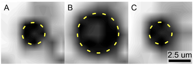
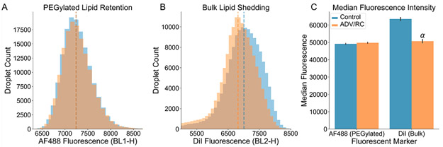
Targeted therapy and molecular imaging using ultrasound have been widely explored using microbubble contrast agents, and more recently, activatable droplet contrast agents that vaporize when exposed to focused ultrasound have been explored. These droplets are coated with a stabilizing, functionalizable shell, typically comprised of fully saturated phospholipids. While the shedding of the lipid shell under ultrasound exposure is a well-studied phenomenon in microbubbles, it has not been fully explored in droplet-based contrast agents, particularly in those that undergo a reversible phase change and recondense following vaporization. Here, we investigate the retention of the lipid shell following repeated transient vaporization events. Two separate fluorescent markers were used to track individual lipid subpopulations: PEGylated lipids, to which targeting ligands are typically bound, and non-PEGylated lipids, which primarily contribute to droplet stability. Following confirmation of the homogeneous surface distribution of each subpopulation of shell lipids using confocal microscopy, high-speed optical imaging provided visual evidence of the ability to repeatedly induce vaporization and recondensation in micron-scale droplets using 5.208 MHz, 3.17 MPa focused ultrasound pulses transmitted from an imaging transducer. Flow cytometry analysis indicated that while PEGylated lipids were fully retained following repeated transient phase change events, 20% of the bulk lipids were shed. While this likely contributed to an observed significant reduction in the average droplet diameter, the selective binding capabilities of droplets functionalized with an RGD peptide, targeted to the integrin αβ, were not affected. These results indicate that repeated droplet activation may promote shifts in the droplet size distribution but will not influence the accuracy of targeting for therapy or molecular imaging.
靶向治疗和分子成像已经广泛使用超声微泡造影剂进行探索,最近,人们还探索了在聚焦超声下蒸发的可激活液滴造影剂。这些液滴涂覆有一层稳定的、功能化的壳,通常由完全饱和的磷脂组成。虽然在超声暴露下脂质壳的脱落是微泡中一个研究得很好的现象,但在基于液滴的造影剂中尚未得到充分研究,特别是在那些经历可逆相变并在蒸发后重新凝结的造影剂中。在这里,我们研究了重复瞬态蒸发事件后脂质壳的保留情况。使用两种不同的荧光标记物来跟踪单个脂质亚群:通常与靶向配体结合的聚乙二醇化脂质,以及主要有助于液滴稳定的非聚乙二醇化脂质。在使用共聚焦显微镜确认每个壳脂质亚群的均匀表面分布之后,高速光学成像提供了可视化证据,证明可以使用 5.208MHz、3.17MPa 的聚焦超声脉冲从成像换能器重复诱导微米级液滴的蒸发和再凝结。流式细胞术分析表明,虽然聚乙二醇化脂质在重复瞬态相变事件后完全保留,但 20%的大部分脂质被脱落。虽然这可能导致观察到的平均液滴直径显著减小,但用靶向整合素 αβ 的 RGD 肽功能化的液滴的选择性结合能力不受影响。这些结果表明,重复的液滴激活可能会促进液滴尺寸分布的变化,但不会影响治疗或分子成像的靶向准确性。