Flower Luke, Carter John-Paul L, Rosales Lopez Juan, Henry Alun Marc
Anaesthesia, University College London Hospitals NHS Foundation Trust, London, UK
Emergency Department, University College London Hospitals NHS Foundation Trust, London, UK.
BMJ Case Rep. 2020 May 17;13(5):e235861. doi: 10.1136/bcr-2020-235861.
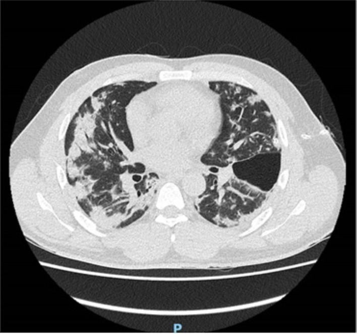
A 36-year-old man was brought to the emergency department with suspected COVID-19, following a 3-week history of cough, fevers and shortness of breath, worsening suddenly in the preceding 4 hours. On presentation he was hypoxaemic, with an SpO of 88% on 15 L/min oxygen, tachycardic and had no audible breath sounds on auscultation of the left hemithorax. Local guidelines recommended that the patient should be initiated on continuous positive airway pressure while investigations were awaited, however given the examination findings an emergency portable chest radiograph was performed. The chest radiograph demonstrated a left-sided tension pneumothorax. This was treated with emergency needle decompression, with good effect, followed by chest drain insertion. A repeat chest radiograph demonstrated lung re-expansion, and the patient was admitted to a COVID-19 specific ward for further observation. This case demonstrates tension pneumothorax as a possible complication of suspected COVID-19 and emphasises the importance of thorough history-taking and clinical examination.
一名36岁男性因疑似感染新型冠状病毒肺炎(COVID-19)被送往急诊科。此前他有3周的咳嗽、发热和呼吸急促病史,在就诊前4小时突然加重。就诊时,他存在低氧血症,吸入15升/分钟氧气时血氧饱和度(SpO)为88%,心动过速,左侧胸廓听诊未闻及呼吸音。当地指南建议在等待检查结果期间应开始对患者进行持续气道正压通气治疗,但鉴于检查结果,进行了急诊便携式胸部X线检查。胸部X线片显示左侧张力性气胸。对此进行了紧急针头减压治疗,效果良好,随后插入胸腔引流管。复查胸部X线片显示肺复张,患者被收入COVID-19专科病房进行进一步观察。该病例表明张力性气胸是疑似COVID-19可能出现的并发症,并强调了全面病史采集和临床检查的重要性。