• 文献检索
  • 文档翻译
  • 深度研究
  • 学术资讯
  • Suppr Zotero 插件Zotero 插件
  • 邀请有礼
  • 套餐&价格
  • 历史记录
应用&插件
Suppr Zotero 插件Zotero 插件浏览器插件Mac 客户端Windows 客户端微信小程序
定价
高级版会员购买积分包购买API积分包
服务
文献检索文档翻译深度研究API 文档MCP 服务
关于我们
关于 Suppr公司介绍联系我们用户协议隐私条款
关注我们

Suppr 超能文献

核心技术专利:CN118964589B侵权必究
粤ICP备2023148730 号-1Suppr @ 2026

文献检索

告别复杂PubMed语法,用中文像聊天一样搜索,搜遍4000万医学文献。AI智能推荐,让科研检索更轻松。

立即免费搜索

文件翻译

保留排版,准确专业,支持PDF/Word/PPT等文件格式,支持 12+语言互译。

免费翻译文档

深度研究

AI帮你快速写综述,25分钟生成高质量综述,智能提取关键信息,辅助科研写作。

立即免费体验

通过原子力显微镜对肝窦内皮细胞进行生物物理纳米表征。

Biophysical nanocharacterization of liver sinusoidal endothelial cells through atomic force microscopy.

作者信息

Zapotoczny Bartlomiej, Braet Filip, Wisse Eddie, Lekka Malgorzata, Szymonski Marek

机构信息

Institute of Nuclear Physics, Polish Academy of Sciences, 31-342, Krakow, Poland.

Faculty of Medicine and Health, School of Medical Sciences (Discipline of Anatomy and Histology), The University of Sydney, Sydney, NSW, 2006, Australia.

出版信息

Biophys Rev. 2020 Jun;12(3):625-636. doi: 10.1007/s12551-020-00699-0. Epub 2020 May 18.

DOI:10.1007/s12551-020-00699-0
PMID:32424787
原文链接:https://pmc.ncbi.nlm.nih.gov/articles/PMC7311612/
Abstract

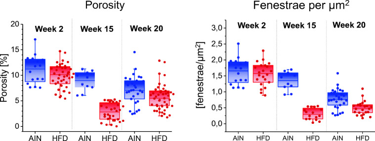
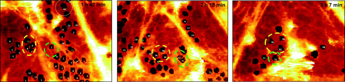
The structural-functional hallmark of the liver sinusoidal endothelium is the presence of fenestrae grouped in sieve plates. Fenestrae are open membrane bound pores supported by a (sub)membranous cytoskeletal lattice. Changes in number and diameter of fenestrae alter bidirectional transport between the sinusoidal blood and the hepatocytes. Their physiological relevance has been shown in different liver disease models. Although the structural organization of fenestrae has been well documented using different electron microscopy approaches, the dynamic nature of those pores remained an enigma until the recent developments in the research field of four dimensional (4-D) AFM. In this contribution we highlight how AFM as a biophysical nanocharacterization tool enhanced our understanding in the dynamic behaviour of liver sinusoidal endothelial fenestrae. Different AFM probing approaches, including spectroscopy, enabled mapping of topography and nanomechanical properties at unprecedented resolution under live cell imaging conditions. This dynamic biophysical characterization approach provided us with novel information on the 'short' life-span, formation, disappearance and closure of hepatic fenestrae. These observations are briefly reviewed against the existing literature.

摘要

肝窦内皮细胞的结构-功能特征是存在聚集在筛板中的窗孔。窗孔是由(亚)膜性细胞骨架晶格支撑的开放的膜结合孔。窗孔数量和直径的变化会改变窦状隙血液与肝细胞之间的双向运输。它们的生理相关性已在不同的肝病模型中得到证实。尽管使用不同的电子显微镜方法对窗孔的结构组织已有充分记录,但直到四维(4-D)原子力显微镜研究领域的最新进展,这些孔的动态性质仍然是个谜。在本论文中,我们强调了原子力显微镜作为一种生物物理纳米表征工具如何增强了我们对肝窦内皮窗孔动态行为的理解。不同的原子力显微镜探测方法,包括光谱学,能够在活细胞成像条件下以前所未有的分辨率绘制形貌和纳米力学特性图。这种动态生物物理表征方法为我们提供了有关肝窗孔“短暂”寿命、形成、消失和闭合的新信息。对照现有文献对这些观察结果进行了简要综述。

https://cdn.ncbi.nlm.nih.gov/pmc/blobs/61fd/7311612/ab979534ff9e/12551_2020_699_Fig6_HTML.jpg
https://cdn.ncbi.nlm.nih.gov/pmc/blobs/61fd/7311612/85b862eaa03a/12551_2020_699_Fig1_HTML.jpg
https://cdn.ncbi.nlm.nih.gov/pmc/blobs/61fd/7311612/9b05312a31b3/12551_2020_699_Fig2_HTML.jpg
https://cdn.ncbi.nlm.nih.gov/pmc/blobs/61fd/7311612/1c1e03c85220/12551_2020_699_Fig3_HTML.jpg
https://cdn.ncbi.nlm.nih.gov/pmc/blobs/61fd/7311612/d75344729420/12551_2020_699_Fig4_HTML.jpg
https://cdn.ncbi.nlm.nih.gov/pmc/blobs/61fd/7311612/efae4495fe67/12551_2020_699_Fig5_HTML.jpg
https://cdn.ncbi.nlm.nih.gov/pmc/blobs/61fd/7311612/ab979534ff9e/12551_2020_699_Fig6_HTML.jpg

相似文献

1
Biophysical nanocharacterization of liver sinusoidal endothelial cells through atomic force microscopy.通过原子力显微镜对肝窦内皮细胞进行生物物理纳米表征。
Biophys Rev. 2020 Jun;12(3):625-636. doi: 10.1007/s12551-020-00699-0. Epub 2020 May 18.
2
Tracking Fenestrae Dynamics in Live Murine Liver Sinusoidal Endothelial Cells.跟踪活体小鼠肝窦内皮细胞窗孔动力学。
Hepatology. 2019 Feb;69(2):876-888. doi: 10.1002/hep.30232. Epub 2019 Jan 3.
3
Actin-spectrin scaffold supports open fenestrae in liver sinusoidal endothelial cells.肌动蛋白-血影蛋白支架支撑肝窦内皮细胞中的开放窗孔。
Traffic. 2019 Dec;20(12):932-942. doi: 10.1111/tra.12700. Epub 2019 Oct 23.
4
Three-dimensional organization of fenestrae labyrinths in liver sinusoidal endothelial cells.肝窦内皮细胞窗孔迷宫的三维组织结构。
Liver Int. 2009 Apr;29(4):603-13. doi: 10.1111/j.1478-3231.2008.01836.x. Epub 2008 Jul 23.
5
Structure and dynamics of the fenestrae-associated cytoskeleton of rat liver sinusoidal endothelial cells.大鼠肝窦内皮细胞窗孔相关细胞骨架的结构与动力学
Hepatology. 1995 Jan;21(1):180-9.
6
Probing the unseen structure and function of liver cells through atomic force microscopy.通过原子力显微镜探究肝细胞的未知结构和功能。
Semin Cell Dev Biol. 2018 Jan;73:13-30. doi: 10.1016/j.semcdb.2017.07.001. Epub 2017 Jul 6.
7
Characterizing liver sinusoidal endothelial cell fenestrae on soft substrates upon AFM imaging and deep learning.在原子力显微镜成像和深度学习中,对软基底上的肝窦内皮细胞窗孔进行特征化描述。
Biochim Biophys Acta Gen Subj. 2020 Dec;1864(12):129702. doi: 10.1016/j.bbagen.2020.129702. Epub 2020 Aug 16.
8
New observations on cytoskeleton and fenestrae in isolated rat liver sinusoidal endothelial cells.对分离的大鼠肝窦内皮细胞中细胞骨架和窗孔的新观察
J Gastroenterol Hepatol. 1995;10 Suppl 1:S3-7. doi: 10.1111/j.1440-1746.1995.tb01792.x.
9
Comparative atomic force and scanning electron microscopy: an investigation on fenestrated endothelial cells in vitro.比较原子力显微镜和扫描电子显微镜:体外有窗孔内皮细胞的研究
J Microsc. 1996 Jan;181(Pt 1):10-7. doi: 10.1046/j.1365-2818.1996.72348.x.
10
Comparative scanning, transmission and atomic force microscopy of the microtubular cytoskeleton in fenestrated liver endothelial cells.窗孔状肝内皮细胞微管细胞骨架的比较扫描、透射和原子力显微镜检查
Scanning Microsc Suppl. 1996;10:225-35; discussion 235-6.

引用本文的文献

1
Liver Sinusoidal Endothelial Cells and Their Regulation of Immunology, Collagenization, and Bioreactivity in Fatty Liver: A Narrative Review.肝窦内皮细胞及其在脂肪肝中对免疫、胶原化和生物活性的调节:一篇叙述性综述
Int J Mol Sci. 2025 Aug 19;26(16):8006. doi: 10.3390/ijms26168006.
2
A specific role for endothelial EPLIN-isoform-regulated actin dynamics in neutrophil transmigration.内皮细胞EPLIN异构体调节的肌动蛋白动力学在中性粒细胞迁移中的特定作用。
Sci Rep. 2025 May 5;15(1):15698. doi: 10.1038/s41598-025-98192-9.
3
Mimicking the Liver Sinusoidal Endothelial Cell Niche In Vitro to Enhance Fenestration in a Genetic Model of Systemic Inflammation.

本文引用的文献

1
Actin-spectrin scaffold supports open fenestrae in liver sinusoidal endothelial cells.肌动蛋白-血影蛋白支架支撑肝窦内皮细胞中的开放窗孔。
Traffic. 2019 Dec;20(12):932-942. doi: 10.1111/tra.12700. Epub 2019 Oct 23.
2
Vascular Endothelial Cell Biology: An Update.血管内皮细胞生物学:最新进展。
Int J Mol Sci. 2019 Sep 7;20(18):4411. doi: 10.3390/ijms20184411.
3
Application of a layered model for determination of the elasticity of biological systems.分层模型在生物系统弹性测定中的应用。
体外模拟肝窦内皮细胞微环境以增强全身炎症遗传模型中的窗孔形成
Cells. 2025 Apr 21;14(8):621. doi: 10.3390/cells14080621.
4
From fixed-dried to wet-fixed to live - comparative super-resolution microscopy of liver sinusoidal endothelial cell fenestrations.从固定干燥到湿固定再到活体——肝窦内皮细胞窗孔的比较超分辨率显微镜观察
Nanophotonics. 2022 Apr 20;11(10):2253-2270. doi: 10.1515/nanoph-2021-0818. eCollection 2022 May.
5
Early and late phases of liver sinusoidal endothelial cell (LSEC) defenestration in mouse model of systemic inflammation.在系统性炎症的小鼠模型中,肝窦内皮细胞(LSEC)窗孔形成的早期和晚期阶段。
Cell Mol Biol Lett. 2024 Nov 11;29(1):139. doi: 10.1186/s11658-024-00655-w.
6
Fenestrated Endothelial Cells across Organs: Insights into Kidney Function and Disease.器官间的窗孔内皮细胞:对肾脏功能和疾病的深入了解。
Int J Mol Sci. 2024 Aug 22;25(16):9107. doi: 10.3390/ijms25169107.
7
Protein disulfide isomerase A1 regulates fenestration dynamics in primary mouse liver sinusoidal endothelial cells (LSECs).蛋白二硫键异构酶 A1 调控原代小鼠肝窦内皮细胞(LSECs)的窗孔动态。
Redox Biol. 2024 Jun;72:103162. doi: 10.1016/j.redox.2024.103162. Epub 2024 Apr 16.
8
Determining Spatial Variability of Elastic Properties for Biological Samples Using AFM.使用原子力显微镜测定生物样品弹性特性的空间变异性。
Micromachines (Basel). 2023 Jan 11;14(1):182. doi: 10.3390/mi14010182.
9
Multimodal on-chip nanoscopy and quantitative phase imaging reveals the nanoscale morphology of liver sinusoidal endothelial cells.多模态片上纳米显微镜和定量相位成像揭示了肝窦内皮细胞的纳米形态。
Proc Natl Acad Sci U S A. 2021 Nov 23;118(47). doi: 10.1073/pnas.2115323118.
10
The wHole Story About Fenestrations in LSEC.肝血窦内皮细胞窗孔的完整故事
Front Physiol. 2021 Sep 13;12:735573. doi: 10.3389/fphys.2021.735573. eCollection 2021.
Micron. 2019 Sep;124:102705. doi: 10.1016/j.micron.2019.102705. Epub 2019 Jun 15.
4
The Effects of Metformin on Age-Related Changes in the Liver Sinusoidal Endothelial Cell.二甲双胍对肝脏窦状内皮细胞衰老相关变化的影响。
J Gerontol A Biol Sci Med Sci. 2020 Jan 20;75(2):278-285. doi: 10.1093/gerona/glz153.
5
Endothelial cell barriers: Transport of molecules between blood and tissues.内皮细胞屏障:血液与组织间的分子转运。
Traffic. 2019 Jun;20(6):390-403. doi: 10.1111/tra.12645. Epub 2019 Apr 24.
6
LSEC Fenestrae Are Preserved Despite Pro-inflammatory Phenotype of Liver Sinusoidal Endothelial Cells in Mice on High Fat Diet.尽管高脂饮食小鼠肝脏窦状内皮细胞具有促炎表型,但肝血窦内皮窗孔仍得以保留。
Front Physiol. 2019 Feb 12;10:6. doi: 10.3389/fphys.2019.00006. eCollection 2019.
7
Actin Depolymerization in Dedifferentiated Liver Sinusoidal Endothelial Cells Promotes Fenestrae Re-Formation.去分化肝窦内皮细胞中的肌动蛋白解聚促进窗孔重新形成。
Hepatol Commun. 2018 Dec 28;3(2):213-219. doi: 10.1002/hep4.1301. eCollection 2019 Feb.
8
Raman spectroscopy-based insight into lipid droplets presence and contents in liver sinusoidal endothelial cells and hepatocytes.基于拉曼光谱的肝窦内皮细胞和肝细胞中脂滴存在和含量的研究。
J Biophotonics. 2019 Apr;12(4):e201800290. doi: 10.1002/jbio.201800290. Epub 2019 Jan 21.
9
Manipulating fenestrations in young and old liver sinusoidal endothelial cells.调控年轻和年老肝窦内皮细胞的窗孔。
Am J Physiol Gastrointest Liver Physiol. 2019 Jan 1;316(1):G144-G154. doi: 10.1152/ajpgi.00179.2018. Epub 2018 Oct 4.
10
Tracking Fenestrae Dynamics in Live Murine Liver Sinusoidal Endothelial Cells.跟踪活体小鼠肝窦内皮细胞窗孔动力学。
Hepatology. 2019 Feb;69(2):876-888. doi: 10.1002/hep.30232. Epub 2019 Jan 3.