Almeida João, Leal Cecília, Figueiredo Luísa
Department of Radiology, Hospital de Santa Marta, Rua de Santa Marta, 1169-024, Lisbon, Portugal.
Insights Imaging. 2020 May 19;11(1):70. doi: 10.1186/s13244-020-00877-4.
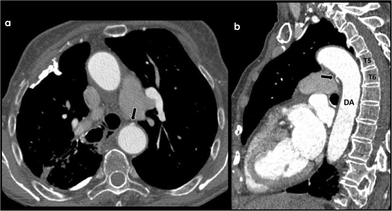
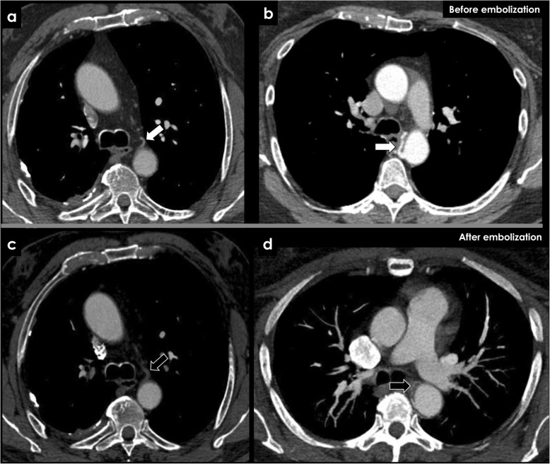
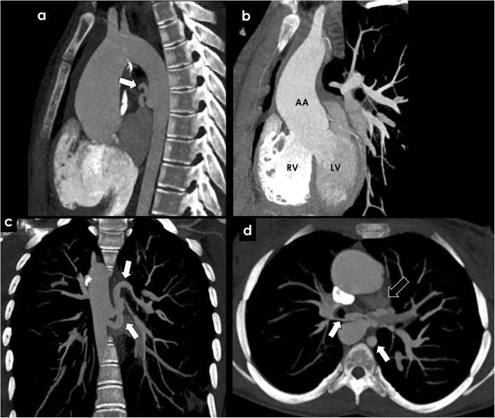
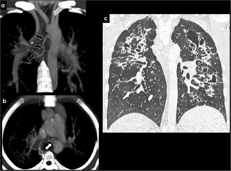
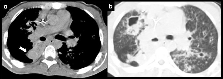
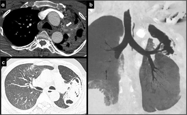
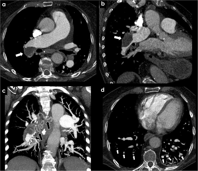
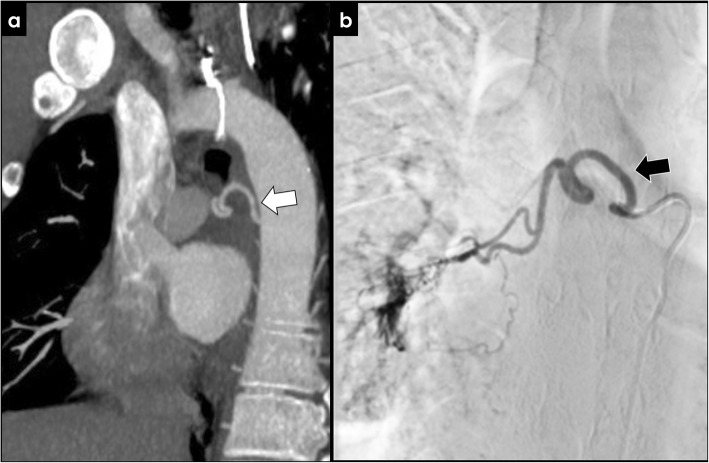
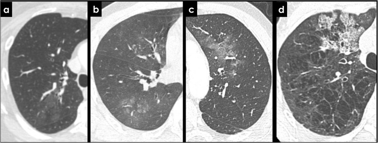
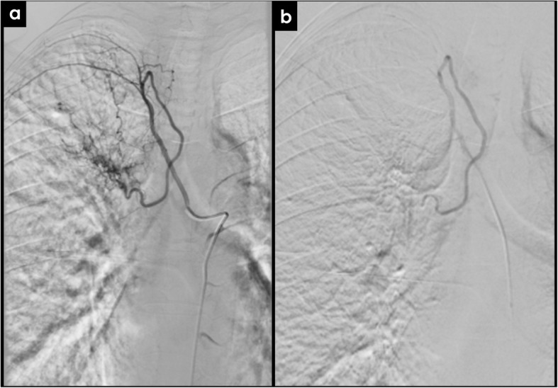
The enlargement of the bronchial arteries occurs in a multitude of congenital and acquired diseases and is responsible for the majority of cases of hemoptysis. In this review, we provide a simplified imaging approach to the evaluation of the bronchial arteries. We highlight the anatomy and function of the bronchial arteries, typical imaging findings, how to recognize bronchial artery dilatation, and its underlying causes. Contrast-enhanced computer tomography plays a major role in diagnosing bronchial artery enlargement and also improves treatment planning. Bronchial artery embolization has proven to be effective in controlling the potential hazardous hemoptysis.
支气管动脉扩张见于多种先天性和后天性疾病,是大多数咯血病例的病因。在本综述中,我们提供一种简化的影像学方法来评估支气管动脉。我们重点介绍支气管动脉的解剖结构和功能、典型的影像学表现、如何识别支气管动脉扩张及其潜在病因。增强计算机断层扫描在诊断支气管动脉扩张中起主要作用,也有助于改善治疗方案的制定。支气管动脉栓塞已被证明在控制潜在的危险性咯血方面有效。