Wasserman Paul L, Wiesler Carissa, Kurra Chandana, Omman Reeba, Taylor Kristin, Puri Ruchir
Department of Radiology, UF Health Jacksonville, 655 West 8th Street, C90, Jacksonville, FL 32209, USA.
Department of Pathology, UF Health Jacksonville, Jacksonville, FL 32209, USA.
Radiol Case Rep. 2020 May 15;15(7):1029-1038. doi: 10.1016/j.radcr.2020.04.025. eCollection 2020 Jul.
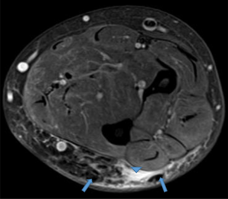
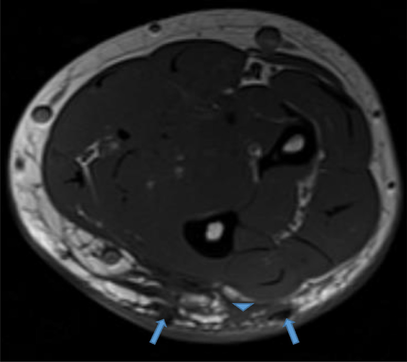
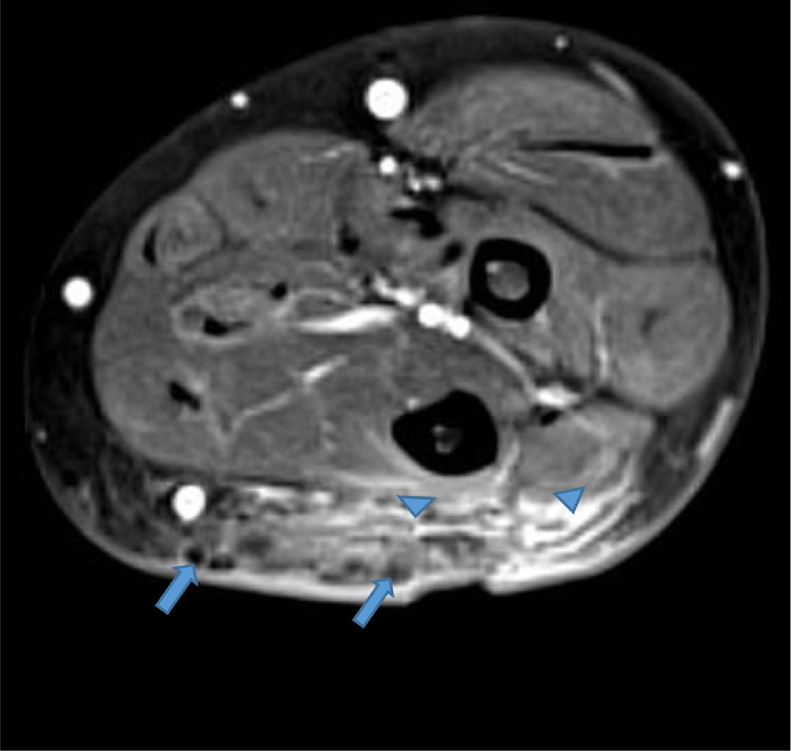
Soft tissue calcifications associated with various connective tissue diseases such as dermatomyositis and scleroderma have been well documented Plaque-like sheets of subcutaneous calcifications presenting as an indurated soft tissue mass in a patient with primary Sjogren syndrome have been rarely documented in the literature. We present the magnetic resonance and conventional radiographic findings of calcinosis cutis and calcinosis circumscripta of a 47-year-old woman with biopsy proven Sjogren syndrome. We also delineate various types of soft tissue calcification, histopathology of calcinosis cutis, and current treatment options. Recognizing the magnetic resonance characteristics of this phenomenon may prove useful to radiologists, especially in the absence of clinical history and conventional radiographs.
与各种结缔组织疾病(如皮肌炎和硬皮病)相关的软组织钙化已有充分记录。原发性干燥综合征患者出现皮下钙化的斑块状片状物,表现为硬结性软组织肿块,这在文献中鲜有记载。我们展示了一名经活检证实为干燥综合征的47岁女性皮肤钙化和局限性钙化的磁共振成像及传统放射学表现。我们还阐述了各种类型的软组织钙化、皮肤钙化的组织病理学以及当前的治疗选择。认识到这种现象的磁共振特征可能对放射科医生有用,尤其是在缺乏临床病史和传统放射照片的情况下。