• 文献检索
  • 文档翻译
  • 深度研究
  • 学术资讯
  • Suppr Zotero 插件Zotero 插件
  • 邀请有礼
  • 套餐&价格
  • 历史记录
应用&插件
Suppr Zotero 插件Zotero 插件浏览器插件Mac 客户端Windows 客户端微信小程序
定价
高级版会员购买积分包购买API积分包
服务
文献检索文档翻译深度研究API 文档MCP 服务
关于我们
关于 Suppr公司介绍联系我们用户协议隐私条款
关注我们

Suppr 超能文献

核心技术专利:CN118964589B侵权必究
粤ICP备2023148730 号-1Suppr @ 2026

文献检索

告别复杂PubMed语法,用中文像聊天一样搜索,搜遍4000万医学文献。AI智能推荐,让科研检索更轻松。

立即免费搜索

文件翻译

保留排版,准确专业,支持PDF/Word/PPT等文件格式,支持 12+语言互译。

免费翻译文档

深度研究

AI帮你快速写综述,25分钟生成高质量综述,智能提取关键信息,辅助科研写作。

立即免费体验

分析聚焦光斑照明方法的超分辨率特性。

Analyzing the super-resolution characteristics of focused-spot illumination approaches.

机构信息

University of Colorado Boulder, Department of Electrical, Computer and Energy Engineering, Boulder,, United States.

University of Colorado Boulder, Department of Applied Mathematics, Boulder, Colorado, United States.

出版信息

J Biomed Opt. 2020 May;25(5):1-13. doi: 10.1117/1.JBO.25.5.056501.

DOI:10.1117/1.JBO.25.5.056501
PMID:32441065
原文链接:https://pmc.ncbi.nlm.nih.gov/articles/PMC7240318/
Abstract

SIGNIFICANCE

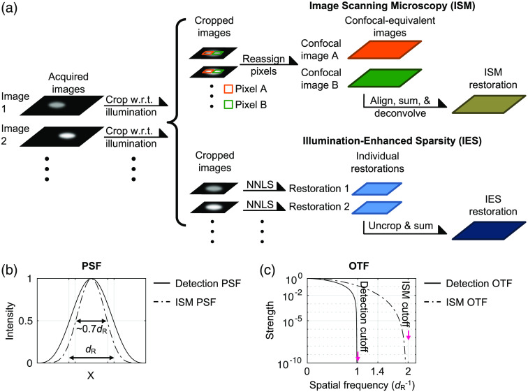
It is commonly assumed that using the objective lens to create a tightly focused light spot for illumination provides a twofold resolution improvement over the Rayleigh resolution limit and that resolution improvement is independent of object properties. Nevertheless, such an assumption has not been carefully examined. We examine this assumption by analyzing the performance of two super-resolution methods, known as image scanning microscopy (ISM) and illumination-enhanced sparsity (IES).

AIM

We aim to identify the fundamental differences between the two methods, and to provide examples that help researchers determine which method to utilize for different imaging conditions.

APPROACH

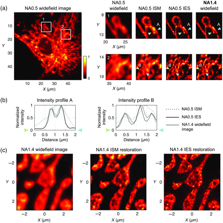
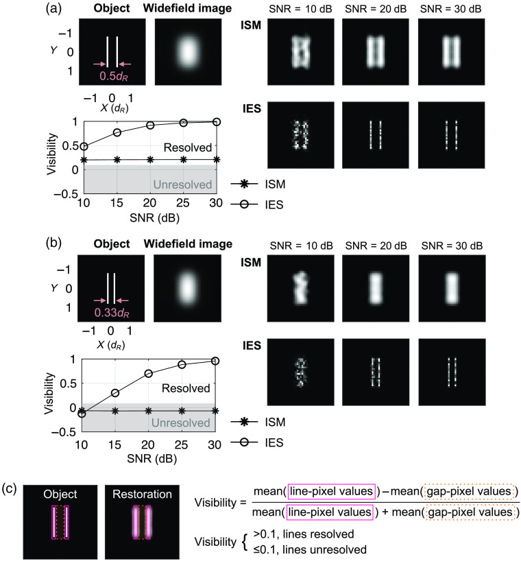
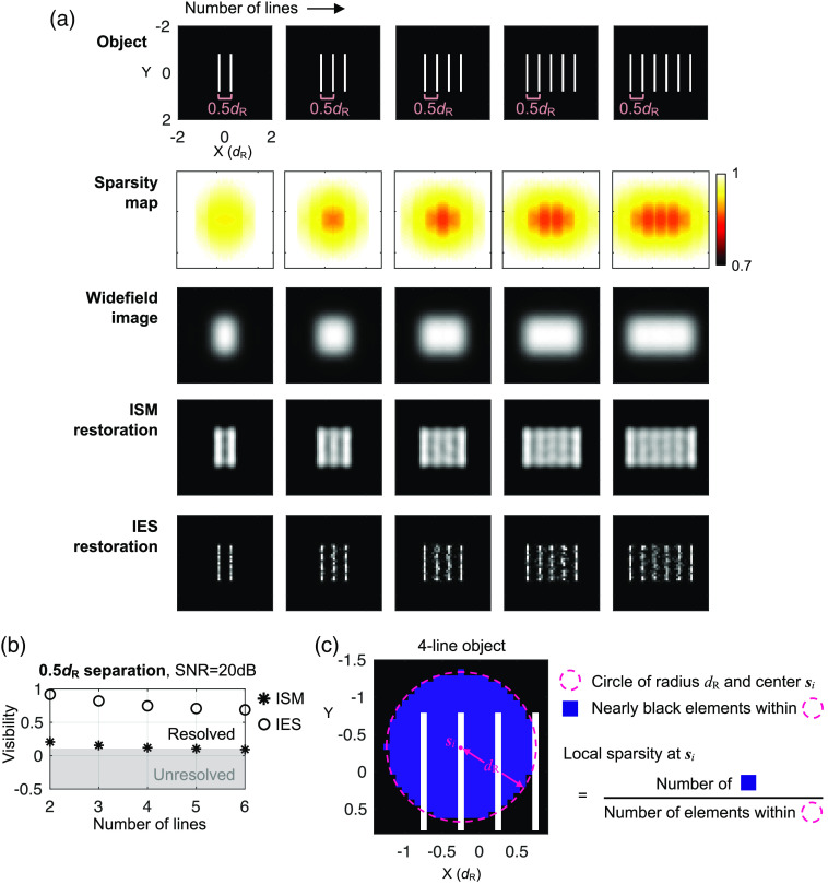
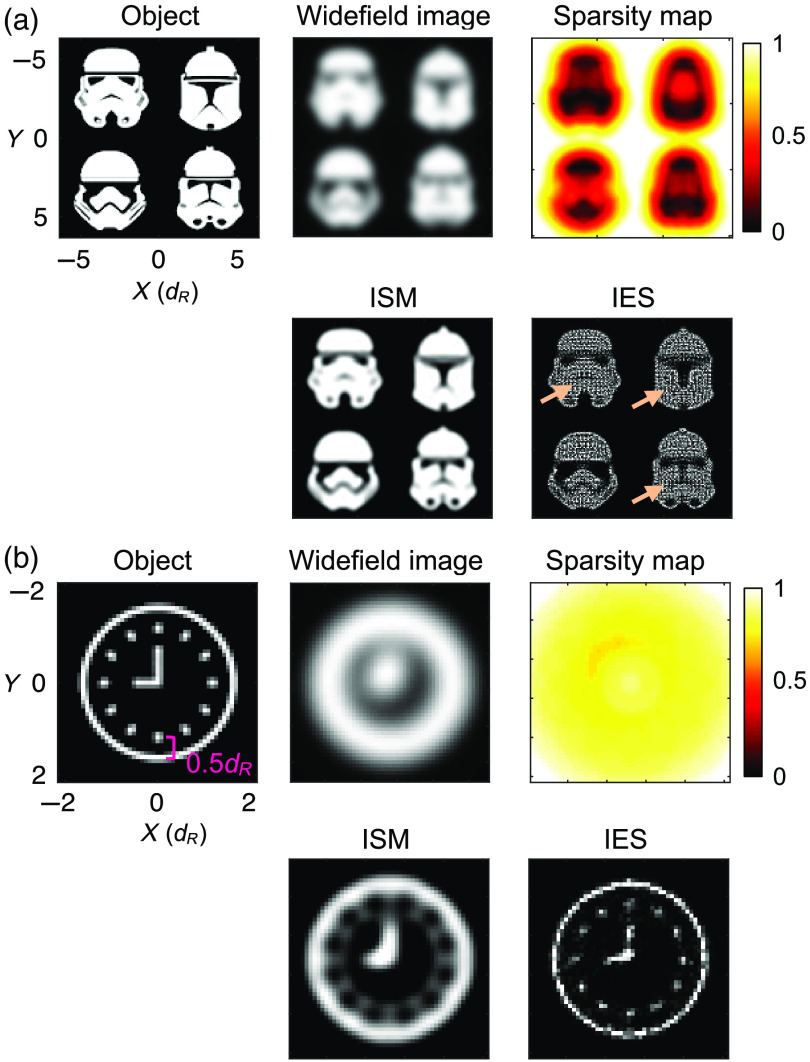
We input the same image datasets into the two methods and analyze their restorations. In numerical simulations, we design objects of distinct brightness and sparsity levels for imaging. We use biological imaging experiments to verify the simulation results.

RESULTS

The resolution of IES often exceeds twice the Rayleigh resolution limit when imaging sparse objects. A decrease in object sparsity negatively affects the resolution improvement in both methods.

CONCLUSIONS

The IES method is superior for imaging sparse objects with its main features being bright and small against a dark, large background. For objects that are largely bright with small dark features, the ISM method is favorable.

摘要

意义

人们普遍认为,使用物镜创建用于照明的紧密聚焦光斑可以提供两倍于瑞利分辨率极限的分辨率提高,并且分辨率提高与物体性质无关。然而,这种假设尚未经过仔细检查。我们通过分析两种超分辨率方法(称为图像扫描显微镜(ISM)和照明增强稀疏性(IES))的性能来检验该假设。

目的

我们旨在确定这两种方法之间的根本区别,并提供有助于研究人员确定在不同成像条件下使用哪种方法的示例。

方法

我们将相同的图像数据集输入到两种方法中,并分析它们的恢复结果。在数值模拟中,我们设计了具有不同亮度和稀疏度水平的物体进行成像。我们使用生物成像实验来验证模拟结果。

结果

当对稀疏物体进行成像时,IES 的分辨率通常超过瑞利分辨率极限的两倍。物体稀疏度的降低会对两种方法的分辨率提高产生负面影响。

结论

IES 方法在对具有明亮和小的背景与暗、大背景的稀疏物体进行成像方面具有优势。对于主要为明亮且带有小暗特征的物体,ISM 方法更为有利。

https://cdn.ncbi.nlm.nih.gov/pmc/blobs/9296/7240318/c746200f8b18/JBO-025-056501-g005.jpg
https://cdn.ncbi.nlm.nih.gov/pmc/blobs/9296/7240318/7ce3cf26ae0c/JBO-025-056501-g001.jpg
https://cdn.ncbi.nlm.nih.gov/pmc/blobs/9296/7240318/636655935be8/JBO-025-056501-g002.jpg
https://cdn.ncbi.nlm.nih.gov/pmc/blobs/9296/7240318/6d2cb5cc82d3/JBO-025-056501-g003.jpg
https://cdn.ncbi.nlm.nih.gov/pmc/blobs/9296/7240318/bd488658f69d/JBO-025-056501-g004.jpg
https://cdn.ncbi.nlm.nih.gov/pmc/blobs/9296/7240318/c746200f8b18/JBO-025-056501-g005.jpg

相似文献

1
Analyzing the super-resolution characteristics of focused-spot illumination approaches.分析聚焦光斑照明方法的超分辨率特性。
J Biomed Opt. 2020 May;25(5):1-13. doi: 10.1117/1.JBO.25.5.056501.
2
Achieving superresolution with illumination-enhanced sparsity.通过增强照明稀疏性实现超分辨率。
Opt Express. 2018 Apr 16;26(8):9850-9865. doi: 10.1364/OE.26.009850.
3
Quantum limits of super-resolution of optical sparse objects via sparsity constraint.基于稀疏约束的光学稀疏物体超分辨率的量子极限
Opt Express. 2012 Oct 8;20(21):23235-52. doi: 10.1364/OE.20.023235.
4
ℓ-regularized maximum likelihood estimation with focused-spot illumination quadruples the diffraction-limited resolution in fluorescence microscopy.
Opt Express. 2020 Dec 21;28(26):39413-39429. doi: 10.1364/OE.411574.
5
Resolution improvement of multifocal structured illumination microscopy with sparse Bayesian learning algorithm.基于稀疏贝叶斯学习算法的多焦点结构照明显微镜分辨率提升
Opt Express. 2018 Nov 26;26(24):31430-31438. doi: 10.1364/OE.26.031430.
6
Resolution and super-resolution.分辨率与超分辨率。
Microsc Res Tech. 2017 Jun;80(6):590-598. doi: 10.1002/jemt.22834. Epub 2017 Feb 9.
7
High-speed super-resolution imaging with compressive imaging-based structured illumination microscopy.基于压缩成像的结构光照明显微镜的高速超分辨率成像。
Opt Express. 2022 Apr 25;30(9):14287-14299. doi: 10.1364/OE.453554.
8
Ultrathin Layered Hyperbolic Metamaterial-Assisted Illumination Nanoscopy.超薄层状双曲超材料辅助照明纳米显微镜技术。
Nano Lett. 2022 Jul 27;22(14):5916-5921. doi: 10.1021/acs.nanolett.2c01932. Epub 2022 Jul 14.
9
Fast 3D super-resolution imaging using a digital micromirror device and binary holography.使用数字微镜器件和二进制全息术进行快速 3D 超分辨率成像。
J Biomed Opt. 2021 Nov;26(11). doi: 10.1117/1.JBO.26.11.116502.
10
Signal, noise and resolution in linear and nonlinear structured-illumination microscopy.线性和非线性结构光照明显微镜中的信号、噪声和分辨率。
J Microsc. 2019 Jan;273(1):3-25. doi: 10.1111/jmi.12753. Epub 2018 Aug 28.

引用本文的文献

1
Imaging plant cells and organs with light-sheet and super-resolution microscopy.利用光片和超分辨率显微镜对植物细胞和器官进行成像。
Plant Physiol. 2022 Feb 4;188(2):683-702. doi: 10.1093/plphys/kiab349.
2
Current and future advances in fluorescence-based visualization of plant cell wall components and cell wall biosynthetic machineries.基于荧光的植物细胞壁成分和细胞壁生物合成机制可视化的当前及未来进展
Biotechnol Biofuels. 2021 Mar 29;14(1):78. doi: 10.1186/s13068-021-01922-0.

本文引用的文献

1
Achieving superresolution with illumination-enhanced sparsity.通过增强照明稀疏性实现超分辨率。
Opt Express. 2018 Apr 16;26(8):9850-9865. doi: 10.1364/OE.26.009850.
2
A joint Richardson-Lucy deconvolution algorithm for the reconstruction of multifocal structured illumination microscopy data.一种用于多焦点结构光照明显微镜数据重建的联合理查森-露西反卷积算法。
Methods Appl Fluoresc. 2015 Jan 16;3(1):014002. doi: 10.1088/2050-6120/3/1/014002.
3
Image scanning microscopy with a quadrant detector.带象限探测器的图像扫描显微镜。
Opt Lett. 2015 Nov 15;40(22):5355-8. doi: 10.1364/OL.40.005355.
4
Super-resolution spinning-disk confocal microscopy using optical photon reassignment.使用光学光子重新分配的超分辨率旋转盘共聚焦显微镜。
Opt Express. 2015 Jun 1;23(11):15003-11. doi: 10.1364/OE.23.015003.
5
Richardson-Lucy deconvolution as a general tool for combining images with complementary strengths.Richardson-Lucy 去卷积作为一种将具有互补优势的图像组合在一起的通用工具。
Chemphyschem. 2014 Mar 17;15(4):794-800. doi: 10.1002/cphc.201300831. Epub 2014 Jan 16.
6
Instant super-resolution imaging in live cells and embryos via analog image processing.通过模拟图像处理实现活细胞和胚胎的即时超分辨率成像。
Nat Methods. 2013 Nov;10(11):1122-6. doi: 10.1038/nmeth.2687. Epub 2013 Oct 6.
7
Superresolution by image scanning microscopy using pixel reassignment.像素重分配的图像扫描显微镜超分辨率。
Opt Lett. 2013 Aug 1;38(15):2889-92. doi: 10.1364/OL.38.002889.
8
Resolution doubling in live, multicellular organisms via multifocal structured illumination microscopy.利用多点结构照明显微镜在活体多细胞生物中实现分辨率加倍。
Nat Methods. 2012 May 13;9(7):749-54. doi: 10.1038/nmeth.2025.
9
Image scanning microscopy.图像扫描显微镜。
Phys Rev Lett. 2010 May 14;104(19):198101. doi: 10.1103/PhysRevLett.104.198101. Epub 2010 May 10.
10
Analysis of confocal laser-microscope optics for 3-D fluorescence correlation spectroscopy.用于三维荧光相关光谱的共聚焦激光显微镜光学分析。
Appl Opt. 1991 Apr 1;30(10):1185-95. doi: 10.1364/AO.30.001185.