• 文献检索
  • 文档翻译
  • 深度研究
  • 学术资讯
  • Suppr Zotero 插件Zotero 插件
  • 邀请有礼
  • 套餐&价格
  • 历史记录
应用&插件
Suppr Zotero 插件Zotero 插件浏览器插件Mac 客户端Windows 客户端微信小程序
定价
高级版会员购买积分包购买API积分包
服务
文献检索文档翻译深度研究API 文档MCP 服务
关于我们
关于 Suppr公司介绍联系我们用户协议隐私条款
关注我们

Suppr 超能文献

核心技术专利:CN118964589B侵权必究
粤ICP备2023148730 号-1Suppr @ 2026

文献检索

告别复杂PubMed语法,用中文像聊天一样搜索,搜遍4000万医学文献。AI智能推荐,让科研检索更轻松。

立即免费搜索

文件翻译

保留排版,准确专业,支持PDF/Word/PPT等文件格式,支持 12+语言互译。

免费翻译文档

深度研究

AI帮你快速写综述,25分钟生成高质量综述,智能提取关键信息,辅助科研写作。

立即免费体验

CareDose 4D 联合 Karl 3D 技术在 COVID-19 低剂量 CT 随访中的应用。

Application of CareDose 4D combined with Karl 3D technology in the low dose computed tomography for the follow-up of COVID-19.

机构信息

Department of Radiology, China Resources & WISCO General Hospital, Wuhan, 430080, China.

Department of Biochemistry and Molecular Biology, China Medical University, Shenyang, 110122, China.

出版信息

BMC Med Imaging. 2020 May 24;20(1):56. doi: 10.1186/s12880-020-00456-5.

DOI:10.1186/s12880-020-00456-5
PMID:32448136
原文链接:https://pmc.ncbi.nlm.nih.gov/articles/PMC7245769/
Abstract

BACKGROUND

Coronavirus disease 2019 (COVID-19) is a highly infectious disease caused by the new coronavirus. Previous studies have shown that the chest CT examination plays an important role in the diagnosis and monitoring of COVID-19. However, some patients with COVID-19 had low white blood cell counts and reduced lymphocyte ratios. Multiple CT examinations may cause radiation damages as well as increase the apoptosis of peripheral blood lymphocytes. A new low-dose CT method should be developed because the regular CT may aggravate the disease.

METHOD

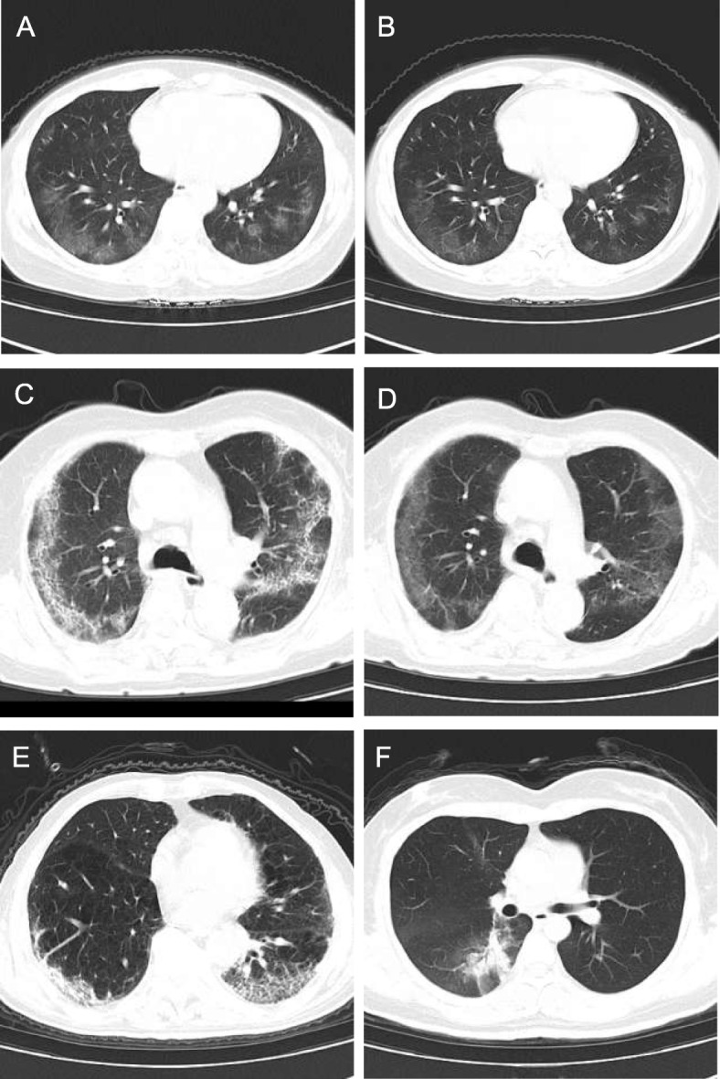
Sixty cases were randomly divided into the study group (n = 30) and control group (n = 30). The lung window was reconstructed by Karl 3D iterative technique in the study group. The image quality was subjectively evaluated by two senior chest group diagnostic physicians using a 5-point double-blind method. The value of CT measurement and its standard deviation (SD) was used as an objective evaluation criteria. The volume of CT dose index (CTDI), dose length product (DLP) and effective dose (ED) from the two groups were compared and analyzed statistically.

RESULT

There was no significant difference in the occurrence rates of ground glass opacities, consolidation, crazy-paving pattern, fiber cable shadow and axial interstitial thickening between the study group and control group (p > 0.05). In addition, no significant difference was found for the subjective score of overall image quality and image noise level (SD) between the two groups (p > 0.05). However, significant differences was found in CTDI, DLP, and ED between the study group and the control group (p < 0.05). The effective dose of the study group was reduced by 76% compared to the control group.

CONCLUSION

CareDose 4D low-dose scanning combined with Karl 3D iterative reconstruction technology can not only greatly reduce the radiation dose, but also provide images that meet the diagnostic criteria of COVID-19, which can be used as a routine method for the follow-up of COVID-19 patients.

摘要

背景

新型冠状病毒引发的 2019 年冠状病毒病(COVID-19)是一种高度传染性疾病。既往研究表明,胸部 CT 检查在 COVID-19 的诊断和监测中发挥着重要作用。然而,部分 COVID-19 患者存在白细胞计数降低和淋巴细胞比值减少的情况。多次 CT 检查可能会造成辐射损伤,增加外周血淋巴细胞凋亡。为避免常规 CT 加重病情,应开发新的低剂量 CT 方法。

方法

60 例患者被随机分为研究组(n=30)和对照组(n=30)。研究组采用卡尔三维迭代技术重建肺窗。由 2 位资深胸部组诊断医师采用 5 分制双盲法对图像质量进行主观评估。采用 CT 测量值及其标准差(SD)作为客观评价标准。对比分析两组 CT 剂量指数(CTDI)、剂量长度乘积(DLP)和有效剂量(ED)值。

结果

研究组和对照组的磨玻璃影、实变、铺路石征、纤维条索影和轴位间质增厚的发生率比较,差异均无统计学意义(p>0.05)。两组的整体图像质量主观评分和图像噪声水平(SD)比较,差异均无统计学意义(p>0.05)。但研究组的 CTDI、DLP 和 ED 与对照组比较,差异均有统计学意义(p<0.05)。与对照组相比,研究组的有效剂量降低了 76%。

结论

CareDose 4D 低剂量扫描联合卡尔三维迭代重建技术不仅能大幅降低辐射剂量,还能提供满足 COVID-19 诊断标准的图像,可作为 COVID-19 患者随访的常规方法。

https://cdn.ncbi.nlm.nih.gov/pmc/blobs/cd27/7245769/c06cca2e73af/12880_2020_456_Fig1_HTML.jpg

相似文献

1
Application of CareDose 4D combined with Karl 3D technology in the low dose computed tomography for the follow-up of COVID-19.CareDose 4D 联合 Karl 3D 技术在 COVID-19 低剂量 CT 随访中的应用。
BMC Med Imaging. 2020 May 24;20(1):56. doi: 10.1186/s12880-020-00456-5.
2
Analysis of clinical features and imaging signs of COVID-19 with the assistance of artificial intelligence.人工智能辅助分析 COVID-19 的临床特征和影像征象。
Eur Rev Med Pharmacol Sci. 2020 Aug;24(15):8210-8218. doi: 10.26355/eurrev_202008_22510.
3
A Comparison of Clinical and Chest CT Findings in Patients With Influenza A (H1N1) Virus Infection and Coronavirus Disease (COVID-19).甲型流感病毒(H1N1)感染与新型冠状病毒肺炎(COVID-19)患者的临床和胸部 CT 表现比较。
AJR Am J Roentgenol. 2020 Nov;215(5):1065-1071. doi: 10.2214/AJR.20.23214. Epub 2020 May 26.
4
Dose-optimised chest computed tomography for diagnosis of Coronavirus Disease 2019 (COVID-19) - Evaluation of image quality and diagnostic impact.用于2019冠状病毒病(COVID-19)诊断的剂量优化胸部计算机断层扫描——图像质量和诊断影响评估
J Radiol Prot. 2020 Sep;40(3):877-891. doi: 10.1088/1361-6498/aba16a.
5
CT Quantitative Analysis and Its Relationship with Clinical Features for Assessing the Severity of Patients with COVID-19.CT 定量分析及其与临床特征的关系,用于评估 COVID-19 患者的严重程度。
Korean J Radiol. 2020 Jul;21(7):859-868. doi: 10.3348/kjr.2020.0293.
6
Dynamic changes of Chest CT follow-up in Coronavirus Disease-19 (COVID-19) pneumonia: relationship to clinical typing.新冠肺炎(COVID-19)肺炎的胸部 CT 随访动态变化:与临床分型的关系。
BMC Med Imaging. 2020 Aug 5;20(1):92. doi: 10.1186/s12880-020-00491-2.
7
[Clinical features and high resolution CT imaging evolution of coronavirus disease 2019].新型冠状病毒肺炎的临床特征及高分辨率CT影像学演变
Zhonghua Jie He He Hu Xi Za Zhi. 2020 Jun 12;43(6):509-515. doi: 10.3760/cma.j.cn112147-20200214-00094.
8
[CT imaging analysis of 33 cases with the 2019 novel coronavirus infection].[33例2019新型冠状病毒感染的CT影像分析]
Zhonghua Yi Xue Za Zhi. 2020 Apr 7;100(13):1007-1011. doi: 10.3760/cma.j.cn112137-20200203-00182.
9
Early Clinical and CT Manifestations of Coronavirus Disease 2019 (COVID-19) Pneumonia.新型冠状病毒肺炎(COVID-19)肺炎的早期临床和 CT 表现。
AJR Am J Roentgenol. 2020 Aug;215(2):338-343. doi: 10.2214/AJR.20.22961. Epub 2020 Mar 17.
10
[Diagnostic imaging findings in COVID-19].[新型冠状病毒肺炎的诊断影像学表现]
Ugeskr Laeger. 2020 Apr 6;182(15).

引用本文的文献

1
Achieving low radiation dose and contrast agents dose in coronary CT angiography at 60-kVp ultra-low tube voltage.在60千伏超低管电压下实现冠状动脉CT血管造影的低辐射剂量和造影剂剂量。
BMC Med Imaging. 2025 Mar 4;25(1):73. doi: 10.1186/s12880-025-01608-1.
2
Reduced-dose deep learning iterative reconstruction for abdominal computed tomography with low tube voltage and tube current.低管电压和管电流下腹部计算机断层扫描的低剂量深度学习迭代重建
BMC Med Inform Decis Mak. 2024 Dec 18;24(1):389. doi: 10.1186/s12911-024-02811-w.
3
Standardization of a CT Protocol for Imaging Patients with Suspected COVID-19-A RACOON Project.

本文引用的文献

1
Comments on "Preliminary estimation of the basic reproduction number of novel Coronavirus (2019-nCoV) in China, from 2019 to 2020: A data-driven Analysis in the early phase of the outbreak".对《2019-2020年中国新型冠状病毒(2019-nCoV)基本再生数的初步估计:疫情早期的数据驱动分析》的评论
Int J Infect Dis. 2020 May;94:72-73. doi: 10.1016/j.ijid.2020.02.024. Epub 2020 Feb 20.
2
Coronavirus Disease 2019 (COVID-19): A Perspective from China.2019 冠状病毒病(COVID-19):来自中国的视角。
Radiology. 2020 Aug;296(2):E15-E25. doi: 10.1148/radiol.2020200490. Epub 2020 Feb 21.
3
Chest CT Findings in Coronavirus Disease-19 (COVID-19): Relationship to Duration of Infection.
用于疑似新型冠状病毒肺炎患者成像的CT方案标准化——一项浣熊计划
Bioengineering (Basel). 2024 Feb 22;11(3):207. doi: 10.3390/bioengineering11030207.
4
Low-Dose Chest CT Protocols for Imaging COVID-19 Pneumonia: Technique Parameters and Radiation Dose.用于新冠肺炎肺炎成像的低剂量胸部CT方案:技术参数与辐射剂量
Life (Basel). 2023 Apr 12;13(4):992. doi: 10.3390/life13040992.
5
Joint application of hybrid iterative reconstruction and adaptive filters on neck-and-shoulder CT imaging: A clinical evaluation.混合迭代重建与自适应滤波器联合应用于颈肩部 CT 成像:一项临床评估。
J Appl Clin Med Phys. 2022 Dec;23(12):e13797. doi: 10.1002/acm2.13797. Epub 2022 Oct 14.
6
Applying a CT texture analysis model trained with deep-learning reconstruction images to iterative reconstruction images in pulmonary nodule diagnosis.将基于深度学习重建图像训练的 CT 纹理分析模型应用于肺结节诊断中的迭代重建图像。
J Appl Clin Med Phys. 2022 Nov;23(11):e13759. doi: 10.1002/acm2.13759. Epub 2022 Aug 23.
7
CHEST CT USAGE IN COVID-19 PNEUMONIA: MULTICENTER STUDY ON RADIATION DOSES AND DIAGNOSTIC QUALITY IN BRAZIL.COVID-19 肺炎的胸部 CT 应用:巴西多中心研究中的辐射剂量和诊断质量
Radiat Prot Dosimetry. 2021 Dec 30;197(3-4):135-145. doi: 10.1093/rpd/ncab171.
8
Overview of Technologies Implemented During the First Wave of the COVID-19 Pandemic: Scoping Review.COVID-19 大流行第一波期间实施的技术概述:范围综述。
J Med Internet Res. 2021 Sep 14;23(9):e29136. doi: 10.2196/29136.
9
The reliability of low-dose chest CT for the initial imaging of COVID-19: comparison of structured findings, categorical diagnoses and dose levels.低剂量胸部 CT 对 COVID-19 初始成像的可靠性:结构发现、分类诊断和剂量水平的比较。
Diagn Interv Radiol. 2021 Sep;27(5):607-614. doi: 10.5152/dir.2021.20802.
10
CT-based radiomics for predicting the rapid progression of coronavirus disease 2019 (COVID-19) pneumonia lesions.基于 CT 的影像组学预测 2019 冠状病毒病(COVID-19)肺炎病变的快速进展。
Br J Radiol. 2021 Jun 1;94(1122):20201007. doi: 10.1259/bjr.20201007. Epub 2021 Apr 21.
胸部计算机断层扫描在 2019 年冠状病毒病(COVID-19)中的表现:与感染持续时间的关系。
Radiology. 2020 Jun;295(3):200463. doi: 10.1148/radiol.2020200463. Epub 2020 Feb 20.
4
Time Course of Lung Changes at Chest CT during Recovery from Coronavirus Disease 2019 (COVID-19).新冠肺炎(COVID-19)康复过程中胸部 CT 肺部变化的时间进程。
Radiology. 2020 Jun;295(3):715-721. doi: 10.1148/radiol.2020200370. Epub 2020 Feb 13.
5
CT Imaging Features of 2019 Novel Coronavirus (2019-nCoV).2019 新型冠状病毒(2019-nCoV)的 CT 影像学特征。
Radiology. 2020 Apr;295(1):202-207. doi: 10.1148/radiol.2020200230. Epub 2020 Feb 4.
6
A novel coronavirus outbreak of global health concern.一场引发全球卫生关注的新型冠状病毒疫情。
Lancet. 2020 Feb 15;395(10223):470-473. doi: 10.1016/S0140-6736(20)30185-9. Epub 2020 Jan 24.
7
CT patterns of fungal pulmonary infections of the lung: comparison of standard-dose and simulated low-dose CT.肺部真菌性肺部感染的 CT 表现:标准剂量与模拟低剂量 CT 的比较。
Eur J Radiol. 2012 Oct;81(10):2860-6. doi: 10.1016/j.ejrad.2011.06.059. Epub 2011 Aug 10.