Jiménez-Lamana Javier, Godin Simon, Aragonès Gerard, Bladé Cinta, Szpunar Joanna, Łobinski Ryszard
Universite de Pau et des Pays de l'Adour, E2S UPPA, CNRS, IPREM UMR, 5254 Pau, France.
Department of Biochemistry and Biotechnology, Nutrigenomics Research Group, Universitat Rovira i Virgili, 43007 Tarragona, Spain.
Nanomaterials (Basel). 2020 May 21;10(5):992. doi: 10.3390/nano10050992.
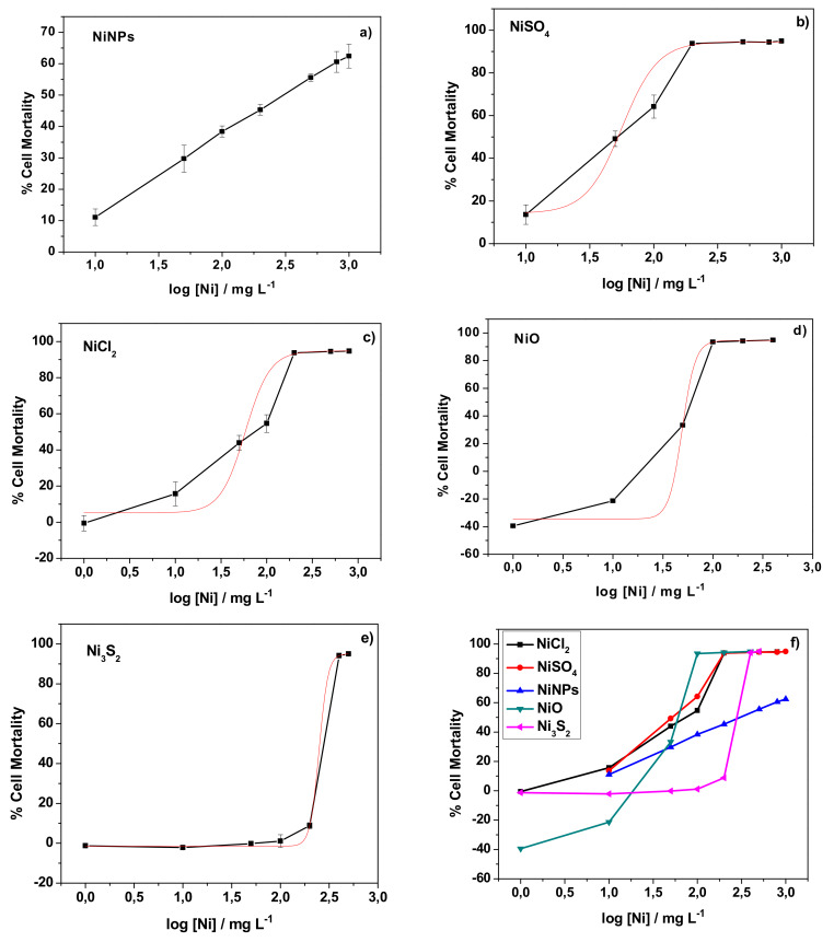
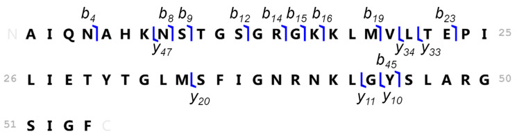
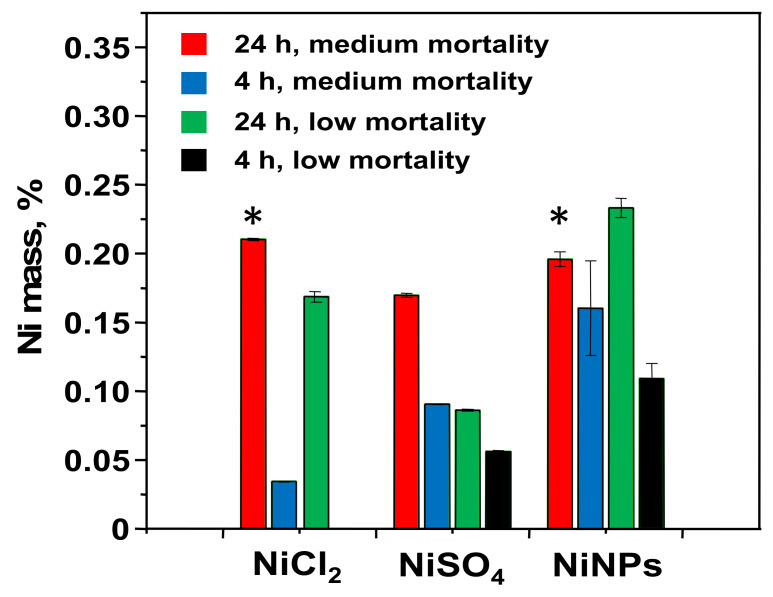
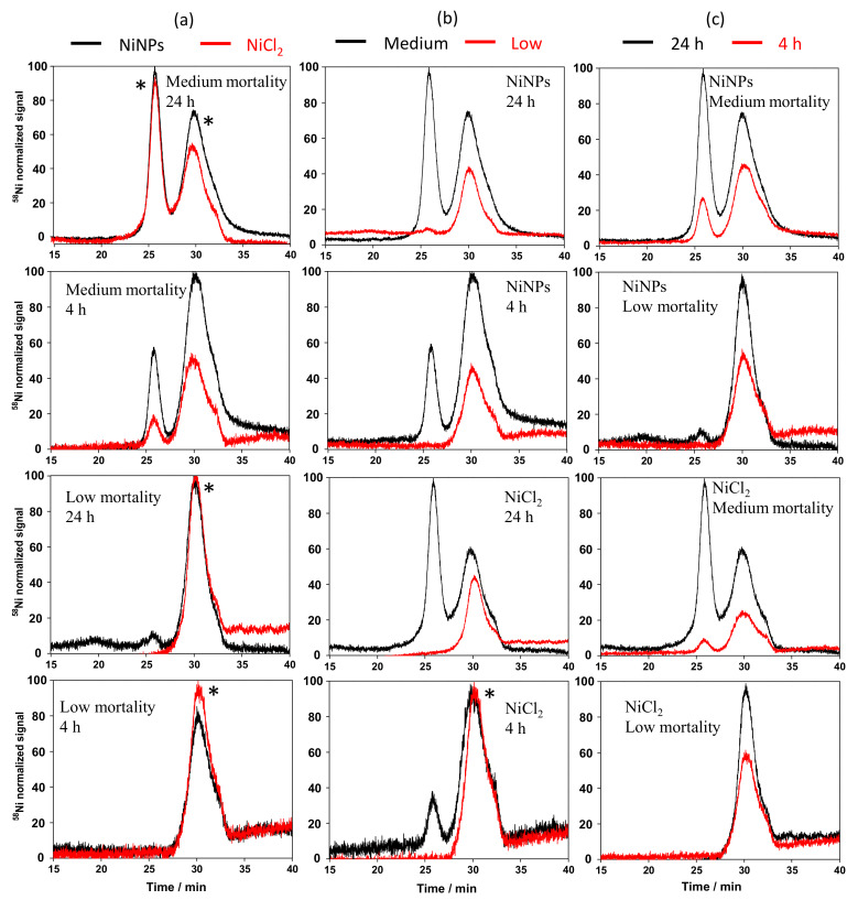
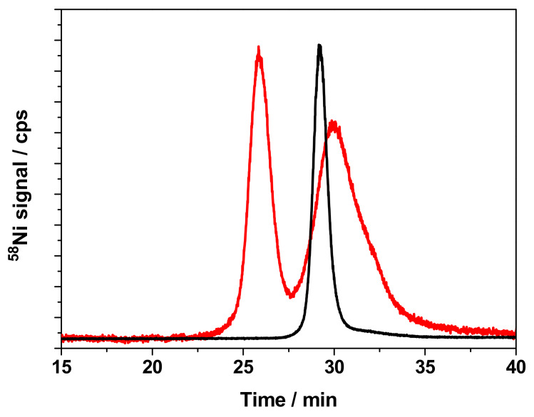
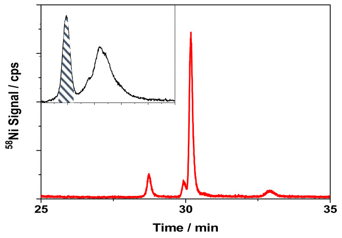
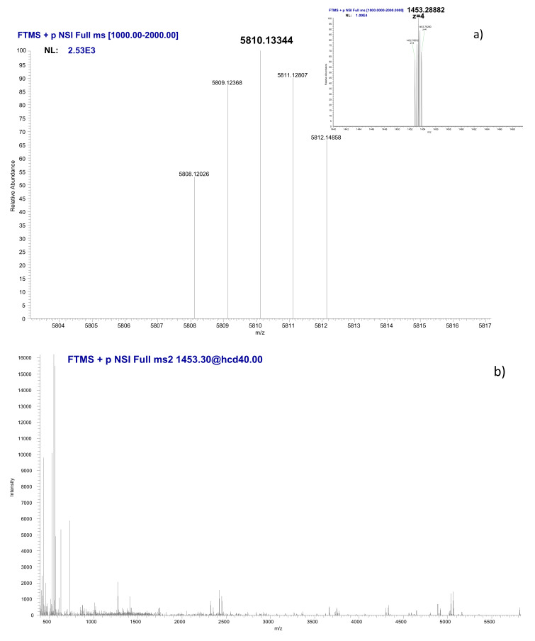
Although nickel allergy and carcinogenicity are well known, their molecular mechanisms are still uncertain, thus demanding studies at the molecular level. The nickel carcinogenicity is known to be dependent on the chemical form of nickel, since only certain nickel compounds can enter the cell. This study investigates, for the first time, the cytotoxicity, cellular uptake, and molecular targets of nickel nanoparticles (NiNPs) in human skin cells in comparison with other chemical forms of nickel. The dose-response curve that was obtained for NiNPs in the cytotoxicity assays showed a linear behavior typical of genotoxic carcinogens. The exposure of keratinocytes to NiNPs leads to the release of Ni ions and its accumulation in the cytosol. A 6 kDa nickel-binding molecule was found to be synthesized by cells exposed to NiNPs at a dose corresponding to medium mortality. This molecule was identified to be tumor-related p63-regulated gene 1 protein.
尽管镍过敏和致癌性已为人熟知,但其分子机制仍不明确,因此需要在分子水平上进行研究。已知镍的致癌性取决于镍的化学形态,因为只有某些镍化合物能够进入细胞。本研究首次调查了镍纳米颗粒(NiNPs)与人皮肤细胞中其他镍化学形态相比的细胞毒性、细胞摄取及分子靶点。在细胞毒性试验中获得的NiNPs剂量反应曲线显示出遗传毒性致癌物典型的线性行为。角质形成细胞暴露于NiNPs会导致镍离子释放并在细胞质中积累。发现一种6 kDa的镍结合分子是由暴露于对应中等死亡率剂量的NiNPs的细胞合成的。该分子被鉴定为肿瘤相关的p63调控基因1蛋白。