• 文献检索
  • 文档翻译
  • 深度研究
  • 学术资讯
  • Suppr Zotero 插件Zotero 插件
  • 邀请有礼
  • 套餐&价格
  • 历史记录
应用&插件
Suppr Zotero 插件Zotero 插件浏览器插件Mac 客户端Windows 客户端微信小程序
定价
高级版会员购买积分包购买API积分包
服务
文献检索文档翻译深度研究API 文档MCP 服务
关于我们
关于 Suppr公司介绍联系我们用户协议隐私条款
关注我们

Suppr 超能文献

核心技术专利:CN118964589B侵权必究
粤ICP备2023148730 号-1Suppr @ 2026

文献检索

告别复杂PubMed语法,用中文像聊天一样搜索,搜遍4000万医学文献。AI智能推荐,让科研检索更轻松。

立即免费搜索

文件翻译

保留排版,准确专业,支持PDF/Word/PPT等文件格式,支持 12+语言互译。

免费翻译文档

深度研究

AI帮你快速写综述,25分钟生成高质量综述,智能提取关键信息,辅助科研写作。

立即免费体验

金属纳米颗粒通过 HIF-1α/miR-29b/MMPs 通路对人表皮角质形成细胞中紧密连接相关蛋白的影响。

Effects of metal nanoparticles on tight junction-associated proteins via HIF-1α/miR-29b/MMPs pathway in human epidermal keratinocytes.

机构信息

Department of Environmental and Occupational Health Sciences, School of Public Health and Information Sciences, University of Louisville, 485 E. Gray Street, Louisville, KY, 40202, USA.

出版信息

Part Fibre Toxicol. 2021 Mar 19;18(1):13. doi: 10.1186/s12989-021-00405-2.

DOI:10.1186/s12989-021-00405-2
PMID:33740985
原文链接:https://pmc.ncbi.nlm.nih.gov/articles/PMC7980342/
Abstract

BACKGROUND

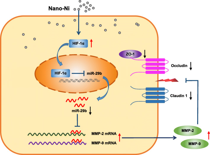
The increasing use of metal nanoparticles in industry and biomedicine raises the risk for unintentional exposure. The ability of metal nanoparticles to penetrate the skin ranges from stopping at the stratum corneum to passing below the dermis and entering the systemic circulation. Despite the potential health risks associated with skin exposure to metal nanoparticles, the mechanisms underlying the toxicity of metal nanoparticles on skin keratinocytes remain unclear. In this study, we proposed that exposure of human epidermal keratinocytes (HaCaT) to metal nanoparticles, such as nickel nanoparticles, dysregulates tight-junction associated proteins by interacting with the HIF-1α/miR-29b/MMPs axis.

METHODS

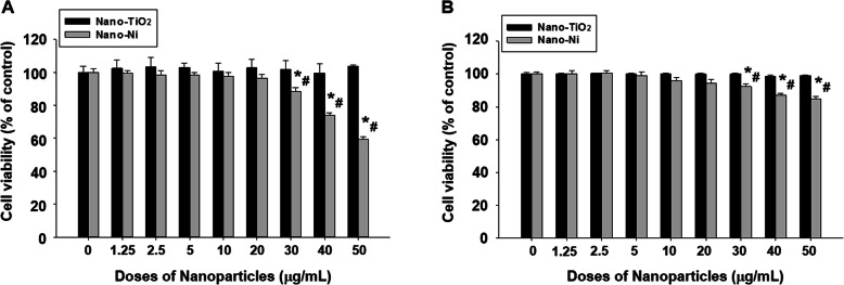
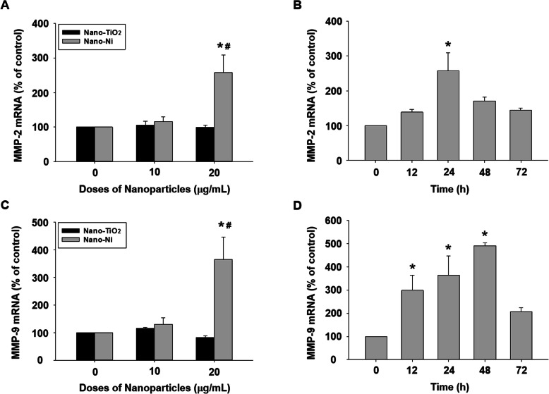
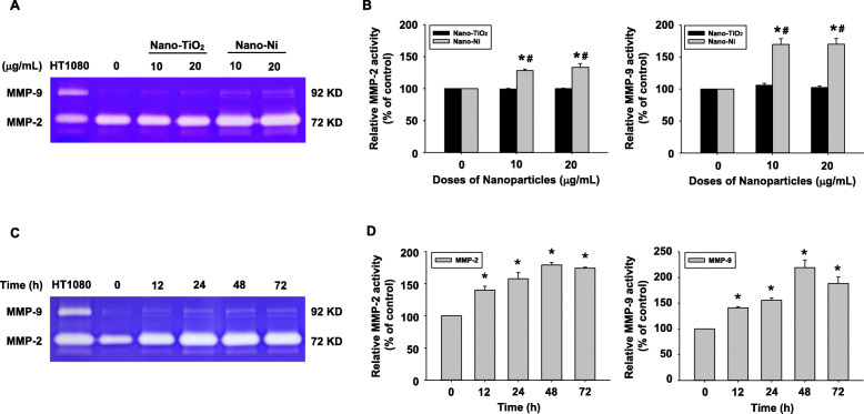
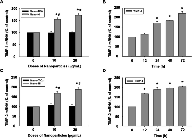
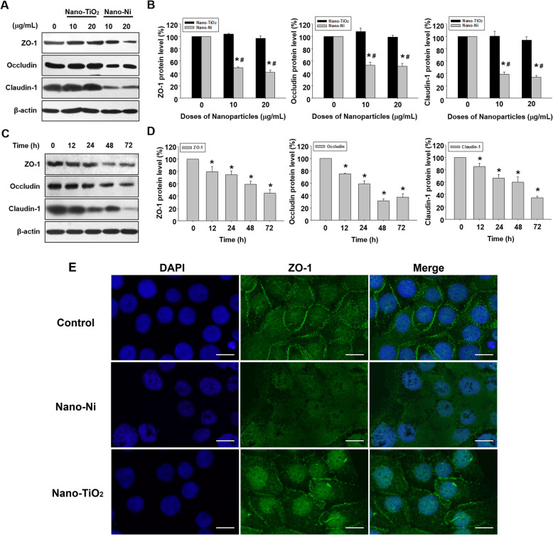
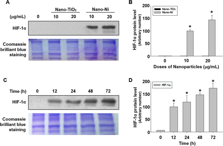
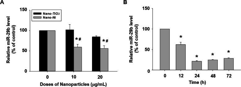
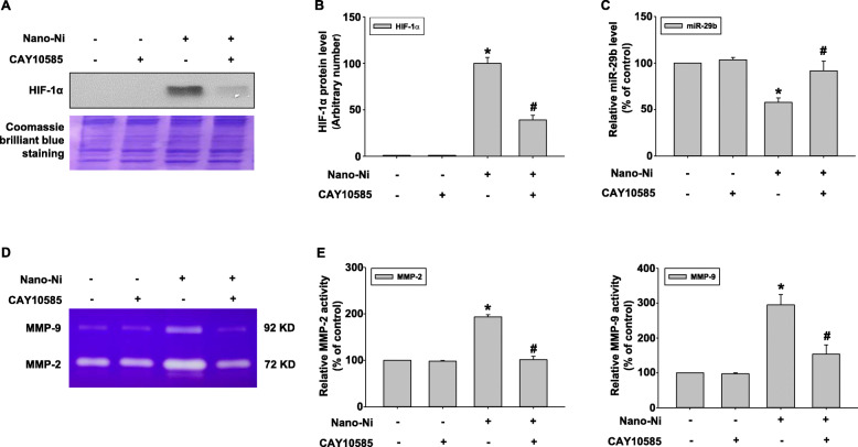
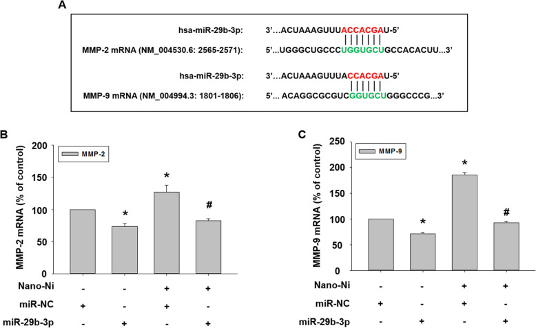
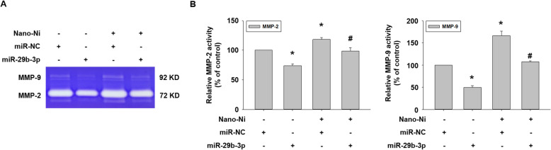
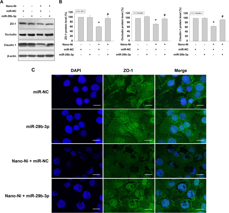
We performed dose-response and time-response studies in HaCaT cells to observe the effects of Nano-Ni or Nano-TiO on the expression and activity of MMP-2 and MMP-9, and on the expression of tight junction-associated proteins, TIMP-1, TIMP-2, miR-29b, and HIF-1α. In the dose-response studies, cells were exposed to 0, 10, or 20 μg/mL of Nano-Ni or Nano-TiO for 24 h. In the time-response studies, cells were exposed to 20 μg/mL of Nano-Ni for 12, 24, 48, or 72 h. After treatment, cells were collected to either assess the expression of mRNAs and miR-29b by real-time PCR or to determine the expression of tight junction-associated proteins and HIF-1α nuclear accumulation by Western blot and/or immunofluorescent staining; the conditioned media were collected to evaluate the MMP-2 and MMP-9 activities by gelatin zymography assay. To further investigate the mechanisms underlying Nano-Ni-induced dysregulation of tight junction-associated proteins, we employed a HIF-1α inhibitor, CAY10585, to perturb HIF-1α accumulation in one experiment, and transfected a miR-29b-3p mimic into the HaCaT cells before Nano-Ni exposure in another experiment. Cells and conditioned media were collected, and the expression and activities of MMPs and the expression of tight junction-associated proteins were determined as described above.

RESULTS

Exposure of HaCaT cells to Nano-Ni resulted in a dose-dependent increase in the expression of MMP-2, MMP-9, TIMP-1, and TIMP-2 and the activities of MMP-2 and MMP-9. However, exposure of cells to Nano-TiO did not cause these effects. Nano-Ni caused a dose-dependent decrease in the expression of miR-29b and tight junction-associated proteins, such as ZO-1, occludin, and claudin-1, while Nano-TiO did not. Nano-Ni also caused a dose-dependent increase in HIF-1α nuclear accumulation. The time-response studies showed that Nano-Ni caused significantly increased expressions of MMP-2 at 24 h, MMP-9 at 12, 24, and 48 h, TIMP-1 from 24 to 72 h, and TIMP-2 from 12 to 72 h post-exposure. The expression of miR-29b and tight junction-associated proteins such as ZO-1, occludin, and claudin-1 decreased as early as 12 h post-exposure, and their levels declined gradually over time. Pretreatment of cells with a HIF-1α inhibitor, CAY10585, abolished Nano-Ni-induced miR-29b down-regulation and MMP-2/9 up-regulation. Introduction of a miR-29b-3p mimic into HaCaT cells by transfection before Nano-Ni exposure ameliorated Nano-Ni-induced increased expression and activity of MMP-2 and MMP-9 and restored Nano-Ni-induced down-regulation of tight junction-associated proteins.

CONCLUSION

Our study herein demonstrated that exposure of human epidermal keratinocytes to Nano-Ni caused increased HIF-1α nuclear accumulation and increased transcription and activity of MMP-2 and MMP-9 and down-regulation of miR-29b and tight junction-associated proteins. Nano-Ni-induced miR-29b down-regulation was through Nano-Ni-induced HIF-1α nuclear accumulation. Restoration of miR-29b level by miR-29b-3p mimic transfection abolished Nano-Ni-induced MMP-2 and MMP-9 activation and down-regulation of tight junction-associated proteins. In summary, our results demonstrated that Nano-Ni-induced dysregulation of tight junction-associated proteins in skin keratinocytes was via HIF-1α/miR-29b/MMPs pathway.

摘要

背景

金属纳米粒子在工业和生物医学中的应用日益广泛,增加了无意暴露的风险。金属纳米粒子穿透皮肤的能力范围从停留在角质层到穿透真皮并进入全身循环。尽管皮肤接触金属纳米粒子存在潜在的健康风险,但金属纳米粒子对皮肤角质形成细胞毒性的机制仍不清楚。在这项研究中,我们提出,暴露于金属纳米粒子(如镍纳米粒子)会通过与 HIF-1α/miR-29b/MMPs 轴相互作用,扰乱紧密连接相关蛋白。

方法

我们在 HaCaT 细胞中进行了剂量反应和时间反应研究,以观察 Nano-Ni 或 Nano-TiO 对 MMP-2 和 MMP-9 的表达和活性以及紧密连接相关蛋白 TIMP-1、TIMP-2、miR-29b 和 HIF-1α 的表达的影响。在剂量反应研究中,细胞暴露于 0、10 或 20μg/mL 的 Nano-Ni 或 Nano-TiO 24 小时。在时间反应研究中,细胞暴露于 20μg/mL 的 Nano-Ni 12、24、48 或 72 小时。处理后,收集细胞以通过实时 PCR 评估 mRNA 和 miR-29b 的表达,或通过 Western blot 和/或免疫荧光染色来确定紧密连接相关蛋白和 HIF-1α 核积累的表达;收集条件培养基以通过明胶酶谱法评估 MMP-2 和 MMP-9 的活性。为了进一步研究 Nano-Ni 诱导的紧密连接相关蛋白失调的机制,我们在一项实验中使用 HIF-1α 抑制剂 CAY10585 扰乱 HIF-1α 的积累,在另一项实验中在暴露于 Nano-Ni 之前转染 HaCaT 细胞 miR-29b-3p 模拟物。收集细胞和条件培养基,如上所述确定 MMPs 的表达和活性以及紧密连接相关蛋白的表达。

结果

暴露于 HaCaT 细胞的 Nano-Ni 导致 MMP-2、MMP-9、TIMP-1 和 TIMP-2 的表达以及 MMP-2 和 MMP-9 的活性呈剂量依赖性增加。然而,暴露于细胞的 Nano-TiO 没有引起这些效应。Nano-Ni 导致 miR-29b 和紧密连接相关蛋白(如 ZO-1、occludin 和 claudin-1)的表达呈剂量依赖性降低,而 Nano-TiO 则没有。Nano-Ni 还导致 HIF-1α 核积累呈剂量依赖性增加。时间反应研究表明,Nano-Ni 在 24 小时引起 MMP-2 的表达显著增加,在 12、24 和 48 小时引起 MMP-9 的表达增加,在 24 至 72 小时引起 TIMP-1 的表达增加,在 12 至 72 小时引起 TIMP-2 的表达增加。暴露于细胞的 miR-29b 和紧密连接相关蛋白(如 ZO-1、occludin 和 claudin-1)的表达早在 12 小时后就开始下降,并且随着时间的推移逐渐下降。用 HIF-1α 抑制剂 CAY10585 预处理细胞可消除 Nano-Ni 诱导的 miR-29b 下调和 MMP-2/9 的上调。在暴露于 Nano-Ni 之前通过转染将 miR-29b-3p 模拟物引入 HaCaT 细胞可改善 Nano-Ni 诱导的 MMP-2 和 MMP-9 的表达和活性增加,并恢复 Nano-Ni 诱导的紧密连接相关蛋白的下调。

结论

本研究表明,暴露于人类表皮角质形成细胞的 Nano-Ni 导致 HIF-1α 核积累增加,MMP-2 和 MMP-9 的转录和活性增加,以及 miR-29b 和紧密连接相关蛋白的下调。Nano-Ni 诱导的 miR-29b 下调是通过 Nano-Ni 诱导的 HIF-1α 核积累引起的。通过 miR-29b-3p 模拟物转染恢复 miR-29b 水平可消除 Nano-Ni 诱导的 MMP-2 和 MMP-9 激活和紧密连接相关蛋白的下调。总之,我们的结果表明,Nano-Ni 诱导的皮肤角质形成细胞中紧密连接相关蛋白的失调是通过 HIF-1α/miR-29b/MMPs 途径实现的。

https://cdn.ncbi.nlm.nih.gov/pmc/blobs/6997/7980342/8cc1a93a0c48/12989_2021_405_Fig12_HTML.jpg
https://cdn.ncbi.nlm.nih.gov/pmc/blobs/6997/7980342/9132fec0a340/12989_2021_405_Fig1_HTML.jpg
https://cdn.ncbi.nlm.nih.gov/pmc/blobs/6997/7980342/84ce44b55e36/12989_2021_405_Fig2_HTML.jpg
https://cdn.ncbi.nlm.nih.gov/pmc/blobs/6997/7980342/05cfa21b627a/12989_2021_405_Fig3_HTML.jpg
https://cdn.ncbi.nlm.nih.gov/pmc/blobs/6997/7980342/d4eaf38b34b2/12989_2021_405_Fig4_HTML.jpg
https://cdn.ncbi.nlm.nih.gov/pmc/blobs/6997/7980342/59813b355456/12989_2021_405_Fig5_HTML.jpg
https://cdn.ncbi.nlm.nih.gov/pmc/blobs/6997/7980342/5a71b1333ec7/12989_2021_405_Fig6_HTML.jpg
https://cdn.ncbi.nlm.nih.gov/pmc/blobs/6997/7980342/d2452f70ef99/12989_2021_405_Fig7_HTML.jpg
https://cdn.ncbi.nlm.nih.gov/pmc/blobs/6997/7980342/74db4cb4e124/12989_2021_405_Fig8_HTML.jpg
https://cdn.ncbi.nlm.nih.gov/pmc/blobs/6997/7980342/2caaf32cf4b2/12989_2021_405_Fig9_HTML.jpg
https://cdn.ncbi.nlm.nih.gov/pmc/blobs/6997/7980342/7b011fbab48f/12989_2021_405_Fig10_HTML.jpg
https://cdn.ncbi.nlm.nih.gov/pmc/blobs/6997/7980342/64e9d8054ae5/12989_2021_405_Fig11_HTML.jpg
https://cdn.ncbi.nlm.nih.gov/pmc/blobs/6997/7980342/8cc1a93a0c48/12989_2021_405_Fig12_HTML.jpg

相似文献

1
Effects of metal nanoparticles on tight junction-associated proteins via HIF-1α/miR-29b/MMPs pathway in human epidermal keratinocytes.金属纳米颗粒通过 HIF-1α/miR-29b/MMPs 通路对人表皮角质形成细胞中紧密连接相关蛋白的影响。
Part Fibre Toxicol. 2021 Mar 19;18(1):13. doi: 10.1186/s12989-021-00405-2.
2
Nickel nanoparticle-induced cell transformation: involvement of DNA damage and DNA repair defect through HIF-1α/miR-210/Rad52 pathway.镍纳米颗粒诱导的细胞转化:通过 HIF-1α/miR-210/Rad52 通路引起的 DNA 损伤和 DNA 修复缺陷。
J Nanobiotechnology. 2021 Nov 17;19(1):370. doi: 10.1186/s12951-021-01117-7.
3
The role of hypoxia inducible factor-1α in the increased MMP-2 and MMP-9 production by human monocytes exposed to nickel nanoparticles.缺氧诱导因子-1α 在镍纳米颗粒暴露下人单核细胞中 MMP-2 和 MMP-9 产生增加中的作用。
Nanotoxicology. 2011 Dec;5(4):568-82. doi: 10.3109/17435390.2010.537791. Epub 2011 Mar 14.
4
Nickel nanoparticles induce epithelial-mesenchymal transition in human bronchial epithelial cells via the HIF-1α/HDAC3 pathway.镍纳米颗粒通过 HIF-1α/HDAC3 通路诱导人支气管上皮细胞上皮-间充质转化。
Nanotoxicology. 2022 Aug-Oct;16(6-8):695-712. doi: 10.1080/17435390.2022.2142169. Epub 2022 Nov 7.
5
Nickel nanoparticles induce autophagy and apoptosis via HIF-1α/mTOR signaling in human bronchial epithelial cells.镍纳米颗粒通过 HIF-1α/mTOR 信号通路诱导人支气管上皮细胞自噬和凋亡。
Environ Pollut. 2023 Jul 15;329:121670. doi: 10.1016/j.envpol.2023.121670. Epub 2023 Apr 18.
6
Up-regulation of Gadd45α after exposure to metal nanoparticles: the role of hypoxia inducible factor 1α.暴露于金属纳米颗粒后Gadd45α的上调:缺氧诱导因子1α的作用
Environ Toxicol. 2015 Apr;30(4):490-9. doi: 10.1002/tox.21926. Epub 2013 Nov 26.
7
The role of miR-21 in nickel nanoparticle-induced MMP-2 and MMP-9 production in mouse primary monocytes: In vitro and in vivo studies.miR-21在镍纳米颗粒诱导小鼠原代单核细胞产生基质金属蛋白酶-2和基质金属蛋白酶-9中的作用:体内外研究
Environ Pollut. 2020 Dec;267:115597. doi: 10.1016/j.envpol.2020.115597. Epub 2020 Sep 4.
8
Comparative mouse lung injury by nickel nanoparticles with differential surface modification.镍纳米颗粒表面修饰差异导致的小鼠肺部损伤比较
J Nanobiotechnology. 2019 Jan 7;17(1):2. doi: 10.1186/s12951-018-0436-0.
9
Calycosin alleviates allergic contact dermatitis by repairing epithelial tight junctions via down-regulating HIF-1α.毛蕊异黄酮通过下调 HIF-1α 修复上皮紧密连接缓解变应性接触性皮炎。
J Cell Mol Med. 2018 Sep;22(9):4507-4521. doi: 10.1111/jcmm.13763. Epub 2018 Jul 11.
10
Matrix metalloproteinase-2 and -9 are induced differently by metal nanoparticles in human monocytes: The role of oxidative stress and protein tyrosine kinase activation.金属纳米颗粒对人单核细胞中基质金属蛋白酶-2和-9的诱导作用不同:氧化应激和蛋白酪氨酸激酶激活的作用
Toxicol Appl Pharmacol. 2008 Dec 1;233(2):276-85. doi: 10.1016/j.taap.2008.08.022. Epub 2008 Sep 16.

引用本文的文献

1
HMGB1 derived from lung epithelial cells after cobalt nanoparticle exposure promotes the activation of lung fibroblasts.钴纳米颗粒暴露后肺上皮细胞来源的高迁移率族蛋白 B1 促进肺成纤维细胞的激活。
Nanotoxicology. 2024 Sep;18(6):565-581. doi: 10.1080/17435390.2024.2404074. Epub 2024 Sep 19.
2
Nanoparticles and Airway Epithelial Cells: Exploring the Impacts and Methodologies in Toxicity Assessment.纳米颗粒与气道上皮细胞:毒性评估中的影响与方法探索。
Int J Mol Sci. 2024 Jul 18;25(14):7885. doi: 10.3390/ijms25147885.
3
The pulmonary effects of nickel-containing nanoparticles: Cytotoxicity, genotoxicity, carcinogenicity, and their underlying mechanisms.

本文引用的文献

1
The role of miR-21 in nickel nanoparticle-induced MMP-2 and MMP-9 production in mouse primary monocytes: In vitro and in vivo studies.miR-21在镍纳米颗粒诱导小鼠原代单核细胞产生基质金属蛋白酶-2和基质金属蛋白酶-9中的作用:体内外研究
Environ Pollut. 2020 Dec;267:115597. doi: 10.1016/j.envpol.2020.115597. Epub 2020 Sep 4.
2
miR-21 mediates nickel nanoparticle-induced pulmonary injury and fibrosis.miR-21 介导镍纳米颗粒诱导的肺损伤和纤维化。
Nanotoxicology. 2020 Nov;14(9):1175-1197. doi: 10.1080/17435390.2020.1808727. Epub 2020 Sep 14.
3
Toxicity Evaluation of TiO Nanoparticles on the 3D Skin Model: A Systematic Review.
含镍纳米颗粒的肺部效应:细胞毒性、遗传毒性、致癌性及其潜在机制。
Environ Sci Nano. 2024 May 1;11(5):1817-1846. doi: 10.1039/d3en00929g. Epub 2024 Mar 21.
4
Shoseiryuto Promotes the Formation of a Tight-Junction Barrier in Cultured Human Bronchial Epithelial Cells.柴芍六君子汤促进人支气管上皮细胞紧密连接屏障的形成。
Evid Based Complement Alternat Med. 2023 Oct 21;2023:4694243. doi: 10.1155/2023/4694243. eCollection 2023.
5
Insights into the toxicological effects of nanomaterials on atherosclerosis: mechanisms involved and influence factors.纳米材料对动脉粥样硬化的毒理学作用的研究进展:涉及的机制和影响因素。
J Nanobiotechnology. 2023 Apr 28;21(1):140. doi: 10.1186/s12951-023-01899-y.
6
Nickel nanoparticles induce autophagy and apoptosis via HIF-1α/mTOR signaling in human bronchial epithelial cells.镍纳米颗粒通过 HIF-1α/mTOR 信号通路诱导人支气管上皮细胞自噬和凋亡。
Environ Pollut. 2023 Jul 15;329:121670. doi: 10.1016/j.envpol.2023.121670. Epub 2023 Apr 18.
7
Allergenicity and Bioavailability of Nickel Nanoparticles Compared to Nickel Microparticles in Mice.与镍微粒相比,镍纳米颗粒在小鼠体内的致敏性和生物利用度
Materials (Basel). 2023 Feb 23;16(5):1834. doi: 10.3390/ma16051834.
8
Dl-3-n-butylphthalide promotes synaptic plasticity by activating the Akt/ERK signaling pathway and reduces the blood-brain barrier leakage by inhibiting the HIF-1α/MMP signaling pathway in vascular dementia model mice.二氢-3-正丁基苯酞通过激活 Akt/ERK 信号通路促进突触可塑性,并通过抑制血管性痴呆模型小鼠中的 HIF-1α/MMP 信号通路降低血脑屏障通透性。
CNS Neurosci Ther. 2023 May;29(5):1392-1404. doi: 10.1111/cns.14112. Epub 2023 Feb 8.
9
Nickel nanoparticles induce epithelial-mesenchymal transition in human bronchial epithelial cells via the HIF-1α/HDAC3 pathway.镍纳米颗粒通过 HIF-1α/HDAC3 通路诱导人支气管上皮细胞上皮-间充质转化。
Nanotoxicology. 2022 Aug-Oct;16(6-8):695-712. doi: 10.1080/17435390.2022.2142169. Epub 2022 Nov 7.
10
A protective role of autophagy in fine airborne particulate matter-induced apoptosis in LN-229 cells.自噬在细颗粒物诱导 LN-229 细胞凋亡中的保护作用。
Toxicology. 2022 Jul;477:153271. doi: 10.1016/j.tox.2022.153271. Epub 2022 Jul 22.
TiO纳米颗粒对3D皮肤模型的毒性评估:一项系统综述。
Front Bioeng Biotechnol. 2020 Jun 10;8:575. doi: 10.3389/fbioe.2020.00575. eCollection 2020.
4
Liraglutide-loaded PLGA/gelatin electrospun nanofibrous mats promote angiogenesis to accelerate diabetic wound healing via the modulation of miR-29b-3p.负载利拉鲁肽的聚乳酸-羟基乙酸共聚物/明胶电纺纳米纤维垫通过调节miR-29b-3p促进血管生成,加速糖尿病伤口愈合。
Biomater Sci. 2020 Aug 7;8(15):4225-4238. doi: 10.1039/d0bm00442a. Epub 2020 Jun 24.
5
Nickel Nanoparticles Induce the Synthesis of a Tumor-Related Polypeptide in Human Epidermal Keratinocytes.镍纳米颗粒诱导人表皮角质形成细胞合成一种肿瘤相关多肽。
Nanomaterials (Basel). 2020 May 21;10(5):992. doi: 10.3390/nano10050992.
6
Dysfunction of pulmonary epithelial tight junction induced by silicon dioxide nanoparticles via the ROS/ERK pathway and protein degradation.二氧化硅纳米颗粒通过 ROS/ERK 通路和蛋白降解引起肺上皮细胞紧密连接功能障碍。
Chemosphere. 2020 Sep;255:126954. doi: 10.1016/j.chemosphere.2020.126954. Epub 2020 May 3.
7
Tight junctions in the development of asthma, chronic rhinosinusitis, atopic dermatitis, eosinophilic esophagitis, and inflammatory bowel diseases.紧密连接在哮喘、慢性鼻-鼻窦炎、特应性皮炎、嗜酸性食管炎和炎症性肠病的发病机制中的作用。
J Leukoc Biol. 2020 May;107(5):749-762. doi: 10.1002/JLB.5MR0120-230R. Epub 2020 Feb 28.
8
The past, present and future perspectives of matrix metalloproteinase inhibitors.基质金属蛋白酶抑制剂的过去、现在和未来展望。
Pharmacol Ther. 2020 Mar;207:107465. doi: 10.1016/j.pharmthera.2019.107465. Epub 2019 Dec 18.
9
Skin barrier damage after exposure to paraphenylenediamine.接触对苯二胺后皮肤屏障损伤。
J Allergy Clin Immunol. 2020 Feb;145(2):619-631.e2. doi: 10.1016/j.jaci.2019.11.023. Epub 2019 Nov 26.
10
Silica nanoparticles induce spermatocyte cell autophagy through microRNA-494 targeting AKT in GC-2spd cells.硅纳米颗粒通过 microRNA-494 靶向 AKT 诱导 GC-2spd 细胞精母细胞细胞自噬。
Environ Pollut. 2019 Dec;255(Pt 1):113172. doi: 10.1016/j.envpol.2019.113172. Epub 2019 Sep 11.