Department of Brain and Cognitive Sciences, Cell Science Research Center, Royan Institute for Stem Cell Biology and Technology, ACECR, Tehran, Iran.
Department of Stem Cells and Developmental Biology, Cell Science Research Center, Royan Institute for Stem Cell Biology and Technology, ACECR, Tehran, Iran.
Stem Cell Res Ther. 2020 May 27;11(1):203. doi: 10.1186/s13287-020-01702-x.
Retinal and/or optic nerve injury is one of the leading causes of blindness due to retinal ganglion cell (RGC) degeneration. There have been extensive efforts to suppress this neurodegeneration. Various somatic tissue-derived mesenchymal stem cells (MSCs) demonstrated significant neuroprotective and axogenic effects on RGCs. An alternative source of MSCs could be human embryonic stem cells (ES-MSCs), which proliferate faster, express lower levels of inflammatory cytokines, and are capable of immune modulation. It has been demonstrated that MSCs secrete factors or extracellular vesicles that may heal the injury. However, possible therapeutic effects and underlying mechanism of human ES-MSC extracellular vesicles (EVs) on optic nerve injury have not been assessed.
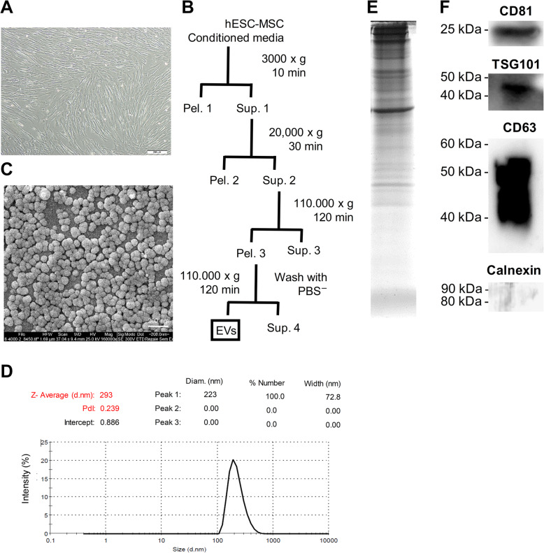
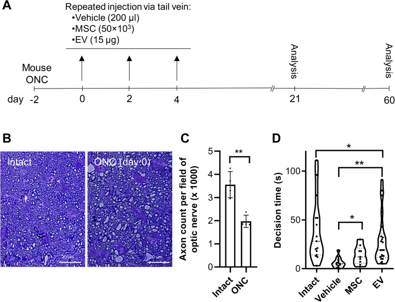
EVs were isolated from human ES-MSCs. Then, ES-MSC EV was applied to an optic nerve crush (ONC) mouse model. Immunohistofluorescence, retro- and anterograde tracing of RGCs, Western blot, tauopathy in RGCs, and function assessments were performed during 2-month post-treatment to evaluate ONC improvement and underlying mechanism of human ES-MSC EV in in vivo.
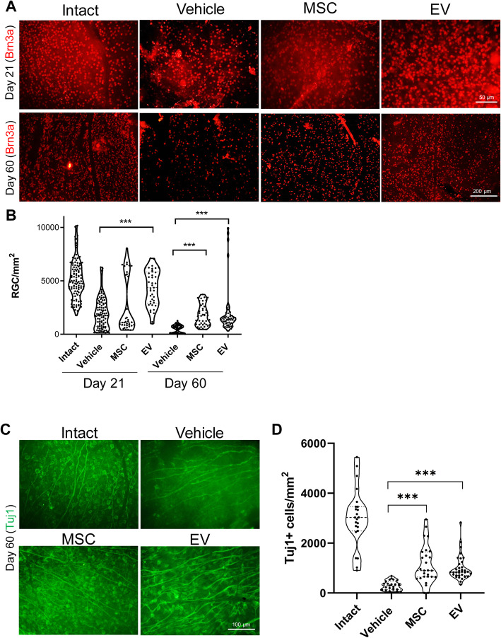
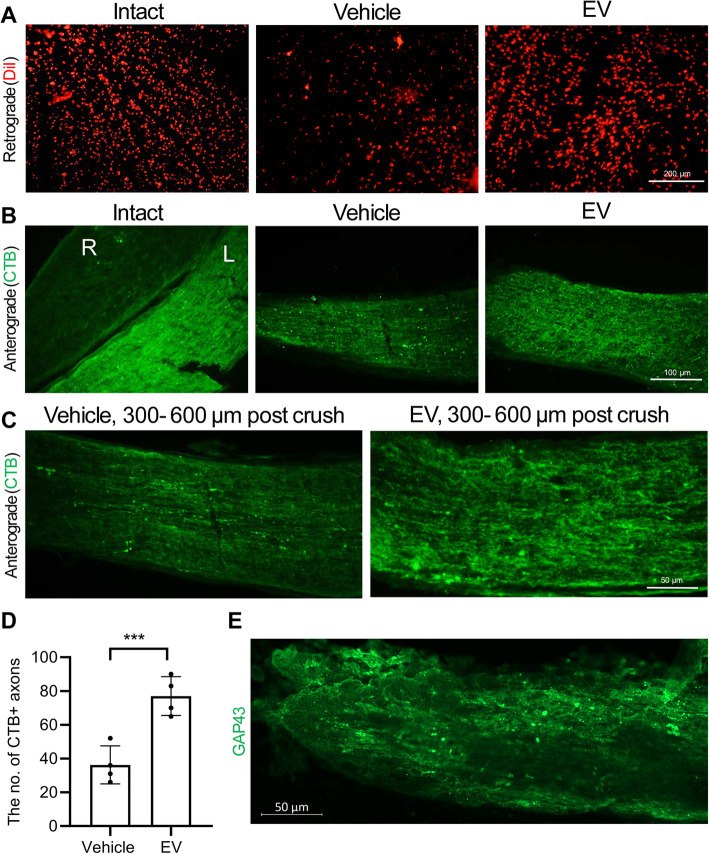
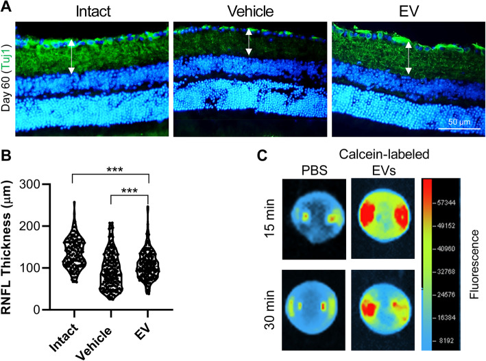
We found that the ES-MSC EV significantly improved Brn3a+ RGCs survival and retro- and anterograde tracing of RGCs, while preventing retinal nerve fiber layer (RNFL) degenerative thinning compared to the vehicle group. The EVs also significantly promoted GAP43+ axon counts in the optic nerve and improved cognitive visual behavior. Furthermore, cis p-tau, a central mediator of neurodegeneration in the injured RGCs, is detectable after the ONC at the early stages demonstrated tauopathy in RGCs. Notably, after EV treatment cis p-tau was downregulated.
Our findings propose that human ES-MSC EVs, as an off-the-shelf and cell-free product, may have profound clinical implications in treating injured RGCs and degenerative ocular disease. Moreover, the possible mechanisms of human ES-MSC EV are related to the rescue of tauopathy process of RGC degeneration.
视网膜和/或视神经损伤是由于视网膜神经节细胞 (RGC) 变性导致失明的主要原因之一。人们一直在努力抑制这种神经退行性变。各种源自体组织的间充质干细胞 (MSC) 对 RGC 表现出显著的神经保护和轴突生成作用。MSC 的另一种来源可能是人类胚胎干细胞 (ES-MSC),它增殖更快,炎症细胞因子表达水平更低,并且能够进行免疫调节。已经证明 MSC 分泌的因子或细胞外囊泡可能有助于修复损伤。然而,尚未评估人类 ES-MSC 细胞外囊泡 (EV) 对视神经损伤的可能治疗效果和潜在机制。
从人类 ES-MSC 中分离 EV。然后,将 ES-MSC EV 应用于视神经挤压 (ONC) 小鼠模型。在治疗后 2 个月进行免疫组织荧光、RGC 逆行和顺行追踪、Western blot、RGC 中的 tau 病变和功能评估,以评估 ONC 改善和人类 ES-MSC EV 在体内的潜在机制。
我们发现,与载体组相比,ES-MSC EV 显著改善了 Brn3a+RGC 存活以及 RGC 的逆行和顺行追踪,同时防止了视网膜神经纤维层 (RNFL) 的退行性变薄。EV 还显著促进了视神经中 GAP43+轴突计数,并改善了认知视觉行为。此外,cis p-tau 是损伤 RGC 中神经退行性变的中枢介质,在早期 ONC 后即可检测到,表明 RGC 中存在 tau 病变。值得注意的是,在 EV 治疗后 cis p-tau 下调。
我们的研究结果表明,作为一种现成的无细胞产品,人类 ES-MSC EV 可能在治疗受损的 RGC 和退行性眼病方面具有深远的临床意义。此外,人类 ES-MSC EV 的可能机制与 RGC 变性的 tau 病变过程的挽救有关。