• 文献检索
  • 文档翻译
  • 深度研究
  • 学术资讯
  • Suppr Zotero 插件Zotero 插件
  • 邀请有礼
  • 套餐&价格
  • 历史记录
应用&插件
Suppr Zotero 插件Zotero 插件浏览器插件Mac 客户端Windows 客户端微信小程序
定价
高级版会员购买积分包购买API积分包
服务
文献检索文档翻译深度研究API 文档MCP 服务
关于我们
关于 Suppr公司介绍联系我们用户协议隐私条款
关注我们

Suppr 超能文献

核心技术专利:CN118964589B侵权必究
粤ICP备2023148730 号-1Suppr @ 2026

文献检索

告别复杂PubMed语法,用中文像聊天一样搜索,搜遍4000万医学文献。AI智能推荐,让科研检索更轻松。

立即免费搜索

文件翻译

保留排版,准确专业,支持PDF/Word/PPT等文件格式,支持 12+语言互译。

免费翻译文档

深度研究

AI帮你快速写综述,25分钟生成高质量综述,智能提取关键信息,辅助科研写作。

立即免费体验

间质干细胞衍生的小细胞外囊泡促进 DBA/2J 型青光眼遗传小鼠模型的神经保护作用。

Mesenchymal Stem Cell-Derived Small Extracellular Vesicles Promote Neuroprotection in a Genetic DBA/2J Mouse Model of Glaucoma.

机构信息

Section of Retinal Ganglion Cell Biology, Laboratory of Retinal Cell and Molecular Biology, National Eye Institute, National Institutes of Health, Bethesda, Maryland, United States.

Neuroscience and Ophthalmology, Institute of Inflammation and Ageing, College of Medical and Dental Sciences, University of Birmingham, Birmingham, United Kingdom.

出版信息

Invest Ophthalmol Vis Sci. 2018 Nov 1;59(13):5473-5480. doi: 10.1167/iovs.18-25310.

DOI:10.1167/iovs.18-25310
PMID:30452601
原文链接:https://pmc.ncbi.nlm.nih.gov/articles/PMC6735616/
Abstract

PURPOSE

To determine if bone marrow-derived stem cell (BMSC) small extracellular vesicles (sEV) promote retinal ganglion cell (RGC) neuroprotection in the genetic DBA/2J mouse model of glaucoma for 12 months.

METHODS

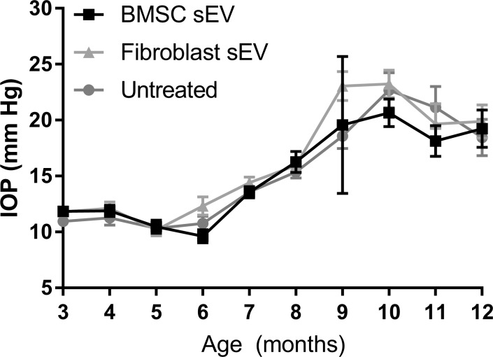
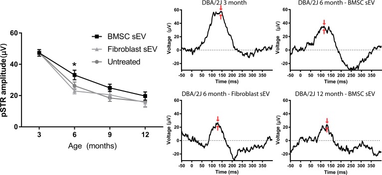
BMSC sEV and control fibroblast-derived sEV were intravitreally injected into 3-month-old DBA/2J mice once a month for 9 months. IOP and positive scotopic threshold responses were measured from 3 months: IOP was measured monthly and positive scotopic threshold responses were measured every 3 months. RGC neuroprotection was determined in wholemounts stained with RNA binding protein with multiple splicing (RBPMS), whereas axonal damage was assessed using paraphenylenediamine staining.

RESULTS

As expected, DBA/2J mice developed chronic ocular hypertension beginning at 6 months. The delivery of BMSC sEV, but not fibroblast sEV, provided significant neuroprotective effects for RBPMS+ RGC while significantly reducing the number of degenerating axons seen in the optic nerve. BMSC sEV significantly preserved RGC function in 6-month-old mice, but provided no benefit at 9 and 12 months.

CONCLUSIONS

BMSC sEV are an effective neuroprotective treatment in a chronic model of ocular hypertension for 1 year, preserving RGC numbers and protecting against axonal degeneration.

摘要

目的

在遗传性 DBA/2J 青光眼小鼠模型中,确定骨髓来源的干细胞(BMSC)小细胞外囊泡(sEV)是否能促进视网膜神经节细胞(RGC)的神经保护作用,时间为 12 个月。

方法

将 BMSC sEV 和对照成纤维细胞衍生的 sEV 每月一次经玻璃体内注射到 3 个月大的 DBA/2J 小鼠中,共注射 9 个月。IOP 和暗适应阈值反应从 3 个月开始测量:IOP 每月测量一次,暗适应阈值反应每 3 个月测量一次。用 RNA 结合蛋白多剪接(RBPMS)对全层片进行染色,以确定 RGC 神经保护作用,而轴突损伤则用对苯二胺染色进行评估。

结果

正如预期的那样,DBA/2J 小鼠从 6 个月开始就出现了慢性眼压升高。BMSC sEV 的递送,但不是成纤维细胞 sEV 的递送,为 RBPMS+RGC 提供了显著的神经保护作用,同时显著减少了视神经中可见的变性轴突数量。BMSC sEV 显著地保留了 6 个月大的小鼠的 RGC 功能,但在 9 个月和 12 个月时没有获益。

结论

BMSC sEV 是一种有效的神经保护治疗方法,可在慢性眼压升高模型中持续 1 年,既能保护 RGC 数量,又能防止轴突退化。

https://cdn.ncbi.nlm.nih.gov/pmc/blobs/714d/6735616/ca9eca673c3b/i1552-5783-59-13-5473-f04.jpg
https://cdn.ncbi.nlm.nih.gov/pmc/blobs/714d/6735616/6c9151870e36/i1552-5783-59-13-5473-f01.jpg
https://cdn.ncbi.nlm.nih.gov/pmc/blobs/714d/6735616/4eb6bfc428a4/i1552-5783-59-13-5473-f02.jpg
https://cdn.ncbi.nlm.nih.gov/pmc/blobs/714d/6735616/fa412dd93825/i1552-5783-59-13-5473-f03.jpg
https://cdn.ncbi.nlm.nih.gov/pmc/blobs/714d/6735616/ca9eca673c3b/i1552-5783-59-13-5473-f04.jpg

相似文献

1
Mesenchymal Stem Cell-Derived Small Extracellular Vesicles Promote Neuroprotection in a Genetic DBA/2J Mouse Model of Glaucoma.间质干细胞衍生的小细胞外囊泡促进 DBA/2J 型青光眼遗传小鼠模型的神经保护作用。
Invest Ophthalmol Vis Sci. 2018 Nov 1;59(13):5473-5480. doi: 10.1167/iovs.18-25310.
2
Mesenchymal Stem Cell-Derived Small Extracellular Vesicles Promote Neuroprotection in Rodent Models of Glaucoma.间质干细胞衍生的小细胞外囊泡促进青光眼啮齿动物模型的神经保护作用。
Invest Ophthalmol Vis Sci. 2018 Feb 1;59(2):702-714. doi: 10.1167/iovs.17-22855.
3
Erythropoietin promotes survival of retinal ganglion cells in DBA/2J glaucoma mice.促红细胞生成素可促进DBA/2J青光眼小鼠视网膜神经节细胞的存活。
Invest Ophthalmol Vis Sci. 2007 Mar;48(3):1212-8. doi: 10.1167/iovs.06-0757.
4
Assessment of inner retina dysfunction and progressive ganglion cell loss in a mouse model of glaucoma.评估青光眼小鼠模型中内视网膜功能障碍和节细胞进行性丧失。
Exp Eye Res. 2014 May;122:40-9. doi: 10.1016/j.exer.2014.02.022. Epub 2014 Mar 12.
5
Viral delivery of multiple miRNAs promotes retinal ganglion cell survival and functional preservation after optic nerve crush injury.病毒递送多种 miRNA 可促进视神经挤压损伤后视网膜神经节细胞的存活和功能保存。
Exp Eye Res. 2020 Aug;197:108071. doi: 10.1016/j.exer.2020.108071. Epub 2020 Jun 20.
6
Anti-inflammatory effect of pigment epithelium-derived factor in DBA/2J mice.色素上皮衍生因子对DBA/2J小鼠的抗炎作用。
Mol Vis. 2009;15:438-50. Epub 2009 Feb 27.
7
Retinal ganglion cell protection by 17-beta-estradiol in a mouse model of inherited glaucoma.17-β-雌二醇对遗传性青光眼小鼠模型视网膜神经节细胞的保护作用
Dev Neurobiol. 2007 Apr;67(5):603-16. doi: 10.1002/dneu.20373.
8
Longitudinal evaluation of retinal ganglion cell function and IOP in the DBA/2J mouse model of glaucoma.在DBA/2J青光眼小鼠模型中对视网膜神经节细胞功能和眼压的纵向评估。
Invest Ophthalmol Vis Sci. 2007 Oct;48(10):4564-72. doi: 10.1167/iovs.07-0483.
9
Reduced retina microglial activation and improved optic nerve integrity with minocycline treatment in the DBA/2J mouse model of glaucoma.在DBA/2J青光眼小鼠模型中,米诺环素治疗可降低视网膜小胶质细胞活化并改善视神经完整性。
Invest Ophthalmol Vis Sci. 2008 Apr;49(4):1437-46. doi: 10.1167/iovs.07-1337.
10
Autophagy deficiency protects against ocular hypertension and neurodegeneration in experimental and spontanous glaucoma mouse models.自噬缺陷可预防实验性和自发性青光眼小鼠模型中的眼压升高和神经退行性变。
Cell Death Dis. 2023 Aug 24;14(8):554. doi: 10.1038/s41419-023-06086-3.

引用本文的文献

1
Rekindling Vision: Innovative Strategies for Treating Retinal Degeneration.重燃光明:治疗视网膜变性的创新策略
Int J Mol Sci. 2025 Apr 25;26(9):4078. doi: 10.3390/ijms26094078.
2
Targeted Neuroprotection of Retinal Ganglion Cells Via AAV2-hSyn-NGF Gene Therapy in Glaucoma Models.青光眼模型中通过腺相关病毒2-人突触蛋白-神经生长因子基因疗法对视网膜神经节细胞进行靶向神经保护
Invest Ophthalmol Vis Sci. 2025 Apr 1;66(4):48. doi: 10.1167/iovs.66.4.48.
3
Astrocyte-Derived Extracellular Vesicles Alleviate Optic Nerve Injury Through Remodeling of Retinal Microenvironmental Homeostasis.

本文引用的文献

1
Stem Cell Ophthalmology Treatment Study: bone marrow derived stem cells in the treatment of Retinitis Pigmentosa.干细胞眼科治疗研究:骨髓源性干细胞治疗视网膜色素变性
Stem Cell Investig. 2018 Jun 6;5:18. doi: 10.21037/sci.2018.04.02. eCollection 2018.
2
hucMSC derived exosomes promote functional recovery in spinal cord injury mice via attenuating inflammation.hucMSC 来源的外泌体通过减轻炎症促进脊髓损伤小鼠的功能恢复。
Mater Sci Eng C Mater Biol Appl. 2018 Aug 1;89:194-204. doi: 10.1016/j.msec.2018.04.006. Epub 2018 Apr 10.
3
Age-related Changes in Eye, Brain and Visuomotor Behavior in the DBA/2J Mouse Model of Chronic Glaucoma.
星形胶质细胞衍生的细胞外囊泡通过重塑视网膜微环境稳态减轻视神经损伤。
Invest Ophthalmol Vis Sci. 2025 Apr 1;66(4):16. doi: 10.1167/iovs.66.4.16.
4
Advances in Regenerative Medicine, Cell Therapy, and 3D Bioprinting for Glaucoma and Retinal Diseases.青光眼和视网膜疾病的再生医学、细胞治疗及3D生物打印进展
Adv Exp Med Biol. 2025 Mar 26. doi: 10.1007/5584_2025_854.
5
Mesenchymal stem cells and mesenchymal stem cell-derived exosomes: a promising strategy for treating retinal degenerative diseases.间充质干细胞和间充质干细胞衍生的外泌体:一种治疗视网膜退行性疾病的有前景的策略。
Mol Med. 2025 Feb 21;31(1):75. doi: 10.1186/s10020-025-01120-w.
6
Extracellular Vesicles and Glaucoma: Opportunities and Challenges.细胞外囊泡与青光眼:机遇与挑战
Curr Eye Res. 2025 Feb 3:1-10. doi: 10.1080/02713683.2025.2459888.
7
Mesenchymal stem cell-derived extracellular vesicles for cell-free therapy of ocular diseases.间充质干细胞衍生的细胞外囊泡用于眼部疾病的无细胞治疗
Extracell Vesicles Circ Nucl Acids. 2022 Apr 24;3(2):102-117. doi: 10.20517/evcna.2022.08. eCollection 2022.
8
A perspective from the National Eye Institute Extracellular Vesicle Workshop: Gaps, needs, and opportunities for studies of extracellular vesicles in vision research.美国国立眼科研究所细胞外囊泡研讨会的观点:视觉研究中细胞外囊泡研究的差距、需求与机遇
J Extracell Vesicles. 2024 Dec;13(12):e70023. doi: 10.1002/jev2.70023.
9
Extracellular vesicles as emerging players in glaucoma: Mechanisms, biomarkers, and therapeutic targets.细胞外囊泡在青光眼中成为新角色:机制、生物标志物及治疗靶点
Vision Res. 2025 Jan;226:108522. doi: 10.1016/j.visres.2024.108522. Epub 2024 Nov 23.
10
Stem Cell-Derived Exosomes: Natural Intercellular Messengers with Versatile Mechanisms for the Treatment of Diabetic Retinopathy.干细胞衍生的外泌体:具有多种机制的天然细胞间信使,可用于治疗糖尿病视网膜病变。
Int J Nanomedicine. 2024 Oct 24;19:10767-10784. doi: 10.2147/IJN.S475234. eCollection 2024.
年龄相关的眼睛、大脑和视动行为变化在慢性青光眼的 DBA/2J 小鼠模型中。
Sci Rep. 2018 Mar 15;8(1):4643. doi: 10.1038/s41598-018-22850-4.
4
Mesenchymal stem cell-derived extracellular vesicles: novel frontiers in regenerative medicine.间质干细胞衍生的细胞外囊泡:再生医学的新前沿。
Stem Cell Res Ther. 2018 Mar 9;9(1):63. doi: 10.1186/s13287-018-0791-7.
5
Mesenchymal Stem Cell-Derived Small Extracellular Vesicles Promote Neuroprotection in Rodent Models of Glaucoma.间质干细胞衍生的小细胞外囊泡促进青光眼啮齿动物模型的神经保护作用。
Invest Ophthalmol Vis Sci. 2018 Feb 1;59(2):702-714. doi: 10.1167/iovs.17-22855.
6
Intravenously delivered mesenchymal stem cell-derived exosomes target M2-type macrophages in the injured spinal cord.静脉注射的间充质干细胞衍生外泌体靶向损伤脊髓中的M2型巨噬细胞。
PLoS One. 2018 Jan 2;13(1):e0190358. doi: 10.1371/journal.pone.0190358. eCollection 2018.
7
Stem Cell Ophthalmology Treatment Study: bone marrow derived stem cells in the treatment of non-arteritic ischemic optic neuropathy (NAION).干细胞眼科治疗研究:骨髓源性干细胞治疗非动脉炎性前部缺血性视神经病变(NAION)
Stem Cell Investig. 2017 Nov 23;4:94. doi: 10.21037/sci.2017.11.05. eCollection 2017.
8
JUN is important for ocular hypertension-induced retinal ganglion cell degeneration.JUN对于高眼压诱导的视网膜神经节细胞变性很重要。
Cell Death Dis. 2017 Jul 20;8(7):e2945. doi: 10.1038/cddis.2017.338.
9
Systemic Administration of Exosomes Released from Mesenchymal Stromal Cells Attenuates Apoptosis, Inflammation, and Promotes Angiogenesis after Spinal Cord Injury in Rats.间质干细胞来源的外泌体系统给药可减轻大鼠脊髓损伤后的细胞凋亡、炎症反应,并促进血管生成。
J Neurotrauma. 2017 Dec 15;34(24):3388-3396. doi: 10.1089/neu.2017.5063. Epub 2017 Aug 18.
10
DBA/2J mouse model for experimental glaucoma: pitfalls and problems.实验性青光眼的 DBA/2J 小鼠模型:陷阱和问题。
Clin Exp Ophthalmol. 2017 Dec;45(9):911-922. doi: 10.1111/ceo.12992. Epub 2017 Jun 13.