• 文献检索
  • 文档翻译
  • 深度研究
  • 学术资讯
  • Suppr Zotero 插件Zotero 插件
  • 邀请有礼
  • 套餐&价格
  • 历史记录
应用&插件
Suppr Zotero 插件Zotero 插件浏览器插件Mac 客户端Windows 客户端微信小程序
定价
高级版会员购买积分包购买API积分包
服务
文献检索文档翻译深度研究API 文档MCP 服务
关于我们
关于 Suppr公司介绍联系我们用户协议隐私条款
关注我们

Suppr 超能文献

核心技术专利:CN118964589B侵权必究
粤ICP备2023148730 号-1Suppr @ 2026

文献检索

告别复杂PubMed语法,用中文像聊天一样搜索,搜遍4000万医学文献。AI智能推荐,让科研检索更轻松。

立即免费搜索

文件翻译

保留排版,准确专业,支持PDF/Word/PPT等文件格式,支持 12+语言互译。

免费翻译文档

深度研究

AI帮你快速写综述,25分钟生成高质量综述,智能提取关键信息,辅助科研写作。

立即免费体验

视神经挤压和间充质干细胞移植后神经元的长期存活、再生和短暂的靶重新连接。

Long-term neuronal survival, regeneration, and transient target reconnection after optic nerve crush and mesenchymal stem cell transplantation.

机构信息

Instituto de Biofísica Carlos Chagas Filho, Universidade Federal do Rio de Janeiro, Rio de Janeiro, RJ, 21941-902, Brazil.

National Institute of Science and Technology for Regenerative Medicine-REGENERE, Rio de Janeiro, Brazil.

出版信息

Stem Cell Res Ther. 2019 Apr 17;10(1):121. doi: 10.1186/s13287-019-1226-9.

DOI:10.1186/s13287-019-1226-9
PMID:30995945
原文链接:https://pmc.ncbi.nlm.nih.gov/articles/PMC6472105/
Abstract

BACKGROUND

Retina and/or optic nerve injury may cause irreversible blindness, due to degeneration of retinal ganglion cells. We and others have previously shown that the intravitreal injection of mesenchymal stem cells (MSCs) protects injured retinal ganglion cells and stimulates their regeneration after optic nerve injury, but the long-term effects of this therapy are still unknown.

METHODS

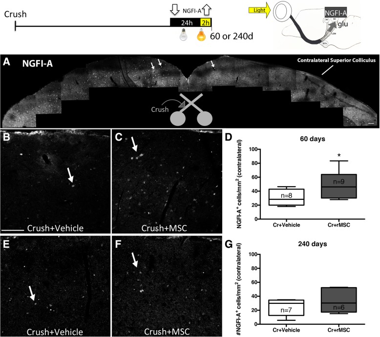
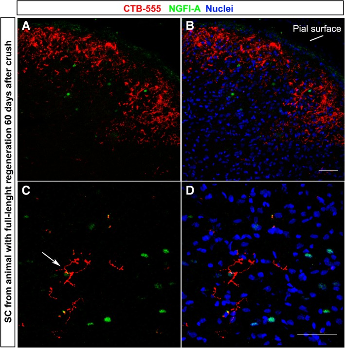
We injected rat MSC (rMSC) intravitreally in adult (3-5 months) Lister Hooded rats of either sex after optic nerve crush. Retinal ganglion cell survival, axonal regeneration, and reconnection were analyzed 60 and 240 days after crush by immunohistochemistry for Tuj1, anterograde labeling with cholera-toxin B and by immunohistochemistry for nerve growth factor-induced gene A (NGFI-A, driven by light stimulation) in the superior colliculus after a cycle of light deprivation-stimulation. Visual behaviors (optokinetic reflex, looming response, and preference for dark) were analyzed 70 days after crush.

RESULTS

rMSC treatment doubled the number of surviving retinal ganglion cells, preferentially of a larger subtype, and of axons regenerating up to 0.5 mm. Some axons regenerated to the lateral geniculate nucleus and superior colliculus. NGFI-A+ cells were doubled in rMSC-treated animals 60 days after crush, but equivalent to vehicle-injected animals 240 days after crush, suggesting that newly formed synapses degenerated. Animals did not recover visual behaviors.

CONCLUSIONS

We conclude that rMSC-induced neuroprotection is sustained at longer time points. Although rMSCs promoted long-term neuroprotection and long-distance axon regeneration, the reconnection of retinal ganglion cells with their targets was transitory, indicating that they need additional stimuli to make stable reconnections.

摘要

背景

视网膜和/或视神经损伤可能导致视网膜神经节细胞变性而造成不可逆转的失明。我们和其他人之前已经表明,间质干细胞(MSCs)的玻璃体内注射可保护受伤的视网膜神经节细胞,并在视神经损伤后刺激其再生,但这种治疗的长期效果尚不清楚。

方法

我们在成年(3-5 个月)Lister Hooded 大鼠视神经挤压后,将大鼠 MSC(rMSC)玻璃体内注射。通过免疫组织化学法检测 Tuj1、霍乱毒素 B 的顺行标记以及光剥夺-刺激后上丘中的神经生长因子诱导基因 A(NGFI-A,由光刺激驱动),分别在挤压后 60 天和 240 天分析视网膜神经节细胞的存活、轴突再生和再连接。在挤压后 70 天分析视觉行为(视动反射、突现反应和对暗的偏好)。

结果

rMSC 治疗使存活的视网膜神经节细胞数量增加了一倍,优先是较大的亚型,并且再生的轴突长达 0.5mm。一些轴突再生到外侧膝状体核和上丘。rMSC 处理动物在挤压后 60 天,NGFI-A+细胞增加了一倍,但在挤压后 240 天与载体注射动物相当,表明新形成的突触退化。动物没有恢复视觉行为。

结论

我们的结论是,rMSC 诱导的神经保护作用在较长时间内持续存在。尽管 rMSCs 促进了长期的神经保护和长距离轴突再生,但视网膜神经节细胞与靶细胞的重新连接是短暂的,这表明它们需要额外的刺激来建立稳定的连接。

https://cdn.ncbi.nlm.nih.gov/pmc/blobs/4964/6472105/c37928a2a73e/13287_2019_1226_Fig6_HTML.jpg
https://cdn.ncbi.nlm.nih.gov/pmc/blobs/4964/6472105/275563b8ffad/13287_2019_1226_Fig1_HTML.jpg
https://cdn.ncbi.nlm.nih.gov/pmc/blobs/4964/6472105/f27e2b264b4d/13287_2019_1226_Fig2_HTML.jpg
https://cdn.ncbi.nlm.nih.gov/pmc/blobs/4964/6472105/601ebb7f7863/13287_2019_1226_Fig3_HTML.jpg
https://cdn.ncbi.nlm.nih.gov/pmc/blobs/4964/6472105/f17bda57c7a3/13287_2019_1226_Fig4_HTML.jpg
https://cdn.ncbi.nlm.nih.gov/pmc/blobs/4964/6472105/04960c90b738/13287_2019_1226_Fig5_HTML.jpg
https://cdn.ncbi.nlm.nih.gov/pmc/blobs/4964/6472105/c37928a2a73e/13287_2019_1226_Fig6_HTML.jpg

相似文献

1
Long-term neuronal survival, regeneration, and transient target reconnection after optic nerve crush and mesenchymal stem cell transplantation.视神经挤压和间充质干细胞移植后神经元的长期存活、再生和短暂的靶重新连接。
Stem Cell Res Ther. 2019 Apr 17;10(1):121. doi: 10.1186/s13287-019-1226-9.
2
Distribution of mesenchymal stem cells and effects on neuronal survival and axon regeneration after optic nerve crush and cell therapy.视神经挤压伤及细胞治疗后间充质干细胞的分布及其对神经元存活和轴突再生的影响
PLoS One. 2014 Oct 27;9(10):e110722. doi: 10.1371/journal.pone.0110722. eCollection 2014.
3
Intravitreal delivery of human NgR-Fc decoy protein regenerates axons after optic nerve crush and protects ganglion cells in glaucoma models.玻璃体内注射人NgR-Fc诱饵蛋白可使视神经挤压伤后轴突再生,并在青光眼模型中保护神经节细胞。
Invest Ophthalmol Vis Sci. 2015 Feb 5;56(2):1357-66. doi: 10.1167/iovs.14-15472.
4
Human mesenchymal stem cell therapy promotes retinal ganglion cell survival and target reconnection after optic nerve crush in adult rats.人骨髓间充质干细胞治疗促进成年大鼠视神经挤压后视网膜神经节细胞存活和靶重新连接。
Stem Cell Res Ther. 2021 Jan 19;12(1):69. doi: 10.1186/s13287-020-02130-7.
5
Regenerative Responses and Axon Pathfinding of Retinal Ganglion Cells in Chronically Injured Mice.慢性损伤小鼠视网膜神经节细胞的再生反应与轴突寻路
Invest Ophthalmol Vis Sci. 2017 Mar 1;58(3):1743-1750. doi: 10.1167/iovs.16-19873.
6
Traditional two-dimensional mesenchymal stem cells (MSCs) are better than spheroid MSCs on promoting retinal ganglion cells survival and axon regeneration.传统二维间充质干细胞(MSCs)比球形体 MSCs 更能促进视网膜神经节细胞存活和轴突再生。
Exp Eye Res. 2019 Aug;185:107699. doi: 10.1016/j.exer.2019.107699. Epub 2019 Jun 13.
7
Effects of a combinatorial treatment with gene and cell therapy on retinal ganglion cell survival and axonal outgrowth after optic nerve injury.基因和细胞联合治疗对视神经损伤后视网膜神经节细胞存活和轴突生长的影响。
Gene Ther. 2020 Feb;27(1-2):27-39. doi: 10.1038/s41434-019-0089-0. Epub 2019 Jun 26.
8
Sustained effect of bone marrow mononuclear cell therapy in axonal regeneration in a model of optic nerve crush.骨髓单个核细胞疗法对视神经挤压模型中轴突再生的持续作用。
Brain Res. 2014 Oct 31;1587:54-68. doi: 10.1016/j.brainres.2014.08.070. Epub 2014 Sep 6.
9
Intravitreally transplanted dental pulp stem cells promote neuroprotection and axon regeneration of retinal ganglion cells after optic nerve injury.玻璃体内移植牙髓干细胞促进视神经损伤后视网膜神经节细胞的神经保护和轴突再生。
Invest Ophthalmol Vis Sci. 2013 Nov 15;54(12):7544-56. doi: 10.1167/iovs.13-13045.
10
Attenuation of Axonal Degeneration by Calcium Channel Inhibitors Improves Retinal Ganglion Cell Survival and Regeneration After Optic Nerve Crush.钙通道抑制剂减轻轴突退变可改善视神经挤压后视网膜神经节细胞的存活与再生。
Mol Neurobiol. 2017 Jan;54(1):72-86. doi: 10.1007/s12035-015-9676-2. Epub 2016 Jan 5.

引用本文的文献

1
Neuroprotective Effect of Methylene Blue in a Rat Model of Traumatic Optic Neuropathy.亚甲蓝在创伤性视神经病变大鼠模型中的神经保护作用
Pharmaceuticals (Basel). 2025 Jun 19;18(6):920. doi: 10.3390/ph18060920.
2
Restorative potential of ciliary body cells in a retinal ganglion cell degeneration model.视网膜神经节细胞变性模型中睫状体细胞的修复潜力
Sci Rep. 2025 May 3;15(1):15503. doi: 10.1038/s41598-025-00283-0.
3
Optic Nerve Crush Does Not Induce Retinal Ganglion Cell Loss in the Contralateral Eye.视神经挤压不会导致对侧眼视网膜神经节细胞丢失。

本文引用的文献

1
Return of function after CNS axon regeneration: Lessons from injury-responsive intrinsically photosensitive and alpha retinal ganglion cells.中枢神经系统轴突再生后的功能恢复:损伤反应性固有光敏性和α视网膜神经节细胞的启示。
Prog Retin Eye Res. 2019 Jul;71:57-67. doi: 10.1016/j.preteyeres.2018.11.006. Epub 2018 Nov 17.
2
Human Wharton's jelly mesenchymal stem cells protect axotomized rat retinal ganglion cells via secretion of anti-inflammatory and neurotrophic factors.人牙髓间充质干细胞通过分泌抗炎和神经营养因子保护大鼠视神经切断后的视网膜神经节细胞。
Sci Rep. 2018 Nov 2;8(1):16299. doi: 10.1038/s41598-018-34527-z.
3
Nerve Growth Factor Role on Retinal Ganglion Cell Survival and Axon Regrowth: Effects of Ocular Administration in Experimental Model of Optic Nerve Injury.
Invest Ophthalmol Vis Sci. 2025 Mar 3;66(3):49. doi: 10.1167/iovs.66.3.49.
4
Beyond conventional therapies: MSCs in the battle against nerve injury.超越传统疗法:间充质干细胞在对抗神经损伤中的作用
Regen Ther. 2025 Jan 13;28:280-291. doi: 10.1016/j.reth.2024.12.017. eCollection 2025 Mar.
5
Mesenchymal stem cells for repairing glaucomatous optic nerve.用于修复青光眼性视神经的间充质干细胞
Int J Ophthalmol. 2024 Apr 18;17(4):748-760. doi: 10.18240/ijo.2024.04.20. eCollection 2024.
6
Multimodal evaluation of the effects of low-intensity ultrasound on cerebral blood flow after traumatic brain injury in mice.多模态评价低强度超声对创伤性脑损伤后小鼠脑血流的影响。
BMC Neurosci. 2024 Feb 13;25(1):8. doi: 10.1186/s12868-024-00849-0.
7
Optic nerve regeneration: Potential treatment approaches.视神经再生:潜在的治疗方法。
Curr Opin Pharmacol. 2024 Feb;74:102428. doi: 10.1016/j.coph.2023.102428. Epub 2024 Jan 3.
8
Interplay between mesenchymal stromal cells and the immune system after transplantation: implications for advanced cell therapy in the retina.移植后间充质基质细胞与免疫系统之间的相互作用:对视网膜高级细胞治疗的影响。
Neural Regen Res. 2024 Mar;19(3):542-547. doi: 10.4103/1673-5374.380876.
9
A hypothermia mimetic molecule (zr17-2) reduces ganglion cell death and electroretinogram distortion in a rat model of intraorbital optic nerve crush (IONC).一种低温模拟分子(zr17-2)可减少眶内视神经挤压(IONC)大鼠模型中的神经节细胞死亡和视网膜电图畸变。
Front Pharmacol. 2023 Jan 23;14:1112318. doi: 10.3389/fphar.2023.1112318. eCollection 2023.
10
In vitro induction and intraocular application in oxygen-induced retinopathy of adipose-derived mesenchymal stem cells.在体诱导和眼内应用于氧诱导的视网膜病变脂肪间充质干细胞。
Mol Vis. 2022 Dec 21;28:432-440. eCollection 2022.
神经生长因子对视网膜神经节细胞存活和轴突再生的作用:眼内给药对视神经损伤实验模型的影响。
Mol Neurobiol. 2019 Feb;56(2):1056-1069. doi: 10.1007/s12035-018-1154-1. Epub 2018 Jun 4.
4
Optic Nerve Regeneration After Crush Remodels the Injury Site: Molecular Insights From Imaging Mass Spectrometry.视神经挤压伤后再生重塑损伤部位:成像质谱技术的分子见解。
Invest Ophthalmol Vis Sci. 2018 Jan 1;59(1):212-222. doi: 10.1167/iovs.17-22509.
5
Optic nerve regeneration in mammals: Regenerated or spared axons?哺乳动物的视神经再生:再生轴突还是保留轴突?
Exp Neurol. 2017 Oct;296:83-88. doi: 10.1016/j.expneurol.2017.07.008. Epub 2017 Jul 14.
6
Vision Loss after Intravitreal Injection of Autologous "Stem Cells" for AMD.玻璃体内注射自体“干细胞”治疗年龄相关性黄斑变性后的视力丧失
N Engl J Med. 2017 Mar 16;376(11):1047-1053. doi: 10.1056/NEJMoa1609583.
7
Bone Marrow-Derived Mesenchymal Stem Cells-Derived Exosomes Promote Survival of Retinal Ganglion Cells Through miRNA-Dependent Mechanisms.骨髓间充质干细胞衍生的外泌体通过 miRNA 依赖的机制促进视网膜神经节细胞的存活。
Stem Cells Transl Med. 2017 Apr;6(4):1273-1285. doi: 10.1002/sctm.16-0428. Epub 2017 Jan 26.
8
Time-Dependent Nerve Growth Factor Signaling Changes in the Rat Retina During Optic Nerve Crush-Induced Degeneration of Retinal Ganglion Cells.视神经挤压诱导视网膜神经节细胞变性期间大鼠视网膜中随时间变化的神经生长因子信号转导变化
Int J Mol Sci. 2017 Jan 5;18(1):98. doi: 10.3390/ijms18010098.
9
Diagnosis and clinical features of common optic neuropathies.常见视神经病变的诊断和临床特征。
Lancet Neurol. 2016 Dec;15(13):1355-1367. doi: 10.1016/S1474-4422(16)30237-X.
10
Retinal regeneration mechanisms linked to multiple cancer molecules: A therapeutic conundrum.与多种癌症分子相关的视网膜再生机制:治疗难题。
Prog Retin Eye Res. 2017 Jan;56:19-31. doi: 10.1016/j.preteyeres.2016.08.003. Epub 2016 Aug 30.