Faculty of Physical Education and Physiotherapy, Opole University of Technology, 45-758 Opole, Prószkowska 76, Poland.
Sport Club for Integration at the Academy of Physical Education in Warsaw, Poland.
Biomed Res Int. 2020 May 4;2020:6584832. doi: 10.1155/2020/6584832. eCollection 2020.
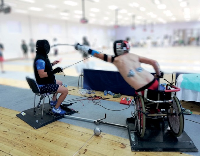
The objective of the present study was to determine the structure of the movement pattern performed during a wheelchair fencing lunge that is executed in response to visual and sensory stimuli. In addition, a comparison was made between fencers in the categories A and B of disability. In addition, the analysis involved the correlation between the duration of the sensorimotor response and the value of the bioelectric signal recorded in selected muscles. Seven Paralympic team athletes specializing in wheelchair fencing (3 in category A and 4 in category B) participated in the research. The fencers perform at international level competitions and are multiple medalists of the Paralympic Games. In the study, a wireless system for sEMG and accelerometer signal measurement was employed to test the intervals between the initiation of the lunge attack and its termination defined by the touch of the weapon on the coach's torso. The electrodes were placed on 9 key muscles responsible for the effectiveness of the executed attack: DEL, TRI, BC, ECR FCR, LD, and EAO. The significant intergroup difference in the muscle activation was found to be 0.333 s for category A fencers and 0.522 s for category A fencers at = 0.039 applies to the (LD LT) muscle, which demonstrates its significance as a postural muscle in the structure of the examined movement pattern. In terms of the values of EMG, a tendency for higher MVC (%) values in most muscles for category A competitors was recorded. The (DL RT) muscle with an intergroup difference of MVC-114.63 for cat. A and 67.50 for cat. B at = 0.039 turned out to play a significant role. The results prove the role of postural muscles: and on the effectiveness of the attacks executed in wheelchair fencing.
本研究的目的是确定在对视觉和感觉刺激做出反应时执行的轮椅击剑弓步运动模式的结构。此外,还比较了残疾类别 A 和 B 的击剑运动员。此外,分析还涉及传感器运动响应的持续时间与在选定肌肉中记录的生物电信号值之间的相关性。七名专门从事轮椅击剑的残奥会运动员(A 类 3 名,B 类 4 名)参加了这项研究。这些击剑运动员参加国际级别的比赛,是残奥会的多枚奖牌获得者。在研究中,使用了一个用于测量 sEMG 和加速度计信号的无线系统,以测试从弓步攻击开始到武器触碰到教练躯干结束的间隔。电极放置在 9 个关键肌肉上,这些肌肉负责执行攻击的有效性:DEL、TRI、BC、ECR FCR、LD 和 EAO。研究发现,A 类运动员的肌肉激活的组间差异显著,为 0.333 秒,B 类运动员的肌肉激活的组间差异为 0.522 秒,差异有统计学意义(P<0.05)。这适用于 LD 和 LT 肌肉,表明它们在被检查的运动模式结构中作为姿势肌肉的重要性。就 EMG 值而言,记录到 A 类运动员的大多数肌肉的 MVC(%)值较高的趋势。A 类运动员的 MVC-114.63 与 B 类运动员的 67.50 之间存在差异的 DL 和 RT 肌肉,差异有统计学意义(P<0.05)。结果证明了姿势肌肉的作用:和 对轮椅击剑中执行的攻击的有效性。