• 文献检索
  • 文档翻译
  • 深度研究
  • 学术资讯
  • Suppr Zotero 插件Zotero 插件
  • 邀请有礼
  • 套餐&价格
  • 历史记录
应用&插件
Suppr Zotero 插件Zotero 插件浏览器插件Mac 客户端Windows 客户端微信小程序
定价
高级版会员购买积分包购买API积分包
服务
文献检索文档翻译深度研究API 文档MCP 服务
关于我们
关于 Suppr公司介绍联系我们用户协议隐私条款
关注我们

Suppr 超能文献

核心技术专利:CN118964589B侵权必究
粤ICP备2023148730 号-1Suppr @ 2026

文献检索

告别复杂PubMed语法,用中文像聊天一样搜索,搜遍4000万医学文献。AI智能推荐,让科研检索更轻松。

立即免费搜索

文件翻译

保留排版,准确专业,支持PDF/Word/PPT等文件格式,支持 12+语言互译。

免费翻译文档

深度研究

AI帮你快速写综述,25分钟生成高质量综述,智能提取关键信息,辅助科研写作。

立即免费体验

多参数动脉自旋标记和扩散加权磁共振成像在Ⅱ级和Ⅲ级胶质瘤鉴别中的应用

Multi-parametric arterial spin labelling and diffusion-weighted magnetic resonance imaging in differentiation of grade II and grade III gliomas.

作者信息

Razek Ahmed Abdel Khalek Abdel, El-Serougy Lamiaa Galal, Abdelsalam Mohamed A, Gaballa Gada Mohamed, Talaat Mona Mohamed

机构信息

Department of Diagnostic Radiology, Mansoura Faculty of Medicine, Mansoura, Egypt.

Department of Neurology, Mansoura Faculty of Medicine, Mansoura, Egypt.

出版信息

Pol J Radiol. 2020 Feb 21;85:e110-e117. doi: 10.5114/pjr.2020.93397. eCollection 2020.

DOI:10.5114/pjr.2020.93397
PMID:32467745
原文链接:https://pmc.ncbi.nlm.nih.gov/articles/PMC7247019/
Abstract

PURPOSE

To assess arterial spin labelling (ASL) perfusion and diffusion MR imaging (DWI) in the differentiation of grade II from grade III gliomas.

MATERIAL AND METHODS

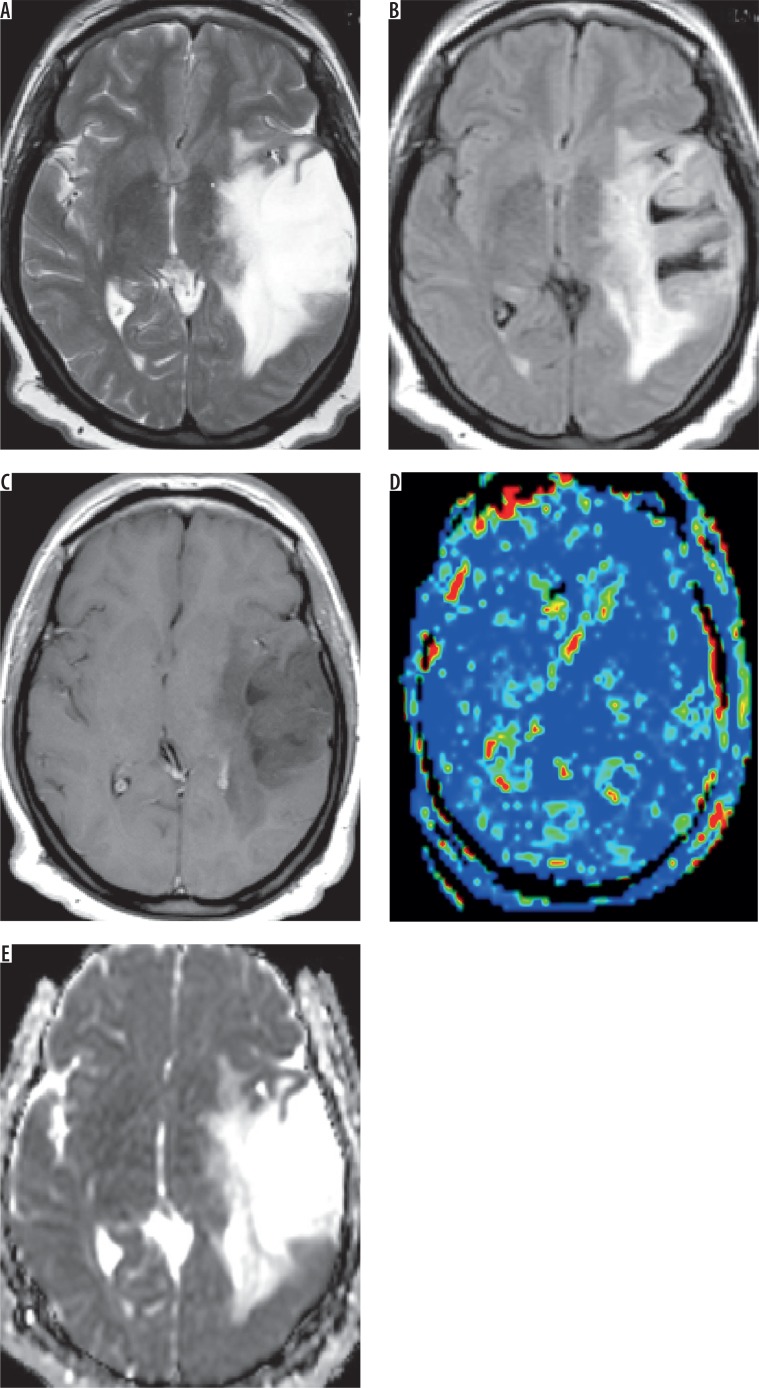
A prospective cohort study was done on 36 patients (20 male and 16 female) with diffuse gliomas, who underwent ASL and DWI. Diffuse gliomas were classified into grade II and grade III. Calculation of tumoural blood flow (TBF) and apparent diffusion coefficient (ADC) of the tumoral and peritumoural regions was made. The ROC curve was drawn to differentiate grade II from grade III gliomas.

RESULTS

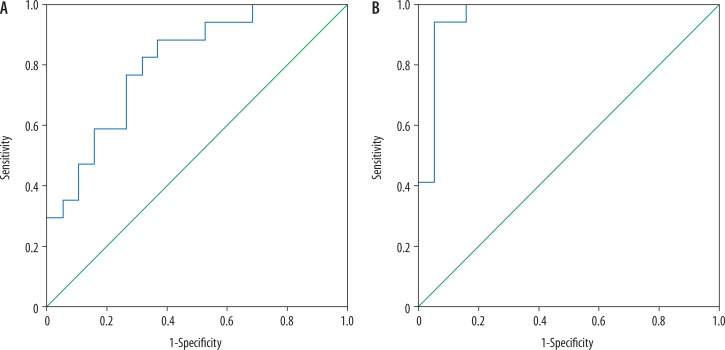
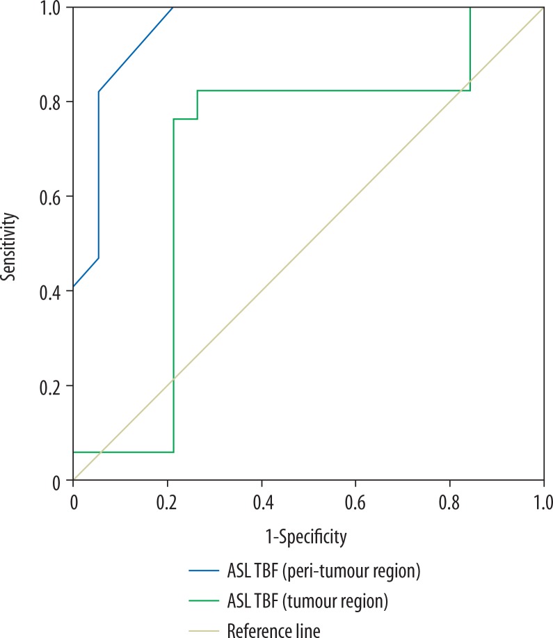
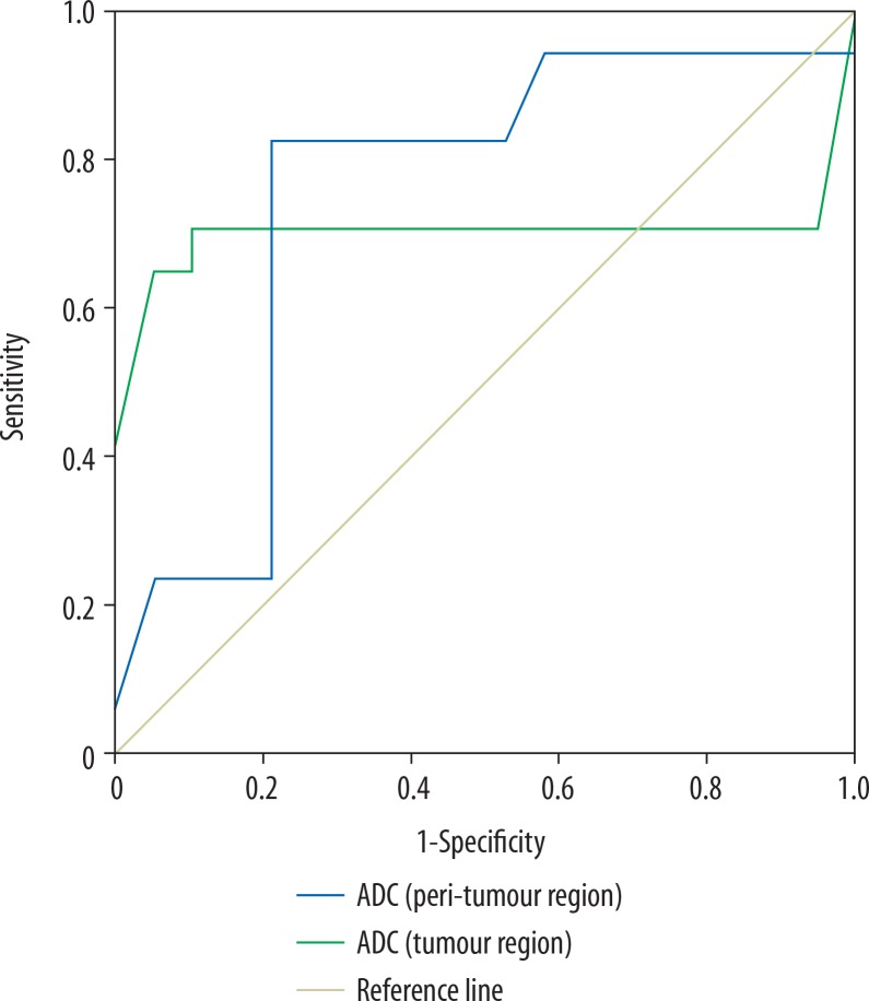
There was a significant difference in TBF of tumoural and peritumoural regions of grade II and III gliomas ( = 0.02 and =0.001, respectively). Selection of 26.1 and 14.8 ml/100 g/min as the cut-off for TBF of tumoural and peritumoural regions differentiated between both groups with area under curve (AUC) of 0.69 and 0.957, and accuracy of 77.8% and 88.9%, respectively. There was small but significant difference in the ADC of tumoural and peritumoural regions between grade II and III gliomas ( = 0.02 for both). The selection of 1.06 and 1.36 × 10 mm/s as the cut-off of ADC of tumoural and peritumoural regions was made, to differentiate grade II from III with AUC of 0.701 and 0.748, and accuracy of 80.6% and 80.6%, respectively. Combined TBF and ADC of tumoural regions revealed an AUC of 0.808 and accuracy of 72.7%. Combined TBF and ADC for peritumoural regions revealed an AUC of 0.96 and accuracy of 94.4%.

CONCLUSION

TBF and ADC of tumoural and peritumoural regions are accurate non-invasive methods of differentiation of grade II from grade III gliomas.

摘要

目的

评估动脉自旋标记(ASL)灌注和扩散磁共振成像(DWI)在鉴别Ⅱ级和Ⅲ级胶质瘤中的作用。

材料与方法

对36例弥漫性胶质瘤患者(20例男性,16例女性)进行了前瞻性队列研究,这些患者均接受了ASL和DWI检查。弥漫性胶质瘤分为Ⅱ级和Ⅲ级。计算肿瘤及瘤周区域的肿瘤血流量(TBF)和表观扩散系数(ADC)。绘制ROC曲线以鉴别Ⅱ级和Ⅲ级胶质瘤。

结果

Ⅱ级和Ⅲ级胶质瘤的肿瘤及瘤周区域TBF存在显著差异(分别为 = 0.02和 = 0.001)。选择26.1和14.8 ml/100 g/min作为肿瘤及瘤周区域TBF的截断值,两组间的区分度曲线下面积(AUC)分别为0.69和0.957,准确率分别为77.8%和88.9%。Ⅱ级和Ⅲ级胶质瘤的肿瘤及瘤周区域ADC存在微小但显著的差异(两者均为 = 0.02)。选择1.06和1.36×10 mm/s作为肿瘤及瘤周区域ADC的截断值,以鉴别Ⅱ级和Ⅲ级胶质瘤,AUC分别为0.701和0.748,准确率分别为80.6%和80.6%。肿瘤区域的联合TBF和ADC显示AUC为0.808,准确率为72.7%。瘤周区域的联合TBF和ADC显示AUC为0.96,准确率为94.4%。

结论

肿瘤及瘤周区域的TBF和ADC是鉴别Ⅱ级和Ⅲ级胶质瘤的准确无创方法。

https://cdn.ncbi.nlm.nih.gov/pmc/blobs/87c7/7247019/a98ad8816b7b/PJR-85-40023-g004.jpg
https://cdn.ncbi.nlm.nih.gov/pmc/blobs/87c7/7247019/f913e15ce66b/PJR-85-40023-g001.jpg
https://cdn.ncbi.nlm.nih.gov/pmc/blobs/87c7/7247019/edff680833b0/PJR-85-40023-g002.jpg
https://cdn.ncbi.nlm.nih.gov/pmc/blobs/87c7/7247019/0d46710c09ec/PJR-85-40023-g003.jpg
https://cdn.ncbi.nlm.nih.gov/pmc/blobs/87c7/7247019/a98ad8816b7b/PJR-85-40023-g004.jpg

相似文献

1
Multi-parametric arterial spin labelling and diffusion-weighted magnetic resonance imaging in differentiation of grade II and grade III gliomas.多参数动脉自旋标记和扩散加权磁共振成像在Ⅱ级和Ⅲ级胶质瘤鉴别中的应用
Pol J Radiol. 2020 Feb 21;85:e110-e117. doi: 10.5114/pjr.2020.93397. eCollection 2020.
2
Multi-parametric MR imaging using pseudo-continuous arterial-spin labeling and diffusion-weighted MR imaging in differentiating subtypes of parotid tumors.使用伪连续动脉自旋标记和弥散加权磁共振成像对腮腺肿瘤进行多参数磁共振成像鉴别诊断。
Magn Reson Imaging. 2019 Nov;63:55-59. doi: 10.1016/j.mri.2019.08.005. Epub 2019 Aug 15.
3
Combination of three-dimensional arterial spin labeling and stretched-exponential model in grading of gliomas.三维动脉自旋标记与拉伸指数模型相结合在胶质瘤分级中的应用
Medicine (Baltimore). 2019 Jun;98(25):e16012. doi: 10.1097/MD.0000000000016012.
4
Intravoxel incoherent motion diffusion-weighted imaging analysis of diffusion and microperfusion in grading gliomas and comparison with arterial spin labeling for evaluation of tumor perfusion.体素内不相干运动扩散加权成像分析胶质瘤分级中的扩散与微灌注,并与动脉自旋标记法比较以评估肿瘤灌注
J Magn Reson Imaging. 2016 Sep;44(3):620-32. doi: 10.1002/jmri.25191. Epub 2016 Feb 16.
5
Multi-parametric arterial spin labeling and diffusion-weighted imaging of paranasal sinuses masses.鼻窦肿块的多参数动脉自旋标记和弥散加权成像。
Oral Radiol. 2023 Apr;39(2):321-328. doi: 10.1007/s11282-022-00640-z. Epub 2022 Jul 28.
6
Measurements of diagnostic examination performance using quantitative apparent diffusion coefficient and proton MR spectroscopic imaging in the preoperative evaluation of tumor grade in cerebral gliomas.使用定量表观扩散系数和质子磁共振波谱成像对脑胶质瘤术前肿瘤分级进行诊断检查性能的测量。
Eur J Radiol. 2011 Nov;80(2):462-70. doi: 10.1016/j.ejrad.2010.07.017. Epub 2010 Aug 13.
7
Perfusion and diffusion MRI signatures in histologic and genetic subtypes of WHO grade II-III diffuse gliomas.世界卫生组织II - III级弥漫性胶质瘤组织学和基因亚型中的灌注及扩散磁共振成像特征
J Neurooncol. 2017 Aug;134(1):177-188. doi: 10.1007/s11060-017-2506-9. Epub 2017 May 25.
8
Efficacy of three-dimensional arterial spin labeling and how it compares against that of contrast enhanced magnetic resonance imaging in preoperative grading of brain gliomas.三维动脉自旋标记技术的效能及其与对比增强磁共振成像在脑胶质瘤术前分级中的比较。
Environ Toxicol. 2023 Jul;38(7):1723-1731. doi: 10.1002/tox.23800. Epub 2023 Apr 11.
9
Arterial spin labeling and diffusion-weighted MR imaging: Utility in differentiating idiopathic orbital inflammatory pseudotumor from orbital lymphoma.动脉自旋标记和弥散加权磁共振成像:在鉴别特发性眼眶炎性假瘤与眼眶淋巴瘤中的应用。
Clin Imaging. 2021 Mar;71:63-68. doi: 10.1016/j.clinimag.2020.10.057. Epub 2020 Nov 6.
10
Dynamic Contrast-Enhanced Perfusion MRI and Diffusion-Weighted Imaging in Grading of Gliomas.动态对比增强灌注磁共振成像与扩散加权成像在胶质瘤分级中的应用
J Neuroimaging. 2015 Sep-Oct;25(5):792-8. doi: 10.1111/jon.12239. Epub 2015 Apr 13.

引用本文的文献

1
A Novel System for Precise Grading of Glioma.一种用于胶质瘤精确分级的新型系统。
Bioengineering (Basel). 2022 Oct 7;9(10):532. doi: 10.3390/bioengineering9100532.
2
Multimodal MRI-Based Radiomic Nomogram for the Early Differentiation of Recurrence and Pseudoprogression of High-Grade Glioma.基于多模态 MRI 的放射组学列线图用于高级别胶质瘤复发与假性进展的早期鉴别。
Biomed Res Int. 2022 Sep 30;2022:4667117. doi: 10.1155/2022/4667117. eCollection 2022.
3
Can Apparent Diffusion Coefficient Predict the Grade, Genotype, or Proliferation Index of Oligodendrogliomas.

本文引用的文献

1
Multi-parametric MR imaging using pseudo-continuous arterial-spin labeling and diffusion-weighted MR imaging in differentiating subtypes of parotid tumors.使用伪连续动脉自旋标记和弥散加权磁共振成像对腮腺肿瘤进行多参数磁共振成像鉴别诊断。
Magn Reson Imaging. 2019 Nov;63:55-59. doi: 10.1016/j.mri.2019.08.005. Epub 2019 Aug 15.
2
Clinical Applications of Arterial Spin Labeling in Brain Tumors.动脉自旋标记在脑肿瘤中的临床应用
J Comput Assist Tomogr. 2019 Jul/Aug;43(4):525-532. doi: 10.1097/RCT.0000000000000873.
3
Assessment of lacrimal glands in thyroid eye disease with diffusion-weighted magnetic resonance imaging.
表观扩散系数能否预测少突胶质细胞瘤的分级、基因型或增殖指数?
Asian J Neurosurg. 2021 Dec 18;16(4):752-758. doi: 10.4103/ajns.AJNS_520_20. eCollection 2021 Oct-Dec.
4
Radiotherapy in adult low-grade glioma: nationwide trends in treatment and outcomes.成人低级别胶质瘤的放射治疗:治疗和结局的全国趋势。
Clin Transl Oncol. 2021 Mar;23(3):628-637. doi: 10.1007/s12094-020-02458-9. Epub 2020 Jul 20.
5
Association of dynamic susceptibility magnetic resonance imaging at initial tumor diagnosis with the prognosis of different molecular glioma subtypes.初始肿瘤诊断时的动态磁敏感加权磁共振成像与不同分子亚型胶质瘤预后的相关性
Neurol Sci. 2020 Dec;41(12):3625-3632. doi: 10.1007/s10072-020-04474-7. Epub 2020 May 28.
利用扩散加权磁共振成像评估甲状腺眼病中的泪腺
Pol J Radiol. 2019 Mar 8;84:e142-e146. doi: 10.5114/pjr.2019.84096. eCollection 2019.
4
Differentiating Glioblastomas from Solitary Brain Metastases Using Arterial Spin Labeling Perfusion- and Diffusion Tensor Imaging-Derived Metrics.利用动脉自旋标记灌注和弥散张量成像衍生指标区分胶质母细胞瘤和单发脑转移瘤。
World Neurosurg. 2019 Jul;127:e593-e598. doi: 10.1016/j.wneu.2019.03.213. Epub 2019 Mar 28.
5
Magnetic resonance spectroscopy of the frontal region in patients with metabolic syndrome: correlation with anthropometric measurement.代谢综合征患者额叶区域的磁共振波谱分析:与人体测量学指标的相关性
Pol J Radiol. 2018 May 15;83:e215-e219. doi: 10.5114/pjr.2018.76024. eCollection 2018.
6
Differentiation of Primary Central Nervous System Lymphoma From Glioblastoma: Quantitative Analysis Using Arterial Spin Labeling and Diffusion Tensor Imaging.原发性中枢神经系统淋巴瘤与胶质母细胞瘤的鉴别:使用动脉自旋标记和扩散张量成像的定量分析
World Neurosurg. 2019 Mar;123:e303-e309. doi: 10.1016/j.wneu.2018.11.155. Epub 2018 Nov 29.
7
Arterial spin labelling and diffusion-weighted magnetic resonance imaging in differentiation of recurrent head and neck cancer from post-radiation changes.动脉自旋标记和扩散加权磁共振成像在复发性头颈癌与放疗后改变的鉴别诊断中的应用
J Laryngol Otol. 2018 Oct;132(10):923-928. doi: 10.1017/S0022215118001743.
8
Diffusion tensor imaging in differentiation of residual head and neck squamous cell carcinoma from post-radiation changes.扩散张量成像在鉴别头颈部残留鳞状细胞癌与放疗后改变中的应用
Magn Reson Imaging. 2018 Dec;54:84-89. doi: 10.1016/j.mri.2018.08.009. Epub 2018 Aug 21.
9
Routine and Advanced Diffusion Imaging Modules of the Salivary Glands.唾液腺的常规与高级扩散成像模块
Neuroimaging Clin N Am. 2018 May;28(2):245-254. doi: 10.1016/j.nic.2018.01.010.
10
Arterial spin labeling perfusion-weighted MR imaging: correlation of tumor blood flow with pathological degree of tumor differentiation, clinical stage and nodal metastasis of head and neck squamous cell carcinoma.动脉自旋标记灌注加权磁共振成像:头颈部鳞状细胞癌肿瘤血流与肿瘤分化病理程度、临床分期及淋巴结转移的相关性
Eur Arch Otorhinolaryngol. 2018 May;275(5):1301-1307. doi: 10.1007/s00405-018-4950-3. Epub 2018 Mar 27.