Department of Pulmonary and Critical Care, The First Affiliated Hospital of Nanchang University, Nanchang, Jiangxi 330006, P.R. China.
Department of Respiratory medicine, The First Affiliated Hospital of Gannan Medical College, Ganzhou, Jiangxi 341000, P.R. China.
Int J Oncol. 2020 Aug;57(2):456-465. doi: 10.3892/ijo.2020.5068. Epub 2020 May 19.
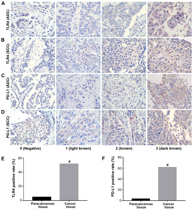
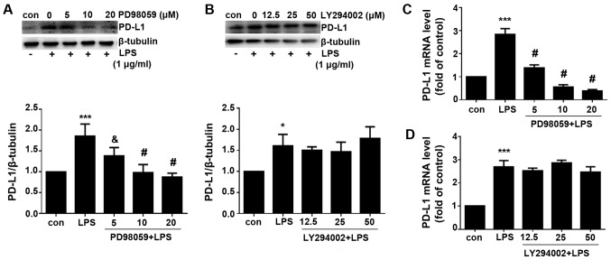
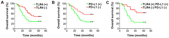
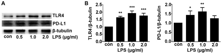
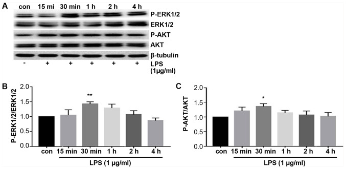
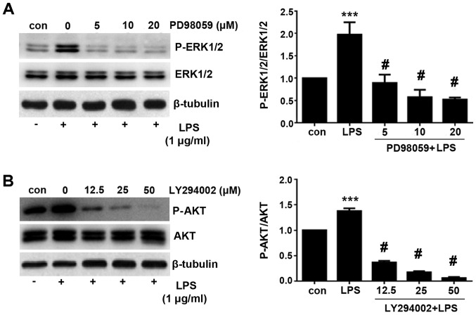
Infection and inflammation serve an important role in tumor development. Toll‑like receptor 4 (TLR4) is a pivotal component of the innate and adaptive immune response during infection and inflammation. Programmed‑death ligand 1 (PD‑L1) is hypothesized as an important factor for non‑small cell lung cancer (NSCLC) immune escape. In the present study, the relationship between TLR4 and PD‑L1, in addition to the associated molecular mechanism, were investigated. TLR4 and PD‑L1 expression in lung cancer tissues were detected using immunohistochemistry, whilst overall patient survival was measured using the Kaplan‑Meier method. The A549 cell line stimulated using lipopolysaccharide (LPS) was applied as the in vitro inflammatory NSCLC model. Associated factors were investigated using reverse transcription‑quantitative PCR and western blotting. Lung cancer tissues exhibited increased PD‑L1 and TLR4 levels compared with those of adjacent para‑cancerous tissues, where there was a positive correlation between TLR4 and PD‑L1 expression. In addition, increased expression of these two proteins was found to be linked with poorer prognoses. Following the stimulation of A549 cells with LPS, TLR4 and PD‑L1 expression levels were revealed to be upregulated in a dose‑dependent manner, where the ERK and PI3K/AKT signaling pathways were found to be activated. Interestingly, in the presence of inhibitors of these two pathways aforementioned, upregulation of PD‑L1 expression was only inhibited by the MEK inhibitor PD98059, which can inhibit ERK activity. These data suggested that the ERK signaling pathway is necessary for the TLR4/PD‑L1 axis. In conclusion, data from the present study suggest that TLR4 and PD‑L1 expression can serve as important prognostic factors for NSCLC, where TLR4 activation may induce PD‑L1 expression through the ERK signaling pathway.
感染和炎症在肿瘤发生发展中起重要作用。Toll 样受体 4(TLR4)是感染和炎症期间固有和适应性免疫反应的关键组成部分。程序性死亡配体 1(PD-L1)被认为是非小细胞肺癌(NSCLC)免疫逃逸的重要因素。在本研究中,检测了 TLR4 与 PD-L1 之间的关系及其相关的分子机制。采用免疫组织化学法检测肺癌组织中 TLR4 和 PD-L1 的表达,采用 Kaplan-Meier 法检测患者的总生存情况。采用脂多糖(LPS)刺激 A549 细胞作为体外炎症性 NSCLC 模型。采用逆转录-定量 PCR 和 Western blot 法检测相关因素。与癌旁组织相比,肺癌组织中 PD-L1 和 TLR4 表达增加,且 TLR4 与 PD-L1 表达呈正相关。此外,发现这两种蛋白的高表达与预后不良有关。用 LPS 刺激 A549 细胞后,TLR4 和 PD-L1 表达水平呈剂量依赖性上调,ERK 和 PI3K/AKT 信号通路被激活。有趣的是,在存在上述两种通路抑制剂的情况下,只有 MEK 抑制剂 PD98059 抑制了 PD-L1 表达的上调,该抑制剂可抑制 ERK 活性。这些数据表明,ERK 信号通路是 TLR4/PD-L1 轴的必要条件。综上所述,本研究数据表明,TLR4 和 PD-L1 表达可作为 NSCLC 的重要预后因素,TLR4 激活可能通过 ERK 信号通路诱导 PD-L1 表达。