Dong Qiangli, Liu Jin, Zeng Lingli, Fan Yiming, Lu Xiaowen, Sun Jinrong, Zhang Liang, Wang Mi, Guo Hua, Zhao Futao, Yan Danfeng, Li Haolun, Guo Weilong, Zhang Yan, Liu Bangshan, Hu Dewen, Li Lingjiang
Department of Psychiatry, The Second Xiangya Hospital, Central South University, Changsha, China.
Mental Health Institute of Central South University, China National Clinical Research Center on Mental Disorders (Xiangya), China National Technology Institute on Mental Disorders, Hunan Technology Institute of Psychiatry, Hunan Key Laboratory of Psychiatry and Mental Health, Changsha, China.
Front Psychiatry. 2020 May 14;11:431. doi: 10.3389/fpsyt.2020.00431. eCollection 2020.
Even with continuous antidepressant treatment, residual symptoms and the risk of relapse can persist in remitted major depressive disorder (MDD) patients. Hence, having a clear recognition of the persistent abnormalities of the underlying neural substrate in MDD through a longitudinal investigation is of great importance.
A total of 127 adult medication-free MDD patients with an acute depressive episode and 118 matched healthy controls (HCs) underwent diffusion tensor imaging. Over a 6-month treatment course, 62 remitted patients underwent a second scan. Remission was defined as a 24-item Hamilton Depression Rating Scale (HAMD) score ≤7 for at least two weeks. Diffusion tensor imaging was performed with a 3.0 T scanner. Differences in whole-brain fractional anisotropy (FA) between MDD patients and HCs were assessed by an independent -test using gender, age, and education as covariates.
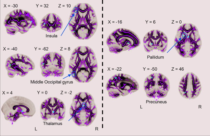
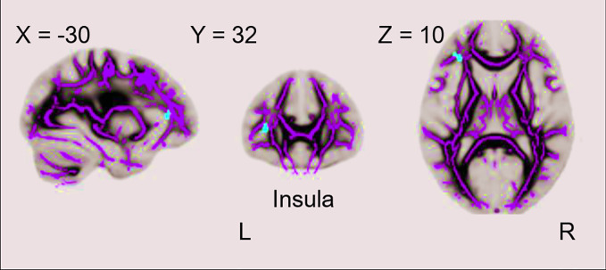
Significant FA reductions in the left insula, left middle occipital gyrus, right thalamus, left pallidum and left precuneus were observed in current MDD (cMDD) patients compared with HCs. Moreover, significant FA reductions in the left insula were observed in remitted (rMDD) patients compared to HCs. However, no significant differences in FA values were found when comparing cMDD and rMDD patients.
The abnormalities in the insula showed state-independent characteristics, while the abnormalities in the middle occipital gyrus, thalamus, pallidum and precuneus seemed to be state-dependent impairments in MDD patients.
即使进行持续的抗抑郁治疗,缓解期的重度抑郁症(MDD)患者仍可能存在残留症状和复发风险。因此,通过纵向研究清晰认识MDD潜在神经基质的持续异常非常重要。
共有127名患有急性抑郁发作的成年未服药MDD患者和118名匹配的健康对照(HCs)接受了扩散张量成像。在为期6个月的治疗过程中,62名缓解患者接受了第二次扫描。缓解定义为汉密尔顿抑郁量表(HAMD)24项评分至少两周≤7分。使用3.0T扫描仪进行扩散张量成像。以性别、年龄和教育程度作为协变量,通过独立样本t检验评估MDD患者和HCs之间全脑分数各向异性(FA)的差异。
与HCs相比,当前MDD(cMDD)患者在左侧岛叶、左侧枕中回、右侧丘脑、左侧苍白球和左侧楔前叶观察到显著的FA降低。此外,与HCs相比,缓解期(rMDD)患者在左侧岛叶也观察到显著的FA降低。然而,比较cMDD和rMDD患者时,FA值没有显著差异。
岛叶的异常表现出与状态无关的特征,而枕中回、丘脑、苍白球和楔前叶的异常似乎是MDD患者与状态相关的损伤。