Flow Cytometry Core Facility, The Blizard Institute, Barts and The London School of Medicine and Dentistry, Queen Mary London University, 4 Newark Street, London, E1 2AT, UK.
Apoptosis. 2020 Aug;25(7-8):548-557. doi: 10.1007/s10495-020-01613-5.
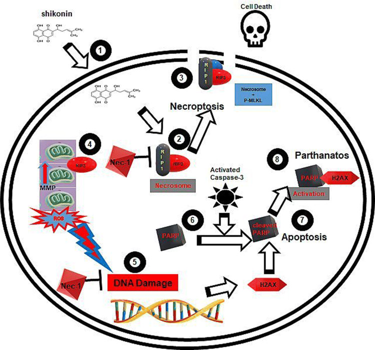
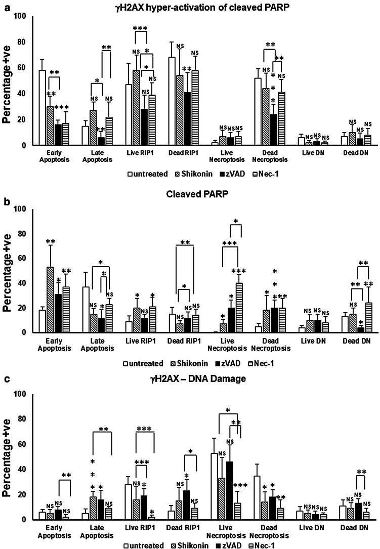
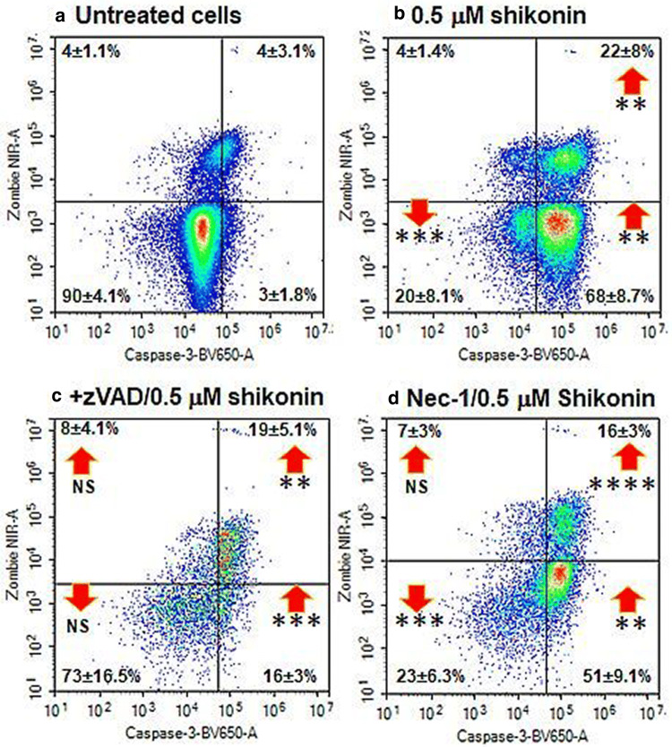
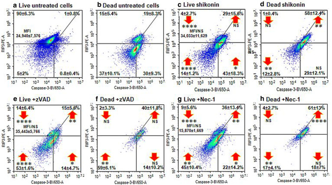
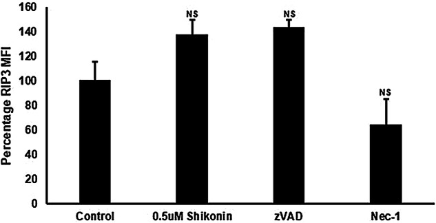
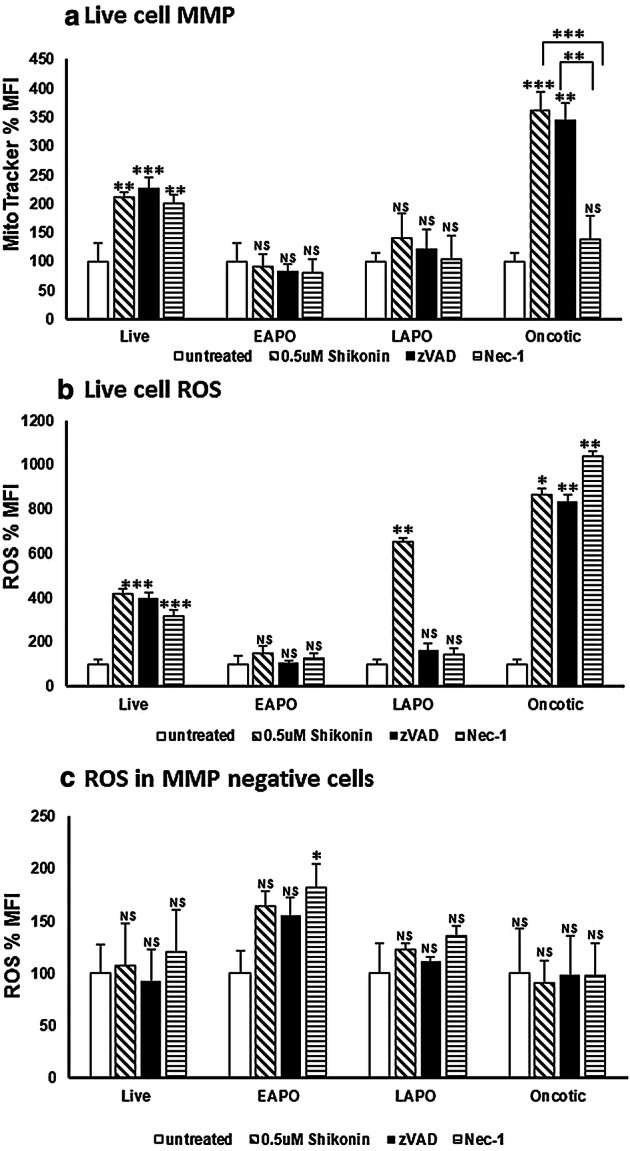
Shikonin induced necroptosis in Jurkat cells were identified flow cytometrically by the up-regulation of RIP3 in live cells and that a proportion of these cells underwent other forms of regulated cell death (RCD) which included parthanatos (< 10%), or cleaved PARP (< 10%) and DNA Damage (> 30%). Live necroptotic cells also possessed functioning mitochondria with hyper-polarized mitochondria membrane potential and generated a fivefold increase in cellular reactive oxygen species (ROS) which was resistant to inhibition by zVAD and necrostatin-1 (Nec-1). After loss of plasma membrane integrity these dead necroptotic cells then showed a higher incidence of parthanatos (> 40%), or cleaved PARP (> 15%) but less DNA Damage (< 15%). Inhibition of shikonin induced apoptosis and necroptosis by zVAD and Nec-1 respectively resulted in live necroptotic cells with an increased incidence of cleaved PARP and reduced levels of DNA Damage respectively. Dead necroptotic cells then showed a reduced incidence of parthanatos and DNA Damage after inhibition by zVAD and Nec-1 respectively. A high proportion of these dead necroptotic cells (30%) which lacked plasma membrane integrity also displayed functioning hyper-polarized mitochondria with high levels of cellular ROS and thus had the capacity to influence the outcome of RCD processes rather than just been the end product of cell death, the necrotic cell. Flow cytometry can thus measure multiple forms of RCD and the level of cellular ROS and MMP which highlights the inter-connection between cell death processes and that a single cell may simultaneously display multiple forms of RCD.
紫草素诱导 Jurkat 细胞发生坏死性凋亡,通过活细胞中 RIP3 的上调来鉴定,其中一部分细胞发生了其他形式的细胞程序性死亡(RCD),包括 PARN 过度激活(<10%)、PARP 切割(<10%)和 DNA 损伤(>30%)。活的坏死性凋亡细胞还具有功能正常的线粒体,其线粒体膜电位高度极化,并产生五倍于细胞活性氧(ROS)的增加,这对 zVAD 和 Nec-1 的抑制具有抗性。在质膜完整性丧失后,这些死亡的坏死性凋亡细胞随后表现出更高比例的 PARN 过度激活(>40%)、PARP 切割(>15%),但 DNA 损伤较少(<15%)。zVAD 和 Nec-1 分别抑制紫草素诱导的细胞凋亡和坏死性凋亡,导致活的坏死性凋亡细胞中 PARP 切割增加,DNA 损伤减少。随后,在 zVAD 和 Nec-1 抑制后,死亡的坏死性凋亡细胞显示出 PARN 过度激活和 DNA 损伤的发生率降低。这些缺乏质膜完整性的死亡坏死性凋亡细胞中,有很高比例(30%)也显示出功能正常的高度极化线粒体,具有高水平的细胞 ROS,因此有能力影响 RCD 过程的结果,而不仅仅是细胞死亡的终产物,即坏死细胞。流式细胞术可以测量多种形式的 RCD 和细胞 ROS 和 MMP 水平,这突出了细胞死亡过程之间的相互联系,并且一个细胞可能同时显示多种形式的 RCD。