Kong Xiufang, Ma Lili, Lv Peng, Cui Xiaomeng, Chen Rongyi, Ji Zongfei, Chen Huiyong, Lin Jiang, Jiang Lindi
Department of Rheumatology, Zhongshan Hospital Affiliated to Fudan University, Shanghai, China.
Department of Radiology, Zhongshan Hospital Affiliated to Fudan University, Shanghai, China.
Arthritis Res Ther. 2020 Jun 5;22(1):131. doi: 10.1186/s13075-020-02203-1.
Takayasu arteritis (TA) is a large vessel vasculitis that can involve pulmonary arteries (PAs). We studied multiple clinical characteristics related to pulmonary artery involvement (PAI) in TA patients.
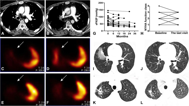
We enrolled 216 patients with TA from a large prospective cohort. PAI was assessed in each patient based on data from magnetic resonance angiography/computed tomography angiography. Pulmonary hypertension, cardiac function, and pulmonary parenchymal lesions were evaluated further in patients with PAI based on echocardiography, the New York Heart Association Functional Classification, and pulmonary computed tomography, respectively. These abnormalities related to PAI were followed up to evaluate treatment effects.
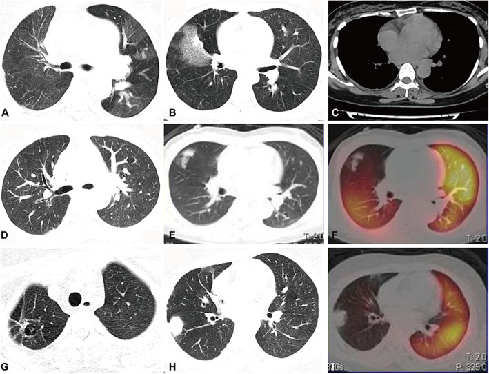
PAI was detected in 56/216 (25.93%) patients, which involved the pulmonary trunk, main PAs, and small vessels in the lungs. Among patients with PAI, 28 (50%) patients were accompanied by pulmonary hypertension, which was graded as 'severe' in 9 (16.07%), 'moderate' in 10 (17.86%), and mild in 9 (16.07%). Twenty-six (46.43%) patients showed advanced NYHA function (III, 20, 35.71%; IV, 6, 10.71%). Furthermore, 21 (37.50%) patients presented with abnormal pulmonary parenchymal lesions in the area corresponding to PAI (e.g. the mosaic sign, infarction, bronchiectasis). During follow-up, two patients died due to heart failure and pulmonary thrombosis. In the remaining patients, the abnormalities mentioned above improved partially after routine treatment.
PAI is common in TA patients. PAI can cause pulmonary hypertension, cardiac insufficiency, and pulmonary parenchymal lesions, which worsen patients' prognosis.
大动脉炎(TA)是一种可累及肺动脉(PA)的大血管血管炎。我们研究了TA患者中与肺动脉受累(PAI)相关的多种临床特征。
我们从一个大型前瞻性队列中纳入了216例TA患者。根据磁共振血管造影/计算机断层扫描血管造影数据评估每位患者的PAI。分别基于超声心动图、纽约心脏协会功能分级和肺部计算机断层扫描,对PAI患者的肺动脉高压、心功能和肺实质病变进行进一步评估。对这些与PAI相关的异常情况进行随访以评估治疗效果。
216例患者中有56例(25.93%)检测到PAI,累及肺动脉主干、主要PA和肺内小血管。在PAI患者中,28例(50%)伴有肺动脉高压,其中9例(16.07%)为“重度”,10例(17.86%)为“中度”,9例(16.07%)为轻度。26例(46.43%)患者表现为纽约心脏协会心功能分级晚期(III级,20例,35.71%;IV级,6例,10.71%)。此外,21例(37.50%)患者在与PAI对应的区域出现肺实质病变异常(如马赛克征、梗死、支气管扩张)。随访期间,2例患者因心力衰竭和肺血栓形成死亡。其余患者经常规治疗后上述异常情况部分改善。
PAI在TA患者中很常见。PAI可导致肺动脉高压、心功能不全和肺实质病变,从而恶化患者预后。