• 文献检索
  • 文档翻译
  • 深度研究
  • 学术资讯
  • Suppr Zotero 插件Zotero 插件
  • 邀请有礼
  • 套餐&价格
  • 历史记录
应用&插件
Suppr Zotero 插件Zotero 插件浏览器插件Mac 客户端Windows 客户端微信小程序
定价
高级版会员购买积分包购买API积分包
服务
文献检索文档翻译深度研究API 文档MCP 服务
关于我们
关于 Suppr公司介绍联系我们用户协议隐私条款
关注我们

Suppr 超能文献

核心技术专利:CN118964589B侵权必究
粤ICP备2023148730 号-1Suppr @ 2026

文献检索

告别复杂PubMed语法,用中文像聊天一样搜索,搜遍4000万医学文献。AI智能推荐,让科研检索更轻松。

立即免费搜索

文件翻译

保留排版,准确专业,支持PDF/Word/PPT等文件格式,支持 12+语言互译。

免费翻译文档

深度研究

AI帮你快速写综述,25分钟生成高质量综述,智能提取关键信息,辅助科研写作。

立即免费体验

混合征是自发性脑出血早期血肿扩大程度的有力预测指标。

Blend Sign Is a Strong Predictor of the Extent of Early Hematoma Expansion in Spontaneous Intracerebral Hemorrhage.

作者信息

Zhang Mingyue, Chen Jie, Zhan Chenyi, Liu Jinjin, Chen Qian, Xia Tianyi, Zhang Tingting, Zhu Dongqin, Chen Chao, Yang Yunjun

机构信息

Department of Radiology, The First Affiliated Hospital of Wenzhou Medical University, Wenzhou, China.

出版信息

Front Neurol. 2020 May 19;11:334. doi: 10.3389/fneur.2020.00334. eCollection 2020.

DOI:10.3389/fneur.2020.00334
PMID:32508731
原文链接:https://pmc.ncbi.nlm.nih.gov/articles/PMC7248383/
Abstract

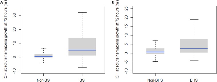
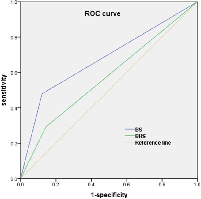
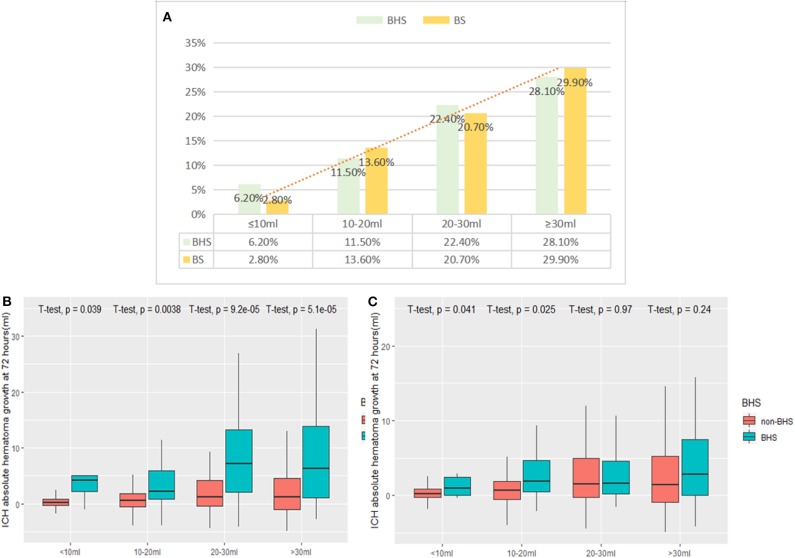
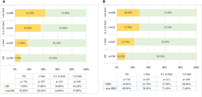
It is unclear which imaging marker is optimal for predicting the extent of hematoma expansion (EHE). We aimed to compare the usefulness of the blend sign (BS) with that of other non-contrast computed tomography (NCCT) markers for predicting the EHE in patients with spontaneous intracerebral hemorrhage (sICH). Patients with sICH admitted to our Neurology Emergency Department between September 2013 and January 2019 were enrolled. The EHE was calculated as the absolute increase in hematoma volume between baseline and follow-up CT (within 72 h). The EHE was categorized into four groups: "no growth," "minimal change" (≤5.1 ml), "moderate change" (5.1-12.5 ml), and "massive change" (>12.5 ml). Univariate and multivariate analyses were performed to investigate the relationship between the NCCT markers [BS, black hole sign (BHS), satellite sign, and island sign] and the EHE. A total of 1,111 sICH patients were included (median age: 60 years; 66.5% males). Multiple linear regression analysis showed that the presence of the BS and BHS was independently associated with the EHE, after adjusting for confounders ( < 0.001 and = 0.003, respectively). The presence of the BS and BHS was positively correlated with growth category ( = 0.285 and = 0.199, both s < 0.001). The BS demonstrated a better predictive performance for the EHE than did the BHS [area under the curve (AUC): 0.67 vs. 0.57; both s < 0.001]. In patients with acute sICH, the BS showed a better performance in predicting the EHE compared with other NCCT markers. This imaging marker may help identify patients at a high risk of significant hematoma expansion and may facilitate its early management.

摘要

目前尚不清楚哪种影像学标志物最适合预测血肿扩大程度(EHE)。我们旨在比较混合征(BS)与其他非增强计算机断层扫描(NCCT)标志物在预测自发性脑出血(sICH)患者EHE方面的效用。纳入了2013年9月至2019年1月期间入住我院神经内科急诊科的sICH患者。EHE计算为基线CT与随访CT(72小时内)之间血肿体积的绝对增加量。EHE分为四组:“无增长”、“微小变化”(≤5.1 ml)、“中度变化”(5.1 - 12.5 ml)和“大量变化”(>12.5 ml)。进行单因素和多因素分析以研究NCCT标志物[BS、黑洞征(BHS)、卫星征和岛征]与EHE之间的关系。共纳入1111例sICH患者(中位年龄:60岁;男性占66.5%)。多元线性回归分析显示,在调整混杂因素后,BS和BHS的存在与EHE独立相关(分别为<0.001和 = 0.003)。BS和BHS的存在与增长类别呈正相关(分别为 = 0.285和 = 0.199,均P<0.001)。与BHS相比,BS对EHE的预测性能更好[曲线下面积(AUC):0.67对0.57;均P<0.001]。在急性sICH患者中,与其他NCCT标志物相比,BS在预测EHE方面表现更好。这种影像学标志物可能有助于识别血肿显著扩大的高危患者,并可能促进其早期管理。

https://cdn.ncbi.nlm.nih.gov/pmc/blobs/5525/7248383/4df4e3aa77d9/fneur-11-00334-g0004.jpg
https://cdn.ncbi.nlm.nih.gov/pmc/blobs/5525/7248383/f7a083f825de/fneur-11-00334-g0001.jpg
https://cdn.ncbi.nlm.nih.gov/pmc/blobs/5525/7248383/d5cf49231545/fneur-11-00334-g0002.jpg
https://cdn.ncbi.nlm.nih.gov/pmc/blobs/5525/7248383/dd9a84c25435/fneur-11-00334-g0003.jpg
https://cdn.ncbi.nlm.nih.gov/pmc/blobs/5525/7248383/4df4e3aa77d9/fneur-11-00334-g0004.jpg

相似文献

1
Blend Sign Is a Strong Predictor of the Extent of Early Hematoma Expansion in Spontaneous Intracerebral Hemorrhage.混合征是自发性脑出血早期血肿扩大程度的有力预测指标。
Front Neurol. 2020 May 19;11:334. doi: 10.3389/fneur.2020.00334. eCollection 2020.
2
Comparison of Spot Sign, Blend Sign and Black Hole Sign for Outcome Prediction in Patients with Intracerebral Hemorrhage.斑点征、混合征和黑洞征对脑出血患者预后预测的比较
J Stroke. 2017 Sep;19(3):333-339. doi: 10.5853/jos.2016.02061. Epub 2017 Sep 29.
3
A comparative study of the blend sign and the black hole sign on CT as a predictor of hematoma expansion in spontaneous intracerebral hemorrhage.CT 上混杂征和黑洞征对自发性脑出血血肿扩大的预测作用的对比研究。
Biosci Trends. 2017;11(6):682-687. doi: 10.5582/bst.2017.01283.
4
Comparison of Black Hole Sign, Satellite Sign, and Iodine Sign to Predict Hematoma Expansion in Patients with Spontaneous Intracerebral Hemorrhage.比较黑洞征、卫星征和碘征在预测自发性脑出血患者血肿扩大中的作用。
Biomed Res Int. 2021 Feb 1;2021:3919710. doi: 10.1155/2021/3919710. eCollection 2021.
5
Determining the Optimal Shape-Related Indicator on Noncontrast Computed Tomography for Predicting Hematoma Expansion in Spontaneous Intracerebral Hemorrhage.确定非增强计算机断层扫描上与形状相关的最佳指标以预测自发性脑出血的血肿扩大
World Neurosurg. 2019 Jan;121:e584-e588. doi: 10.1016/j.wneu.2018.09.172. Epub 2018 Oct 3.
6
Comparison of Ultra-Early Hematoma Growth and Common Noncontrast Computed Tomography Features in Predicting Hematoma Enlargement in Patients with Spontaneous Intracerebral Hemorrhage.超早期血肿增长与普通非增强 CT 特征在预测自发性脑出血患者血肿扩大中的比较。
World Neurosurg. 2020 Feb;134:e75-e81. doi: 10.1016/j.wneu.2019.09.053. Epub 2019 Oct 21.
7
Accuracy of imaging markers on noncontrast computed tomography in predicting intracerebral hemorrhage expansion.非对比 CT 成像标志物预测脑出血扩大的准确性。
Neurol Res. 2020 Nov;42(11):973-979. doi: 10.1080/01616412.2020.1795577. Epub 2020 Jul 22.
8
Noncontrast Computed Tomography Markers as Predictors of Revised Hematoma Expansion in Acute Intracerebral Hemorrhage.非对比计算机断层扫描标志物可预测急性脑出血血肿扩大。
J Am Heart Assoc. 2021 Feb 2;10(3):e018248. doi: 10.1161/JAHA.120.018248. Epub 2021 Jan 28.
9
Comparison of different noncontrast computed tomographic markers for predicting early perihematomal edema expansion in patients with intracerebral hemorrhage.比较不同的非对比计算机断层扫描标志物,以预测脑出血患者早期血肿周围水肿扩大。
J Clin Neurosci. 2023 Jun;112:1-5. doi: 10.1016/j.jocn.2023.03.006. Epub 2023 Apr 1.
10
The Accuracy of the Spot Sign and the Blend Sign for Predicting Hematoma Expansion in Patients with Spontaneous Intracerebral Hemorrhage.斑点征和融合征预测自发性脑出血患者血肿扩大的准确性
Med Sci Monit. 2017 May 12;23:2250-2257. doi: 10.12659/msm.901583.

引用本文的文献

1
Comparative assessment of the spot sign and leakage sign as predictive factors for spontaneous intracranial hematoma expansion.斑点征和渗漏征作为自发性颅内血肿扩大预测因素的比较评估
Emerg Radiol. 2025 Jun 12. doi: 10.1007/s10140-025-02352-3.
2
CT radiomics combined with clinical and radiological factors predict hematoma expansion in hypertensive intracerebral hemorrhage.CT影像组学联合临床及影像学因素预测高血压性脑出血的血肿扩大。
Eur Radiol. 2025 Jan;35(1):6-19. doi: 10.1007/s00330-024-10921-2. Epub 2024 Jul 11.
3
Research advances in predicting the expansion of hypertensive intracerebral hemorrhage based on CT images: an overview.

本文引用的文献

1
Does Noncontrast Computed Tomography Scan Predict Rebleeding After Endoscopic Surgery for Spontaneous Intracerebral Hemorrhage?非增强计算机断层扫描能否预测自发性脑出血内镜手术后再出血?
World Neurosurg. 2019 Jul;127:e965-e971. doi: 10.1016/j.wneu.2019.04.010. Epub 2019 Apr 6.
2
Absolute risk and predictors of the growth of acute spontaneous intracerebral haemorrhage: a systematic review and meta-analysis of individual patient data.急性自发性脑出血增长的绝对风险和预测因素:一项个体患者数据的系统回顾和荟萃分析。
Lancet Neurol. 2018 Oct;17(10):885-894. doi: 10.1016/S1474-4422(18)30253-9. Epub 2018 Aug 14.
3
Computed Tomographic Black Hole Sign Predicts Postoperative Rehemorrhage in Patients with Spontaneous Intracranial Hemorrhage Following Stereotactic Minimally Invasive Surgery.
基于 CT 图像预测高血压性脑出血扩大的研究进展:综述。
PeerJ. 2024 Jun 7;12:e17556. doi: 10.7717/peerj.17556. eCollection 2024.
4
Predictor of the Postoperative Swelling After Craniotomy for Spontaneous Intracerebral Hemorrhage: Sphericity Index as a Novel Parameter.自发性脑出血开颅术后肿胀的预测因素:球形指数作为一个新参数
Korean J Neurotrauma. 2023 Sep 11;19(3):333-347. doi: 10.13004/kjnt.2023.19.e41. eCollection 2023 Sep.
5
Prior knowledge-based precise diagnosis of blend sign from head computed tomography.基于先验知识对头计算机断层扫描混合征进行精确诊断。
Front Neurosci. 2023 Feb 10;17:1112355. doi: 10.3389/fnins.2023.1112355. eCollection 2023.
6
A non-contrast computed tomography-based radiomics nomogram for the prediction of hematoma expansion in patients with deep ganglionic intracerebral hemorrhage.基于非增强计算机断层扫描的影像组学列线图预测深部基底节区脑出血患者血肿扩大情况
Front Neurol. 2022 Oct 11;13:974183. doi: 10.3389/fneur.2022.974183. eCollection 2022.
7
Irregular shape as an independent predictor of prognosis in patients with primary intracerebral hemorrhage.形态不规则可作为预测原发性脑出血患者预后的独立指标。
Sci Rep. 2022 May 20;12(1):8552. doi: 10.1038/s41598-022-12536-3.
8
Advances in computed tomography-based prognostic methods for intracerebral hemorrhage.基于计算机断层扫描的脑出血预后方法的进展。
Neurosurg Rev. 2022 Jun;45(3):2041-2050. doi: 10.1007/s10143-022-01760-0. Epub 2022 Feb 28.
9
Patients with venous thromboembolism after spontaneous intracerebral hemorrhage: a review.自发性脑出血后静脉血栓栓塞症患者:一项综述
Thromb J. 2021 Nov 27;19(1):93. doi: 10.1186/s12959-021-00345-z.
10
Risk of Post-stroke Epilepsy Following Stroke-Associated Acute Symptomatic Seizures.卒中相关性急性症状性癫痫发作后发生卒中后癫痫的风险
Front Aging Neurosci. 2021 Sep 13;13:707732. doi: 10.3389/fnagi.2021.707732. eCollection 2021.
计算机断层扫描黑洞征可预测立体定向微创手术治疗自发性颅内出血患者术后再出血情况。
World Neurosurg. 2018 Dec;120:e153-e160. doi: 10.1016/j.wneu.2018.07.256. Epub 2018 Aug 6.
4
Predicting Intracerebral Hemorrhage Expansion With Noncontrast Computed Tomography: The BAT Score.基于非增强 CT 的脑出血扩大预测:BAT 评分。
Stroke. 2018 May;49(5):1163-1169. doi: 10.1161/STROKEAHA.117.020138. Epub 2018 Apr 18.
5
Accuracy of the Blend Sign on Computed Tomography as a Predictor of Hematoma Growth after Spontaneous Intracerebral Hemorrhage: A Systematic Review.计算机断层扫描上混合征作为自发性脑出血后血肿扩大预测指标的准确性:一项系统评价
J Stroke Cerebrovasc Dis. 2018 Jun;27(6):1705-1710. doi: 10.1016/j.jstrokecerebrovasdis.2018.01.032. Epub 2018 Mar 7.
6
Predictors of hematoma expansion predictors after intracerebral hemorrhage.脑出血后血肿扩大的预测因素
Oncotarget. 2017 Jul 18;8(51):89348-89363. doi: 10.18632/oncotarget.19366. eCollection 2017 Oct 24.
7
Comparison of Spot Sign, Blend Sign and Black Hole Sign for Outcome Prediction in Patients with Intracerebral Hemorrhage.斑点征、混合征和黑洞征对脑出血患者预后预测的比较
J Stroke. 2017 Sep;19(3):333-339. doi: 10.5853/jos.2016.02061. Epub 2017 Sep 29.
8
Island Sign: An Imaging Predictor for Early Hematoma Expansion and Poor Outcome in Patients With Intracerebral Hemorrhage.岛征:脑出血患者早期血肿扩大及预后不良的影像学预测指标
Stroke. 2017 Nov;48(11):3019-3025. doi: 10.1161/STROKEAHA.117.017985. Epub 2017 Oct 10.
9
Blend sign predicts poor outcome in patients with intracerebral hemorrhage.混合征预示脑出血患者预后不良。
PLoS One. 2017 Aug 22;12(8):e0183082. doi: 10.1371/journal.pone.0183082. eCollection 2017.
10
Blood pressure reduction and noncontrast CT markers of intracerebral hemorrhage expansion.血压降低与脑出血扩大的非增强CT标志物
Neurology. 2017 Aug 8;89(6):548-554. doi: 10.1212/WNL.0000000000004210. Epub 2017 Jul 12.