• 文献检索
  • 文档翻译
  • 深度研究
  • 学术资讯
  • Suppr Zotero 插件Zotero 插件
  • 邀请有礼
  • 套餐&价格
  • 历史记录
应用&插件
Suppr Zotero 插件Zotero 插件浏览器插件Mac 客户端Windows 客户端微信小程序
定价
高级版会员购买积分包购买API积分包
服务
文献检索文档翻译深度研究API 文档MCP 服务
关于我们
关于 Suppr公司介绍联系我们用户协议隐私条款
关注我们

Suppr 超能文献

核心技术专利:CN118964589B侵权必究
粤ICP备2023148730 号-1Suppr @ 2026

文献检索

告别复杂PubMed语法,用中文像聊天一样搜索,搜遍4000万医学文献。AI智能推荐,让科研检索更轻松。

立即免费搜索

文件翻译

保留排版,准确专业,支持PDF/Word/PPT等文件格式,支持 12+语言互译。

免费翻译文档

深度研究

AI帮你快速写综述,25分钟生成高质量综述,智能提取关键信息,辅助科研写作。

立即免费体验

基于 CT 图像预测高血压性脑出血扩大的研究进展:综述。

Research advances in predicting the expansion of hypertensive intracerebral hemorrhage based on CT images: an overview.

机构信息

Department of Anesthesiology, Nanan District People's Hospital of Chongqing, Chongqing, China.

Department of Breast and Thyroid Surgery, Chongqing Bishan District Maternal and Child Health Care Hospital, Chongqing, China.

出版信息

PeerJ. 2024 Jun 7;12:e17556. doi: 10.7717/peerj.17556. eCollection 2024.

DOI:10.7717/peerj.17556
PMID:38860211
原文链接:https://pmc.ncbi.nlm.nih.gov/articles/PMC11164062/
Abstract

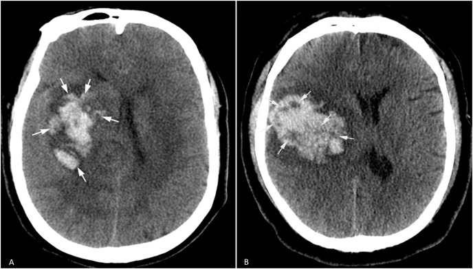
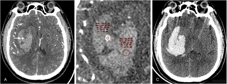
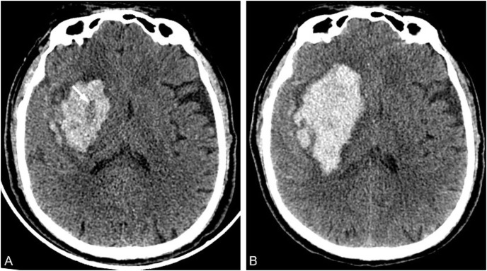
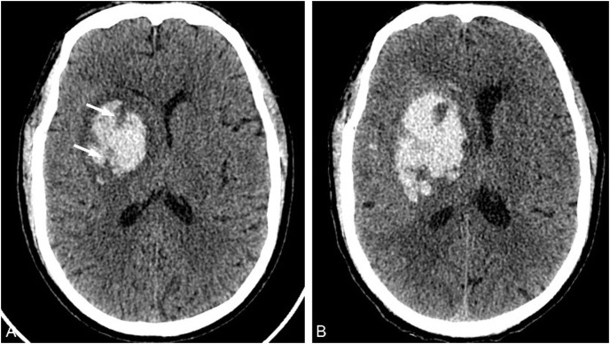
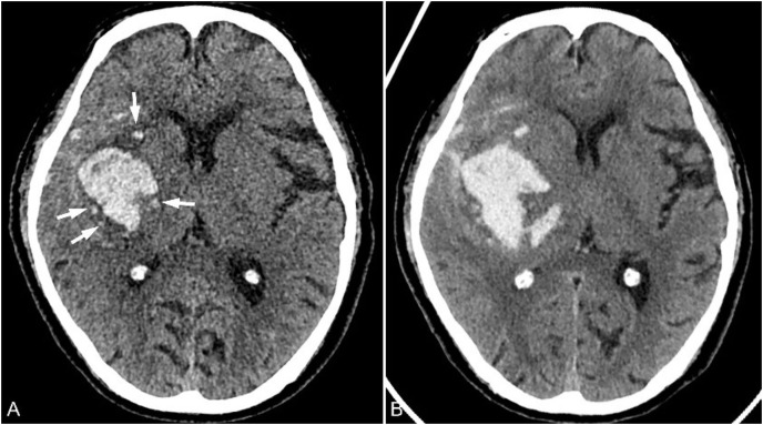
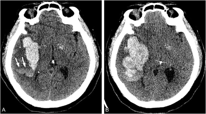
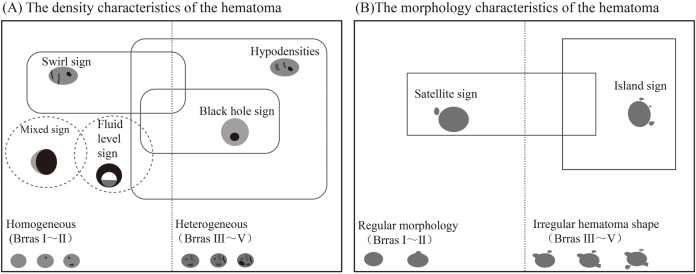
Hematoma expansion (HE) is an important risk factor for death or poor prognosis in patients with hypertensive intracerebral hemorrhage (HICH). Accurately predicting the risk of HE in patients with HICH is of great clinical significance for timely intervention and improving patient prognosis. Many imaging signs reported in literatures showed the important clinical value for predicting HE. In recent years, the development of radiomics and artificial intelligence has provided new methods for HE prediction with high accuracy. Therefore, this article reviews the latest research progress in CT imaging, radiomics, and artificial intelligence of HE, in order to help identify high-risk patients for HE in clinical practice.

摘要

血肿扩大(HE)是高血压性脑出血(HICH)患者死亡或预后不良的重要危险因素。准确预测 HICH 患者发生 HE 的风险,对于及时干预和改善患者预后具有重要的临床意义。文献中报道的许多影像学征象均显示出对预测 HE 的重要临床价值。近年来,影像组学和人工智能的发展为 HE 预测提供了高精度的新方法。因此,本文综述了 HE 的 CT 影像学、影像组学和人工智能的最新研究进展,以期有助于在临床实践中识别出 HE 的高危患者。

https://cdn.ncbi.nlm.nih.gov/pmc/blobs/c37f/11164062/c5bdc8c38d10/peerj-12-17556-g010.jpg
https://cdn.ncbi.nlm.nih.gov/pmc/blobs/c37f/11164062/3f82b5f29f7d/peerj-12-17556-g001.jpg
https://cdn.ncbi.nlm.nih.gov/pmc/blobs/c37f/11164062/25648ac49618/peerj-12-17556-g002.jpg
https://cdn.ncbi.nlm.nih.gov/pmc/blobs/c37f/11164062/963e3890128f/peerj-12-17556-g003.jpg
https://cdn.ncbi.nlm.nih.gov/pmc/blobs/c37f/11164062/322c6f332210/peerj-12-17556-g004.jpg
https://cdn.ncbi.nlm.nih.gov/pmc/blobs/c37f/11164062/5defcf4c5de2/peerj-12-17556-g005.jpg
https://cdn.ncbi.nlm.nih.gov/pmc/blobs/c37f/11164062/615fa357fd53/peerj-12-17556-g006.jpg
https://cdn.ncbi.nlm.nih.gov/pmc/blobs/c37f/11164062/551e00921acb/peerj-12-17556-g007.jpg
https://cdn.ncbi.nlm.nih.gov/pmc/blobs/c37f/11164062/11a5a22eb34c/peerj-12-17556-g008.jpg
https://cdn.ncbi.nlm.nih.gov/pmc/blobs/c37f/11164062/30c58ca296e7/peerj-12-17556-g009.jpg
https://cdn.ncbi.nlm.nih.gov/pmc/blobs/c37f/11164062/c5bdc8c38d10/peerj-12-17556-g010.jpg

相似文献

1
Research advances in predicting the expansion of hypertensive intracerebral hemorrhage based on CT images: an overview.基于 CT 图像预测高血压性脑出血扩大的研究进展:综述。
PeerJ. 2024 Jun 7;12:e17556. doi: 10.7717/peerj.17556. eCollection 2024.
2
Lower Ionized Calcium Predicts Hematoma Expansion and Poor Outcome in Patients with Hypertensive Intracerebral Hemorrhage.低离子钙预测高血压性脑出血患者的血肿扩大及不良预后。
World Neurosurg. 2018 Oct;118:e500-e504. doi: 10.1016/j.wneu.2018.06.223. Epub 2018 Jul 6.
3
Establishment and evaluation of a nomogram model for predicting hematoma expansion in hypertensive intracerebral hemorrhage based on clinical factors and plain CT scan signs.基于临床因素和 CT 平扫征象的预测高血压性脑出血血肿扩大的列线图模型的建立与评估。
Ann Palliat Med. 2021 Dec;10(12):12789-12800. doi: 10.21037/apm-21-3569.
4
Predicting postoperative rehemorrhage in hypertensive intracerebral hemorrhage using noncontrast CT radiomics and clinical data with an interpretable machine learning approach.使用非增强CT影像组学和临床数据以及可解释的机器学习方法预测高血压性脑出血术后再出血
Sci Rep. 2024 Apr 27;14(1):9717. doi: 10.1038/s41598-024-60463-2.
5
CT radiomics combined with clinical and radiological factors predict hematoma expansion in hypertensive intracerebral hemorrhage.CT影像组学联合临床及影像学因素预测高血压性脑出血的血肿扩大。
Eur Radiol. 2025 Jan;35(1):6-19. doi: 10.1007/s00330-024-10921-2. Epub 2024 Jul 11.
6
[Hematoma Expansion within 24 hours of Hypertensive Intracerebral Hemorrhage and Its Association with Signs on Nonenhanced Computed Tomography].[高血压性脑出血24小时内血肿扩大及其与非增强计算机断层扫描征象的相关性]
Zhongguo Yi Xue Ke Xue Yuan Xue Bao. 2019 Aug 30;41(4):472-478. doi: 10.3881/j.issn.1000-503X.11256.
7
A Nomogram Based on CT Radiomics and Clinical Risk Factors for Prediction of Prognosis of Hypertensive Intracerebral Hemorrhage.基于 CT 影像组学和临床危险因素的高血压性脑出血预后预测列线图
Comput Intell Neurosci. 2022 Dec 7;2022:9751988. doi: 10.1155/2022/9751988. eCollection 2022.
8
Prognostic prediction of hypertensive intracerebral hemorrhage using CT radiomics and machine learning.利用 CT 放射组学和机器学习预测高血压性脑出血的预后。
Brain Behav. 2021 May;11(5):e02085. doi: 10.1002/brb3.2085. Epub 2021 Feb 24.
9
External validation study on the value of deep learning algorithm for the prediction of hematoma expansion from noncontrast CT scans.基于非对比 CT 扫描的深度学习算法预测血肿扩大的外部验证研究。
BMC Med Imaging. 2022 Mar 14;22(1):45. doi: 10.1186/s12880-022-00772-y.
10
A reliable grading system for prediction of hematoma expansion in intracerebral hemorrhage in the basal ganglia.一种用于预测基底节脑出血血肿扩大的可靠分级系统。
Biosci Trends. 2018;12(2):193-200. doi: 10.5582/bst.2018.01061.

引用本文的文献

1
Efficacy of Concurrent Utilization of Mannitol and Nimodipine in Treating Hypertensive Intracerebral Hemorrhage and Its Effects on Neurological Function.甘露醇与尼莫地平联合应用治疗高血压性脑出血的疗效及其对神经功能的影响
Psychiatry Clin Psychopharmacol. 2025 Apr 16;35(2):117-123. doi: 10.5152/pcp.2025.24986. eCollection 2025 Jun.
2
Outcomes, neurological function, and inflammation indices following minimally invasive hematoma removal in hypertensive cerebral hemorrhage patients.高血压脑出血患者微创血肿清除术后的结局、神经功能及炎症指标
Am J Transl Res. 2025 Feb 25;17(2):1510-1521. doi: 10.62347/NQYU7306. eCollection 2025.

本文引用的文献

1
Research on predicting hematoma expansion in spontaneous intracerebral hemorrhage based on deep features of the VGG-19 network.基于 VGG-19 网络的深度学习特征预测自发性脑出血血肿扩大的研究。
Postgrad Med J. 2024 Jul 18;100(1186):592-602. doi: 10.1093/postmj/qgae037.
2
CT-based radiomics models predict spontaneous intracerebral hemorrhage expansion and are comparable with CT angiography spot sign.基于CT的影像组学模型可预测自发性脑出血的扩大,且与CT血管造影斑点征相当。
Front Neurol. 2024 Feb 26;15:1332509. doi: 10.3389/fneur.2024.1332509. eCollection 2024.
3
Predicting hematoma expansion in acute spontaneous intracerebral hemorrhage: integrating clinical factors with a multitask deep learning model for non-contrast head CT.
预测急性自发性脑出血中的血肿扩大:将临床因素与用于非增强头部CT的多任务深度学习模型相结合。
Neuroradiology. 2024 Apr;66(4):577-587. doi: 10.1007/s00234-024-03298-y. Epub 2024 Feb 10.
4
Uncertainty-aware deep-learning model for prediction of supratentorial hematoma expansion from admission non-contrast head computed tomography scan.用于从入院时的非增强头部计算机断层扫描预测幕上血肿扩大的不确定性感知深度学习模型。
NPJ Digit Med. 2024 Feb 6;7(1):26. doi: 10.1038/s41746-024-01007-w.
5
CT-based deep learning model for predicting hospital discharge outcome in spontaneous intracerebral hemorrhage.基于CT的深度学习模型预测自发性脑出血患者的出院结局
Eur Radiol. 2024 Jul;34(7):4417-4426. doi: 10.1007/s00330-023-10505-6. Epub 2023 Dec 21.
6
Prediction of early hematoma expansion of spontaneous intracerebral hemorrhage based on deep learning radiomics features of noncontrast computed tomography.基于非增强 CT 深度学习放射组学特征预测自发性脑出血的早期血肿扩大。
Eur Radiol. 2024 May;34(5):2908-2920. doi: 10.1007/s00330-023-10410-y. Epub 2023 Nov 8.
7
Support vector machines.支持向量机
Am J Orthod Dentofacial Orthop. 2023 Nov;164(5):754-757. doi: 10.1016/j.ajodo.2023.08.003.
8
Blend Sign and Haemorrhage Location and Volume Predict Late Recurrence and Mortality in Intracerebral Haemorrhage Patients.混合体征、出血部位及出血量可预测脑出血患者的晚期复发及死亡率。
J Clin Med. 2023 Sep 22;12(19):6131. doi: 10.3390/jcm12196131.
9
Normotensive state during acute phase of hypertensive intracerebral hemorrhage.高血压性脑出血急性期的血压正常状态
J Neurosci Rural Pract. 2023 Jul-Sep;14(3):465-469. doi: 10.25259/JNRP_168_2023. Epub 2023 Jun 8.
10
Radiomic markers of intracerebral hemorrhage expansion on non-contrast CT: independent validation and comparison with visual markers.非增强CT上脑出血扩大的影像组学标志物:独立验证及与视觉标志物的比较
Front Neurosci. 2023 Aug 16;17:1225342. doi: 10.3389/fnins.2023.1225342. eCollection 2023.