Hindi Aseel R, Bede Salwan Y
B.D.S. Department of Oral and Maxillofacial surgery, College of Dentistry, University of Baghdad Bab- Almoadham, Medical City, Baghdad, Iraq.
B.D.S., F.I.B.M.S. Professor. Department of Oral and Maxillofacial Surgery, College of Dentistry, University of Bagh-dad Bab- Almoadham, Medical City, Baghdad, Iraq.
J Clin Exp Dent. 2020 May 1;12(5):e474-e478. doi: 10.4317/jced.56727. eCollection 2020 May.
The aims of this study were to evaluate the effect of implant site preparation in low-density bone using osseodensification method in terms of implant stability changes during the osseous healing period and peri-implant bone density using CBCT.
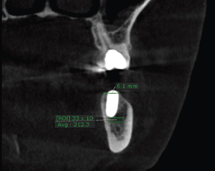
This prospective observational clinical study included 24 patients who received 46 dental implants that were installed in low-density bone using the osseodensification method. CBCT was used to measure the bone density pre- and postoperatively and implant stability was measured using Periotest® immediately after implant insertion and then after 6 weeks and 12 weeks postoperatively. The data were analyzed using paired t-test and the probability value <0.05 was considered statistically significant.
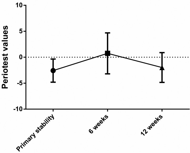
Of the 46 implants, 43 were osseointegrated making the early survival of the implants 93.5%. There was a significant increase in bone density postoperatively; 337.6 ±182.9 compared to 265.3 ±173.9 Hounsfield units preoperatively. The primary implant stability was -2.7 ± 2.13 Periotest values (PTV), at the 6th week it decreased significantly (<0.0001) to become 0.7 (± 4) PTV, and at the 12th week (secondary stability) it increased significantly (<0.0001) to become -2.1 (± 2.8) PTV. The difference between primary and secondary stability was statistically non-significant (=0.0814).
Osseodensification resulted in high primary stability and increased peri-implant bone density but it did not prevent the implant stability drop during the first 6 weeks after insertion of implants. Osseodensification, implant stability, low-density bone.
本研究的目的是通过骨致密化方法评估在低密度骨中进行种植位点预备,对骨愈合期种植体稳定性变化以及使用锥形束计算机断层扫描(CBCT)测量种植体周围骨密度的影响。
这项前瞻性观察性临床研究纳入了24例患者,他们接受了46颗使用骨致密化方法植入低密度骨的牙种植体。使用CBCT测量术前和术后的骨密度,并在种植体植入后立即以及术后6周和12周使用Periotest®测量种植体稳定性。采用配对t检验分析数据,概率值<0.05被认为具有统计学意义。
46颗种植体中,43颗实现了骨结合,种植体早期存活率为93.5%。术后骨密度显著增加;术前为265.3±173.9亨氏单位,术后为337.6±182.9亨氏单位。种植体初始稳定性为-2.7±2.13 Periotest值(PTV),在第6周时显著下降(<0.0001)至0.7(±4)PTV,在第12周(二级稳定性)时显著增加(<0.0001)至-2.1(±2.8)PTV。初始稳定性和二级稳定性之间的差异无统计学意义(=0.0814)。
骨致密化导致了较高的初始稳定性并增加了种植体周围骨密度,但它并不能防止种植体植入后前6周内种植体稳定性的下降。骨致密化、种植体稳定性、低密度骨。