Burgholzer P, Bauer-Marschallinger J, Haltmeier M
Research Center for Non-Destructive Testing (RECENDT), Linz, Austria.
Department of Mathematics, University of Innsbruck, Innsbruck, Austria.
Photoacoustics. 2020 May 21;19:100191. doi: 10.1016/j.pacs.2020.100191. eCollection 2020 Sep.
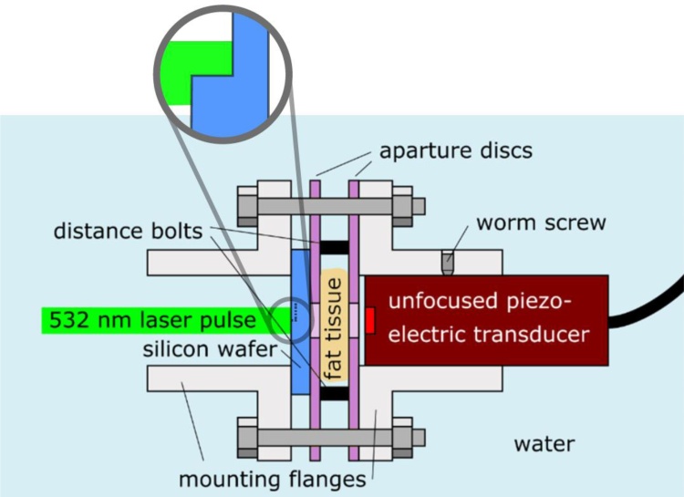
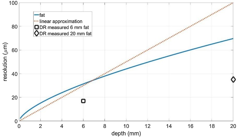
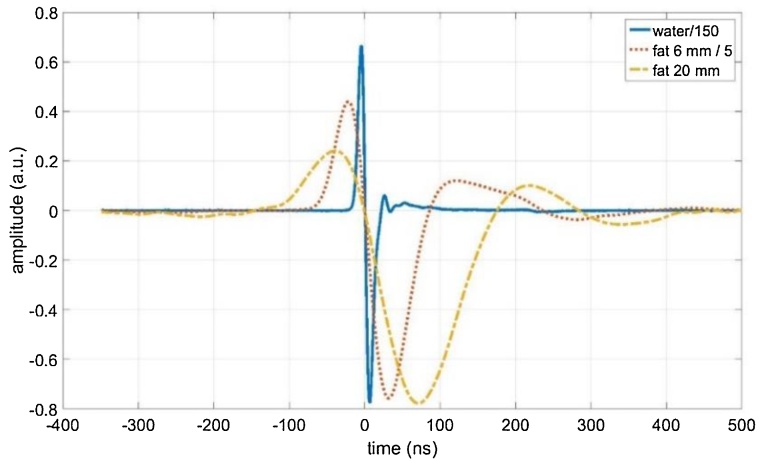
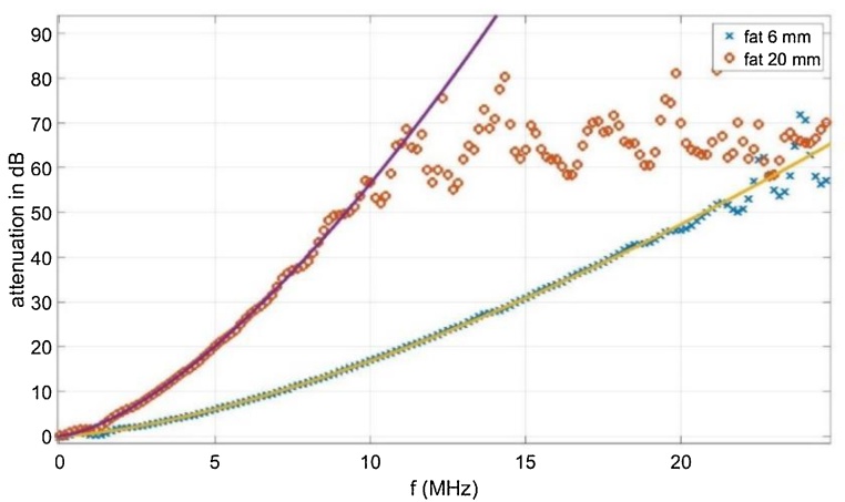
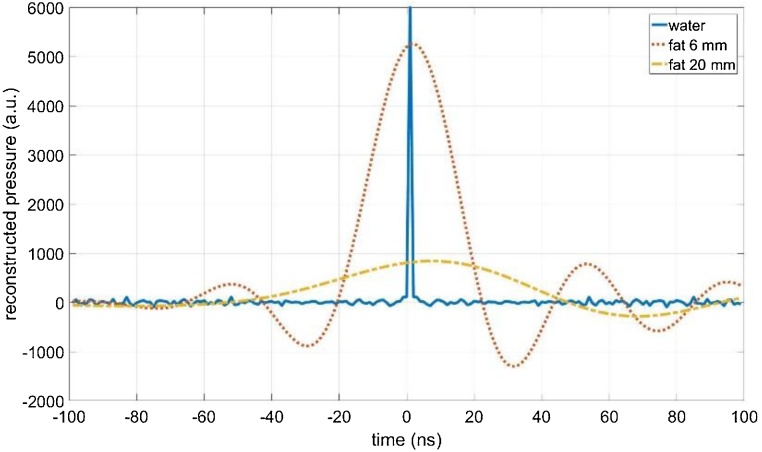
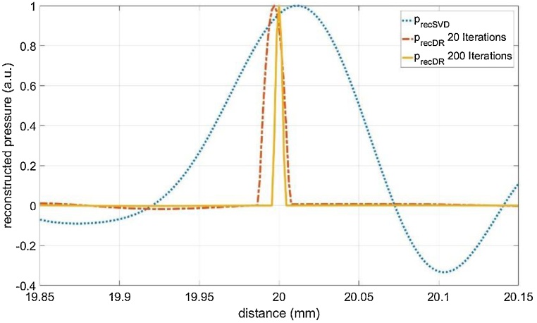
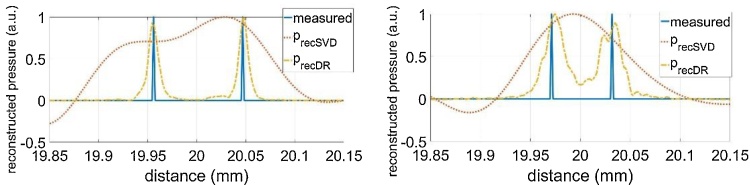
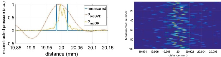
The spatial resolution achievable in photoacoustic imaging decreases with the imaging depth, resulting in blurred images for deeper structures. Apart from technical limitations, the ultimate resolution limit results from the second law of thermodynamics. The attenuation of the optically generated acoustic waves on their way from the imaged structure to the sample surface by scattering and dissipation leads to an increase of entropy. The resulting loss of spatial resolution for structures embedded in attenuating media can be compensated by numerical methods that make use of additional available information. In this article, we demonstrate this using experimental data from plane one-dimensional (1D) acoustic waves propagating in fat tissue. The acoustic waves are optically induced by nanosecond laser pulses and measured with piezoelectric transducers. The experimental results of 1D compensation are also relevant for photoacoustic imaging in 2D or 3D in an acoustically attenuating medium by dividing the reconstruction problem into two steps: First, the ideal signal, which is the solution of the un-attenuated wave equation, is determined by the proposed 1D attenuation compensation for each detector signal. In a second step, any ultrasound reconstruction method for un-attenuated data can be used for image reconstruction. For the reconstruction of a small step milled into a silicon wafer surface, which allows the generation of two photoacoustic pulses with a small time offset, we take advantage of non-negativity and sparsity and inverted the measured, frequency dependent acoustic attenuation of the fat tissue. We were able to improve the spatial resolution for imaging through 20 mm of porcine fat tissue compared to the diffraction limit at the cut-off frequency by at least a factor of two.
光声成像中可实现的空间分辨率会随着成像深度的增加而降低,导致深层结构的图像模糊。除了技术限制外,最终的分辨率极限源于热力学第二定律。光学产生的声波在从成像结构传播到样品表面的过程中,由于散射和耗散而衰减,这会导致熵增加。对于嵌入衰减介质中的结构,由此产生的空间分辨率损失可以通过利用额外可用信息的数值方法来补偿。在本文中,我们使用在脂肪组织中传播的平面一维(1D)声波的实验数据来证明这一点。声波由纳秒激光脉冲光学诱导,并由压电换能器进行测量。一维补偿的实验结果对于在声学衰减介质中进行二维或三维光声成像也具有相关性,方法是将重建问题分为两个步骤:首先,通过为每个探测器信号提出的一维衰减补偿来确定理想信号,即未衰减波动方程的解。第二步,可以使用任何用于未衰减数据的超声重建方法进行图像重建。对于在硅片表面铣削出的一个小台阶的重建,它可以产生两个具有小时间偏移的光声脉冲,我们利用非负性和稀疏性,对测量得到的脂肪组织的频率相关声学衰减进行反演。与截止频率处的衍射极限相比,我们能够将通过20毫米猪脂肪组织成像的空间分辨率提高至少两倍。