• 文献检索
  • 文档翻译
  • 深度研究
  • 学术资讯
  • Suppr Zotero 插件Zotero 插件
  • 邀请有礼
  • 套餐&价格
  • 历史记录
应用&插件
Suppr Zotero 插件Zotero 插件浏览器插件Mac 客户端Windows 客户端微信小程序
定价
高级版会员购买积分包购买API积分包
服务
文献检索文档翻译深度研究API 文档MCP 服务
关于我们
关于 Suppr公司介绍联系我们用户协议隐私条款
关注我们

Suppr 超能文献

核心技术专利:CN118964589B侵权必究
粤ICP备2023148730 号-1Suppr @ 2026

文献检索

告别复杂PubMed语法,用中文像聊天一样搜索,搜遍4000万医学文献。AI智能推荐,让科研检索更轻松。

立即免费搜索

文件翻译

保留排版,准确专业,支持PDF/Word/PPT等文件格式,支持 12+语言互译。

免费翻译文档

深度研究

AI帮你快速写综述,25分钟生成高质量综述,智能提取关键信息,辅助科研写作。

立即免费体验

卵囊吞噬作用的动力学。

Dynamics of Oocyst Phagocytosis by Macrophages.

机构信息

Aix Marseille Univ, IRD, AP-HM, SSA, VITROME, Marseille, France.

IHU-Méditerranée Infection, Marseille, France.

出版信息

Front Cell Infect Microbiol. 2020 May 19;10:207. doi: 10.3389/fcimb.2020.00207. eCollection 2020.

DOI:10.3389/fcimb.2020.00207
PMID:32509593
原文链接:https://pmc.ncbi.nlm.nih.gov/articles/PMC7248298/
Abstract

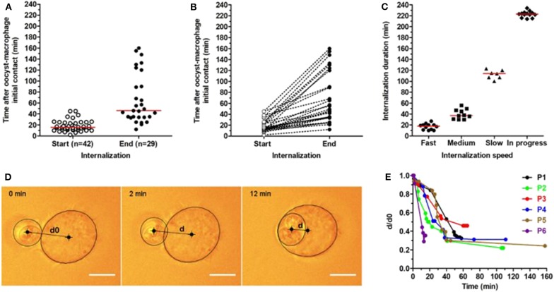
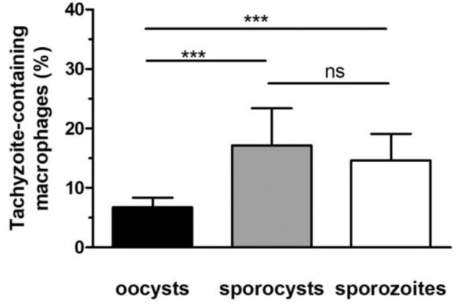
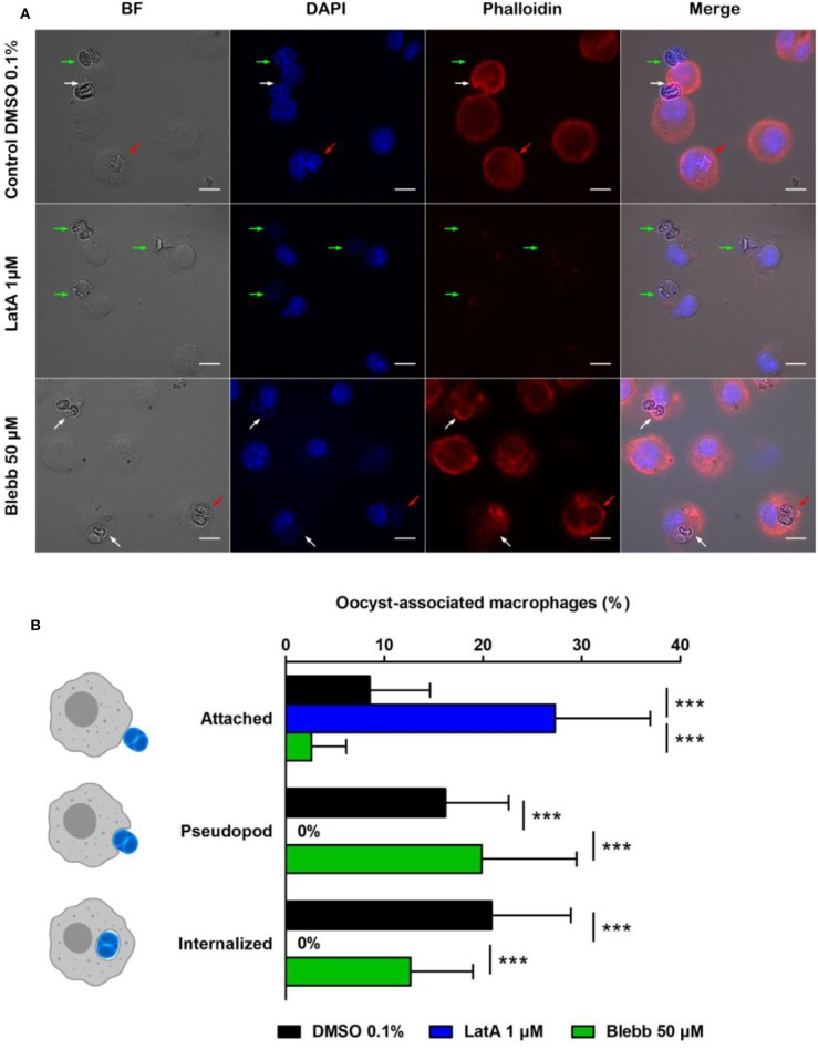
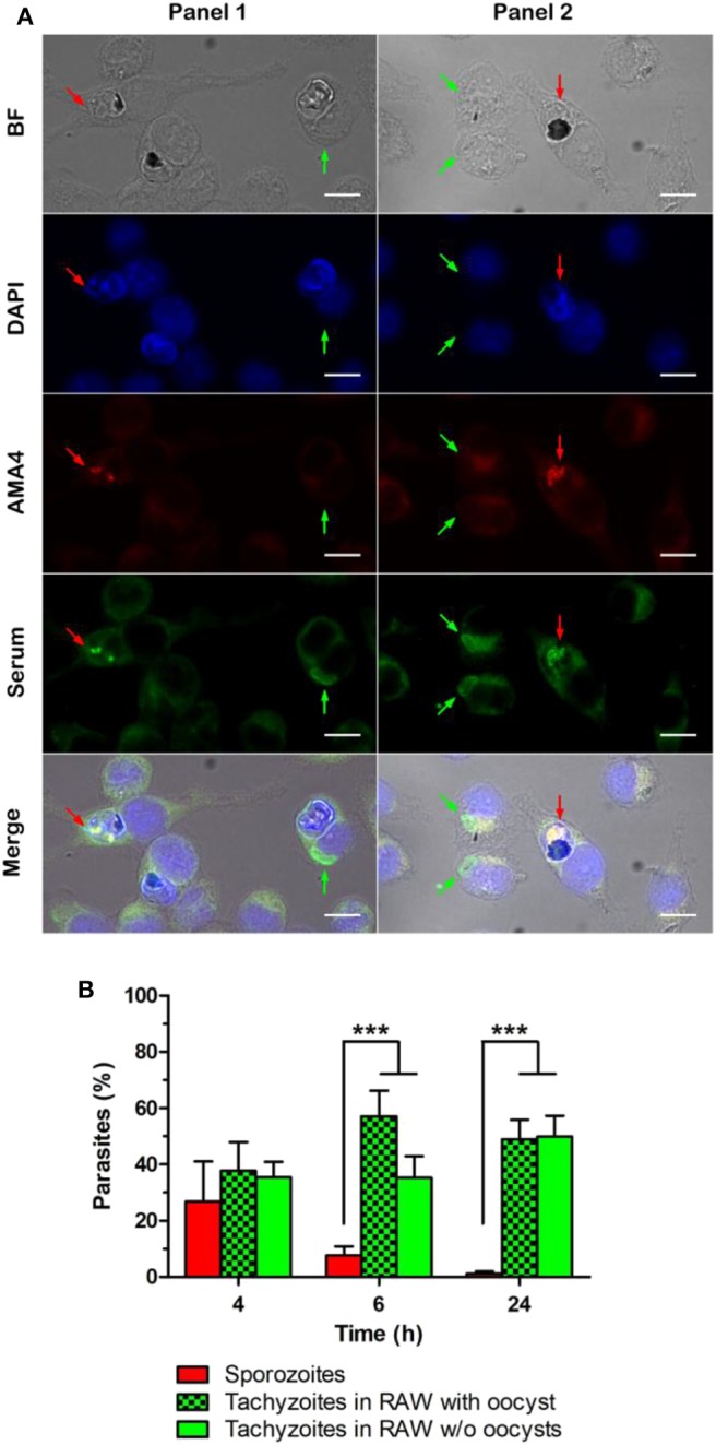
Oocysts are the environmentally resistant stage of the protozoan parasite . They are responsible for foodborne infections in humans and animals worldwide. Infectious oocysts contain sporozoites that have to exit the sporocyst and oocyst walls to initiate replication of the parasite within the host tissues. Given their robustness and resistance to chemical degradation, it is still unclear how the oocyst and sporocyst walls release the sporozoites. This process called excystation is thought to occur in the small intestine as a result of the combined action of digestive agents, yet to be identified. By using an oocyst-macrophage co-culture platform, we previously demonstrated that the excystation of sporozoites and their differentiation into replicative tachyzoites could occur in absence of digestive factors, following phagocytosis by macrophages. Here, we further characterize the dynamics of the oocyst phagocytosis at the single-cell level by using optical tweezers and micropipette aspiration techniques. Our results show that the oocyst internalization kinetics can vary among a given population of macrophages, but similar processes and dynamics could be observed. Most of the cells manipulate oocysts for ~15 min before internalizing them in typically 30 min. This process mainly involves the actin cytoskeleton of the macrophages. Liberated sporozoites within macrophages then differentiate into tachyzoites within 4-6 h following oocyst-macrophage contact. Tachyzoites appear to develop better in macrophages challenged with free sporocysts or sporozoites than with whole oocysts, suggesting that opening of the oocyst wall is one of the most limiting steps for sporozoite excystation completion.

摘要

卵囊是原生动物寄生虫的环境抗性阶段。它们是导致全球人类和动物食源性感染的罪魁祸首。传染性卵囊含有孢子,孢子必须离开孢子囊和卵囊壁,才能在宿主组织内开始寄生虫的复制。鉴于它们的坚固性和对化学降解的抵抗力,目前尚不清楚卵囊和孢子囊壁如何释放孢子。这个过程称为出囊,据认为是在小肠中由于尚未确定的消化剂的共同作用而发生的。通过使用卵囊-巨噬细胞共培养平台,我们之前已经证明,在没有消化因素的情况下,巨噬细胞吞噬后,孢子的出囊和分化为复制性速殖子可以发生。在这里,我们使用光镊和微吸管抽吸技术在单细胞水平上进一步研究了卵囊吞噬的动力学。我们的结果表明,在给定的巨噬细胞群体中,卵囊的内化动力学可能会有所不同,但可以观察到类似的过程和动力学。大多数细胞在将卵囊内化之前,会操纵卵囊约 15 分钟,通常需要 30 分钟。这个过程主要涉及巨噬细胞的肌动蛋白细胞骨架。释放到巨噬细胞内的游离孢子在与卵囊-巨噬细胞接触后 4-6 小时内分化为速殖子。速殖子似乎在受到游离孢子囊或孢子的挑战的巨噬细胞中比在完整的卵囊中发育得更好,这表明卵囊壁的打开是完成孢子出囊的最关键步骤之一。

https://cdn.ncbi.nlm.nih.gov/pmc/blobs/cf70/7248298/dfa27ef47a13/fcimb-10-00207-g0004.jpg
https://cdn.ncbi.nlm.nih.gov/pmc/blobs/cf70/7248298/e5daf32a0b67/fcimb-10-00207-g0001.jpg
https://cdn.ncbi.nlm.nih.gov/pmc/blobs/cf70/7248298/af2977e38746/fcimb-10-00207-g0002.jpg
https://cdn.ncbi.nlm.nih.gov/pmc/blobs/cf70/7248298/8833c50ef571/fcimb-10-00207-g0003.jpg
https://cdn.ncbi.nlm.nih.gov/pmc/blobs/cf70/7248298/dfa27ef47a13/fcimb-10-00207-g0004.jpg

相似文献

1
Dynamics of Oocyst Phagocytosis by Macrophages.卵囊吞噬作用的动力学。
Front Cell Infect Microbiol. 2020 May 19;10:207. doi: 10.3389/fcimb.2020.00207. eCollection 2020.
2
Macrophages facilitate the excystation and differentiation of Toxoplasma gondii sporozoites into tachyzoites following oocyst internalisation.巨噬细胞促进刚地弓形虫卵囊内化后孢子囊的逸出和分化为速殖子。
Sci Rep. 2016 Sep 19;6:33654. doi: 10.1038/srep33654.
3
Generation of Toxoplasma gondii and Hammondia hammondi Oocysts and Purification of Their Sporozoites for Downstream Manipulation.弓形虫和哈氏巴贝斯虫卵囊的生成及囊内子孢子的纯化,以便下游操作。
Methods Mol Biol. 2020;2071:81-98. doi: 10.1007/978-1-4939-9857-9_4.
4
Late Embryogenesis Abundant Proteins Contribute to the Resistance of Toxoplasma gondii Oocysts against Environmental Stresses.晚期胚胎丰富蛋白有助于弓形虫卵囊抵抗环境压力。
mBio. 2023 Apr 25;14(2):e0286822. doi: 10.1128/mbio.02868-22. Epub 2023 Feb 21.
5
Transcriptomic analysis of toxoplasma development reveals many novel functions and structures specific to sporozoites and oocysts.转录组分析揭示了速殖子和卵囊特有的许多新功能和结构。
PLoS One. 2012;7(2):e29998. doi: 10.1371/journal.pone.0029998. Epub 2012 Feb 13.
6
Toxoplasma gondii Oocyst Infectivity Assessed Using a Sporocyst-Based Cell Culture Assay Combined with Quantitative PCR for Environmental Applications.弓形虫卵囊感染性评估:一种基于孢囊的细胞培养测定法结合定量 PCR 技术在环境应用中的应用。
Appl Environ Microbiol. 2019 Oct 1;85(20). doi: 10.1128/AEM.01189-19. Print 2019 Oct 15.
7
Ultrastructure of excystment of Toxoplasma gondii oocysts.刚地弓形虫卵囊脱囊的超微结构
J Protozool. 1978 Nov;25(4):438-43. doi: 10.1111/j.1550-7408.1978.tb04166.x.
8
Global proteomic analysis of the oocyst/sporozoite of Toxoplasma gondii reveals commitment to a host-independent lifestyle.弓形虫卵囊/子孢子的全局蛋白质组分析揭示了其向宿主非依赖性生活方式的转变。
BMC Genomics. 2013 Mar 15;14:183. doi: 10.1186/1471-2164-14-183.
9
Ultrastructure of the oocysts, sporocysts, and sporozoites of Toxoplasma gondii.刚地弓形虫卵囊、子孢子囊和子孢子的超微结构
J Parasitol. 1998 Jun;84(3):505-12.
10
Characterization of an IgG monoclonal antibody targeted to both tissue cyst and sporocyst walls of Toxoplasma gondii.一种靶向刚地弓形虫组织包囊和子孢子囊壁的IgG单克隆抗体的特性分析
Exp Parasitol. 2016 Apr;163:46-56. doi: 10.1016/j.exppara.2016.01.014. Epub 2016 Feb 4.

引用本文的文献

1
Comparative Phosphoproteomic Analysis of Sporulated Oocysts and Tachyzoites of Reveals Stage-Specific Patterns.比较研究孢子化卵囊和速殖子的磷酸化蛋白质组,揭示了阶段特异性模式。
Molecules. 2022 Feb 2;27(3):1022. doi: 10.3390/molecules27031022.
2
Recombinant Ribosomal Protein P2 Modulates the Functions of Murine Macrophages In Vitro and Provides Immunity against Acute Toxoplasmosis In Vivo.重组核糖体蛋白P2体外调节小鼠巨噬细胞功能并在体内提供抗急性弓形虫病免疫力。
Vaccines (Basel). 2021 Apr 7;9(4):357. doi: 10.3390/vaccines9040357.
3
Expanding the Known Repertoire of C-Type Lectin Receptors Binding to Toxoplasma gondii Oocysts Using a Modified High-Resolution Immunofluorescence Assay.

本文引用的文献

1
Structure, composition, and roles of the oocyst and sporocyst walls.卵囊壁和孢子囊壁的结构、组成及作用。
Cell Surf. 2018 Dec 19;5:100016. doi: 10.1016/j.tcsw.2018.100016. eCollection 2019 Dec.
2
Environmental transmission of : Oocysts in water, soil and food.的环境传播:水中、土壤和食物中的卵囊。 (原句中“of”后面缺少具体内容,翻译不太完整准确,推测这里可能是某个病原体之类的,比如“Environmental transmission of [pathogen name] : Oocysts in water, soil and food.” 完整意思为“[病原体名称]的环境传播:水中、土壤和食物中的卵囊” )
Food Waterborne Parasitol. 2019 Apr 1;15:e00049. doi: 10.1016/j.fawpar.2019.e00049. eCollection 2019 Jun.
3
Phagocytosis: Mechanosensing, Traction Forces, and a Molecular Clutch.
利用改良的高分辨率免疫荧光检测方法扩展识别刚地弓形虫卵囊的 C 型凝集素受体的已知谱。
mSphere. 2021 Mar 31;6(2):e01341-20. doi: 10.1128/mSphere.01341-20.
吞噬作用:机械感知、牵引力和分子离合器。
Curr Biol. 2020 Jan 6;30(1):R24-R26. doi: 10.1016/j.cub.2019.11.047.
4
Squeezing in a Meal: Myosin Functions in Phagocytosis.挤时间吃饭:肌球蛋白在吞噬作用中的功能。
Trends Cell Biol. 2020 Feb;30(2):157-167. doi: 10.1016/j.tcb.2019.11.002. Epub 2019 Dec 10.
5
The secreted kinase ROP17 promotes Toxoplasma gondii dissemination by hijacking monocyte tissue migration.分泌激酶 ROP17 通过劫持单核细胞组织迁移来促进刚地弓形虫的传播。
Nat Microbiol. 2019 Nov;4(11):1951-1963. doi: 10.1038/s41564-019-0504-8. Epub 2019 Jul 22.
6
From Entry to Early Dissemination- Initial Encounter With Its Host.从进入到早期传播——与宿主的初次接触。
Front Cell Infect Microbiol. 2019 Mar 5;9:46. doi: 10.3389/fcimb.2019.00046. eCollection 2019.
7
Latrunculin A Accelerates Actin Filament Depolymerization in Addition to Sequestering Actin Monomers.拉曲库林 A 通过隔离肌动蛋白单体来加速肌动蛋白丝的解聚。
Curr Biol. 2018 Oct 8;28(19):3183-3192.e2. doi: 10.1016/j.cub.2018.07.082. Epub 2018 Sep 27.
8
The first detection of Toxoplasma gondii DNA in environmental air samples using gelatine filters, real-time PCR and loop-mediated isothermal (LAMP) assays: qualitative and quantitative analysis.使用明胶滤器、实时聚合酶链反应(PCR)和环介导等温扩增(LAMP)检测法首次在环境空气样本中检测到刚地弓形虫DNA:定性和定量分析
Parasitology. 2017 Nov;144(13):1791-1801. doi: 10.1017/S0031182017001172. Epub 2017 Jul 12.
9
Macrophages facilitate the excystation and differentiation of Toxoplasma gondii sporozoites into tachyzoites following oocyst internalisation.巨噬细胞促进刚地弓形虫卵囊内化后孢子囊的逸出和分化为速殖子。
Sci Rep. 2016 Sep 19;6:33654. doi: 10.1038/srep33654.
10
Controlled One-on-One Encounters between Immune Cells and Microbes Reveal Mechanisms of Phagocytosis.免疫细胞与微生物之间的一对一受控接触揭示了吞噬作用的机制。
Biophys J. 2015 Aug 4;109(3):469-76. doi: 10.1016/j.bpj.2015.06.042.