Frontier Research Institute for Materials Science, Nagoya Institute of Technology, Gokiso, Showa-ku, Nagoya 466-8555, Japan; Department of Molecular Cellular and Developmental Biology, Yale University, PO Box 208103, New Haven, CT 06520, USA.
Department of Genetics, Cell Biology and Development, University of Minnesota, Minneapolis, MN 55455, USA.
Curr Biol. 2018 Oct 8;28(19):3183-3192.e2. doi: 10.1016/j.cub.2018.07.082. Epub 2018 Sep 27.
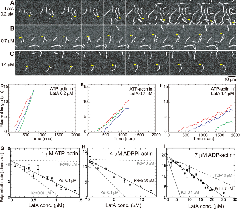
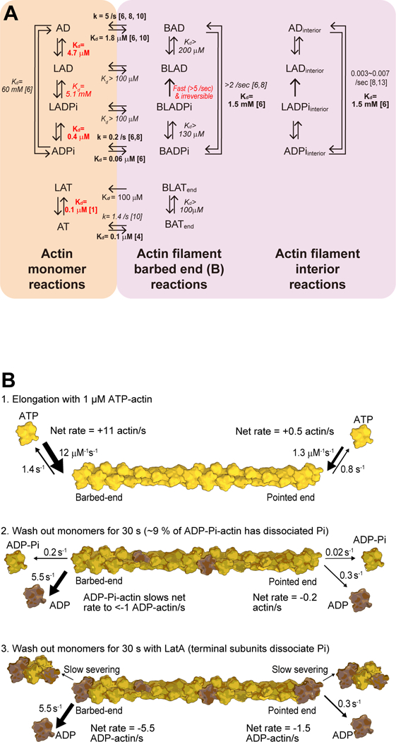
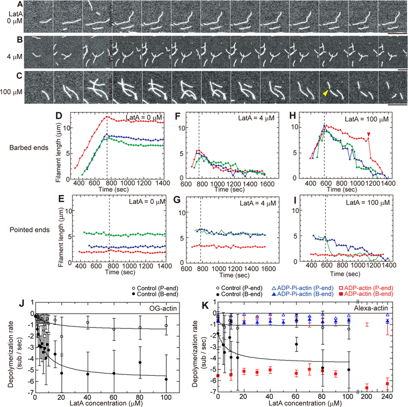
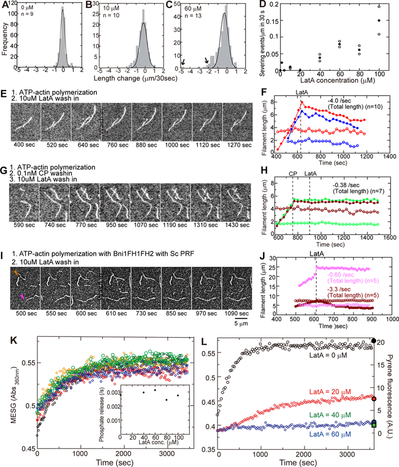
Latrunculin A (LatA), a toxin from the red sea sponge Latrunculia magnifica, is the most widely used reagent to depolymerize actin filaments in experiments on live cells. LatA binds actin monomers and sequesters them from polymerization [1, 2]. Low concentrations of LatA result in rapid (tens of seconds) disassembly of actin filaments in animal [3] and yeast cells [2]. Depolymerization is usually assumed to result from sequestration of actin monomers. Our observations of single-muscle actin filaments by TIRF microscopy showed that LatA bound ATP-actin monomers with a higher affinity (K = 0.1 μM) than ADP-P-actin (K = 0.4 μM) or ADP-actin (K = 4.7 μM). LatA also slowly severed filaments and increased the depolymerization rate at both ends of filaments freshly assembled from ATP-actin to the rates of ADP-actin. This rate plateaued at LatA concentrations >60 μM. LatA did not change the depolymerization rates of ADP- actin filaments or ADP-P-actin filaments generated with 160 mM phosphate in the buffer. LatA did not increase the rate of phosphate release from bulk samples of filaments assembled from ATP-actin. Thermodynamic analysis showed that LatA binds weakly to actin filaments with a K >100 μM. We propose that concentrations of LatA much lower than this K promote phosphate dissociation only from both ends of filaments, resulting in depolymerization limited by the rate of ADP-actin dissociation. Thus, one must consider both rapid actin depolymerization and severing in addition to sequestering actin monomers when interpreting the effects of LatA on cells.
拉他丁 A(LatA)是从红海海绵 Latrunculia magnifica 中提取的一种毒素,是在活细胞实验中使肌动蛋白丝解聚最常用的试剂。LatA 结合肌动蛋白单体并将其与聚合隔离开来[1,2]。低浓度的 LatA 会导致动物[3]和酵母细胞[2]中的肌动蛋白丝迅速(数十秒内)解聚。通常认为解聚是由于肌动蛋白单体的隔离。我们通过 TIRF 显微镜观察单个肌肉肌动蛋白丝的结果表明,LatA 与 ATP-肌动蛋白单体的亲和力(K = 0.1 μM)高于 ADP-P-肌动蛋白(K = 0.4 μM)或 ADP-肌动蛋白(K = 4.7 μM)。LatA 还缓慢地切断纤维,并将刚从 ATP-肌动蛋白组装而成的纤维两端的解聚速率提高到 ADP-肌动蛋白的速率。在 LatA 浓度>60 μM 时,该速率达到平台期。LatA 并未改变缓冲液中含 160 mM 磷酸盐时生成的 ADP-肌动蛋白纤维或 ADP-P-肌动蛋白纤维的解聚速率。LatA 不会增加从 ATP-肌动蛋白组装而成的纤维的块状样品中磷酸盐的释放速率。热力学分析表明,LatA 与肌动蛋白丝的结合力较弱,K>100 μM。我们提出,远低于此 K 的 LatA 浓度仅促进纤维两端磷酸盐的解离,从而导致解聚受到 ADP-肌动蛋白解离速率的限制。因此,在解释 LatA 对细胞的影响时,除了隔离肌动蛋白单体外,还必须考虑肌动蛋白的快速解聚和切断。