Wu Richard Y, Williamson Tyler D, Sahoo Narayan, Nguyen Trang, Ikner Shane M, Liu Amy Y, Wisdom Paul G, Lii MingFu, Hunter Rachel A, Alvarez Paola E, Gunn G Brandon, Frank Steven J, Hojo Yoshifumi, Zhu X Ronald, Gillin Michael T
Departments of Radiation Physics, The University of Texas MD Anderson Cancer Center, Houston, TX, United States.
Departments of Imaging and Radiation Oncology Core, The University of Texas MD Anderson Cancer Center, Houston, TX, United States.
Phys Imaging Radiat Oncol. 2020 Jan;13:44-49. doi: 10.1016/j.phro.2020.03.004. Epub 2020 Mar 26.
Computed tomography (CT) scanning is the basis for radiation treatment planning, but the 50-cm standard scanning field of view (sFOV) may be too small for imaging larger patients. We evaluated the 65-cm high-definition (HD) FOV of a large-bore CT scanner for CT number accuracy, geometric distortion, image quality degradation, and dosimetric accuracy of photon treatment plans.
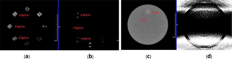
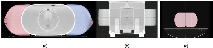
CT number accuracy was tested by placing two 16-cm acrylic phantoms on either side of a 40-cm phantom to simulate a large patient extending beyond the 50-cm-diameter standard scanning FOV. Dosimetric accuracy was tested using anthropomorphic pelvis and thorax phantoms, with additional acrylic body parts on either side of the phantoms. Two volumetric modulated arc therapy beams (a 15-MV and a 6-MV) were used to cover the planning target volumes. Two-dimensional dose distributions were evaluated with GAFChromic film and point dose accuracy was checked with multiple thermoluminescent dosimeter (TLD) capsules placed in the phantoms. Image quality was tested by placing an American College of Radiology accreditation phantom inside the 40-cm phantom.
The HD FOV showed substantial changes in CT numbers, with differences of 314 HU-725 HU at different density levels. The volume of the body parts extending into the HD FOV was distorted. However, TLD-reported doses for all PTVs agreed within ± 3%. Dose agreement in organs at risk were within the passing criteria, and the gamma index pass rate was >97%. Image quality was degraded.
The HD FOV option is adequate for RT simulation and met accreditation standards, although care should be taken during contouring because of reduced image quality.
计算机断层扫描(CT)是放射治疗计划的基础,但50厘米的标准扫描视野(sFOV)对于体型较大的患者成像可能过小。我们评估了大孔径CT扫描仪65厘米高清(HD)视野在CT数值准确性、几何畸变、图像质量下降以及光子治疗计划剂量准确性方面的表现。
通过在一个40厘米的模体两侧放置两个16厘米的丙烯酸模体来测试CT数值准确性,以模拟超出50厘米直径标准扫描视野的大体型患者。使用人体骨盆和胸部模体测试剂量准确性,在模体两侧添加额外的丙烯酸身体部位。使用两个容积调强弧形治疗束(一个15兆伏和一个6兆伏)来覆盖计划靶体积。用GAFChromic胶片评估二维剂量分布,并用放置在模体内的多个热释光剂量计(TLD)胶囊检查点剂量准确性。通过将美国放射学会认证模体放置在40厘米模体内来测试图像质量。
高清视野下CT数值有显著变化,在不同密度水平下差异为314亨氏单位至725亨氏单位。延伸到高清视野内的身体部位体积发生了畸变。然而,所有计划靶体积的TLD报告剂量在±3%范围内一致。危及器官的剂量一致性符合通过标准,伽马指数通过率>97%。图像质量下降。
高清视野选项适用于放疗模拟且符合认证标准,不过由于图像质量降低,在勾画轮廓时应谨慎。