Department of Neurology Neurological Institute Taipei Veterans General Hospital Taipei Taiwan.
School of Medicine National Yang-Ming University Taipei Taiwan.
J Am Heart Assoc. 2020 Jul 7;9(13):e016233. doi: 10.1161/JAHA.120.016233. Epub 2020 Jun 17.
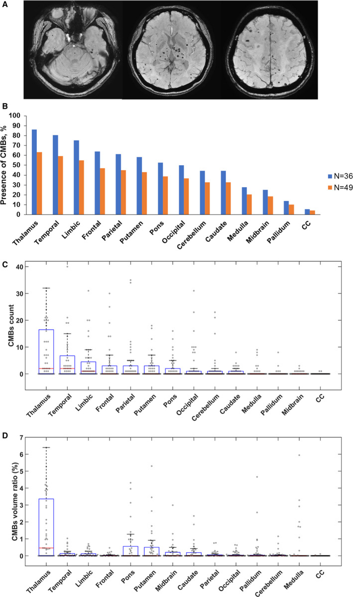
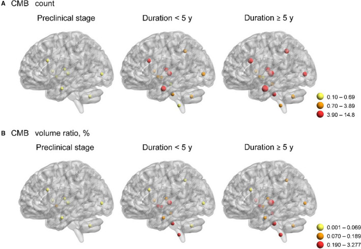
Background Cerebral autosomal dominant arteriopathy with subcortical infarcts and leukoencephalopathy, caused by mutations, is characterized by recurrent ischemic strokes and progressive cognitive decline. It remains unclear whether cerebral microbleeds (CMBs) can serve as a surrogate marker for disease progression in cerebral autosomal dominant arteriopathy with subcortical infarcts and leukoencephalopathy. We aimed to investigate the CMB burdens in mutation carriers at different disease stages and test their associations with cognitive performance. Methods and Results Forty-nine individuals carrying cysteine-altering mutations received brain magnetic resonance imaging with T1-weighted and susceptibility-weighted images. Whole brain images were segmented into 14 regions using Statistical Parametric Mapping and FreeSurfer software, and semiautomatic methods were used to locate and quantify the number and volume of CMBs. In our study participants, the median of CMB counts was 13, with a wide individual variation (range, 0-286). CMBs were most frequently present in thalamus, followed by temporal lobe. In the whole brain, the CMB counts and CMB volume ratios (ie, CMB volume divided by the volume of corresponding brain region) gradually increased as the disease advanced. CMB counts in the thalamus and temporal and frontal lobes increased more rapidly than other brain regions as disease progressed. There were significant associations between Mini-Mental State Examination scores and CMB counts in the frontal lobe, temporal lobe, and pons. Conclusions CMBs may have an influential role in the clinical manifestations of cerebral autosomal dominant arteriopathy with subcortical infarcts and leukoencephalopathy. CMB burdens and their distribution in different brain regions may be capable to serve as a disease marker for monitoring the disease severity of cerebral autosomal dominant arteriopathy with subcortical infarcts and leukoencephalopathy.
由 基因突变引起的脑常染色体显性动脉病伴皮质下梗死和白质脑病,其特征是反复发作的缺血性中风和进行性认知衰退。目前尚不清楚脑微出血(CMB)是否可以作为脑常染色体显性动脉病伴皮质下梗死和白质脑病疾病进展的替代标志物。我们旨在研究不同疾病阶段 突变携带者的 CMB 负担,并测试它们与认知表现的相关性。
49 名携带半胱氨酸改变突变的个体接受了 T1 加权和磁化率加权成像的脑部磁共振成像检查。使用统计参数映射和 FreeSurfer 软件将全脑图像分为 14 个区域,并使用半自动方法定位和量化 CMB 的数量和体积。在我们的研究参与者中,CMB 计数的中位数为 13,个体差异很大(范围为 0-286)。CMB 最常出现在丘脑,其次是颞叶。在整个大脑中,CMB 计数和 CMB 体积比(即 CMB 体积除以相应脑区的体积)随着疾病的进展逐渐增加。随着疾病的进展,丘脑、颞叶和额叶的 CMB 计数增加得更快。在额叶、颞叶和脑桥中,CMB 计数与简易精神状态检查评分之间存在显著相关性。
CMB 可能在脑常染色体显性动脉病伴皮质下梗死和白质脑病的临床表现中具有重要作用。不同脑区的 CMB 负担及其分布可能能够作为监测脑常染色体显性动脉病伴皮质下梗死和白质脑病疾病严重程度的疾病标志物。