• 文献检索
  • 文档翻译
  • 深度研究
  • 学术资讯
  • Suppr Zotero 插件Zotero 插件
  • 邀请有礼
  • 套餐&价格
  • 历史记录
应用&插件
Suppr Zotero 插件Zotero 插件浏览器插件Mac 客户端Windows 客户端微信小程序
定价
高级版会员购买积分包购买API积分包
服务
文献检索文档翻译深度研究API 文档MCP 服务
关于我们
关于 Suppr公司介绍联系我们用户协议隐私条款
关注我们

Suppr 超能文献

核心技术专利:CN118964589B侵权必究
粤ICP备2023148730 号-1Suppr @ 2026

文献检索

告别复杂PubMed语法,用中文像聊天一样搜索,搜遍4000万医学文献。AI智能推荐,让科研检索更轻松。

立即免费搜索

文件翻译

保留排版,准确专业,支持PDF/Word/PPT等文件格式,支持 12+语言互译。

免费翻译文档

深度研究

AI帮你快速写综述,25分钟生成高质量综述,智能提取关键信息,辅助科研写作。

立即免费体验

后巩膜形态作为开角型青光眼生物标志物的特征:双眼间比较研究。

The Shape of Posterior Sclera as a Biometric Signature in Open-angle Glaucoma: An Intereye Comparison Study.

机构信息

Department of Ophthalmology, College of Medicine, The Catholic University of Korea, Incheon St. Mary's Hospital, Incheon.

Department of Ophthalmology, College of medicine, Chuncheon Sacred Heart Hospital, Hallym University, Chuncheon-si, Gangwon-do, Republic of Korea.

出版信息

J Glaucoma. 2020 Oct;29(10):890-898. doi: 10.1097/IJG.0000000000001573.

DOI:10.1097/IJG.0000000000001573
PMID:32555059
原文链接:https://pmc.ncbi.nlm.nih.gov/articles/PMC7647446/
Abstract

PURPOSE

To characterize intereye differences in posterior segment parameters and determine their significance in open-angle glaucoma patients with unilateral damage.

METHODS

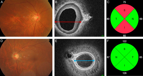
Both eyes from 65 subjects without any nerve damage and 43 patients undergoing treatment for unilateral open-angle glaucoma were included in this study. A 12.0×9.0×2.6 mm volume of the posterior segment in each eye was scanned with swept-source optical coherence tomography. Coronally reconstructed optical coherence tomography images were analyzed to determine the deepest point of the eye (DPE), which we then calculated the distance (Disc-DPE distance), depth (Disc-DPE depth), angle (Disc-DPE angle) from the optic disc center to the DPE. Posterior pole shape was analyzed measuring the posterior pole-cross-sectional area, posterior pole-horizontal width (PP-HW), and posterior pole-vertical width) of the posterior pole. These measurements and their intereye absolute difference (IAD; absolute difference in measurements between the right and left eyes) values were compared between the healthy and unilateral glaucomatous patients.

RESULTS

The posterior sclera measurements, including the Disc-DPE distance, Disc-DPE depth, and posterior pole-cross-sectional area, were significantly different between the unilateral glaucoma eyes and contralateral healthy eyes (P=0.043, P=0.035, and P=0.049, respectively). By contrast, none of the intereye differences in optic nerve head parameters were significant in the unilateral glaucoma patients. In comparison with the IAD values, the baseline intraocular pressure and PP-HW of the posterior segment showed significant differences between the healthy and the unilateral glaucoma patients (P=0.019 and P=0.036, respectively). A multivariate analysis showed that a larger baseline intraocular pressure IAD [odds ratio (OR), 1.381; P=0.009)] and larger PP-HW IAD (OR, 1.324; P=0.032) were significantly associated with the presence of glaucoma.

CONCLUSIONS

Compared with the fellow healthy eyes, glaucomatous eyes had larger and more steeply curved posterior poles, which represent a structural variation of the posterior sclera that might be associated with glaucomatous optic neuropathy.

摘要

目的

描述双眼后部参数的差异,并确定其在单侧损害的开角型青光眼患者中的意义。

方法

本研究纳入了 65 名无神经损伤的受试者和 43 名接受单侧开角型青光眼治疗的患者的双眼。使用扫频源光学相干断层扫描仪对每只眼的 12.0×9.0×2.6mm 后节体积进行扫描。对冠状重建的光学相干断层扫描图像进行分析,以确定眼球最深处(DPE),然后计算从视盘中心到 DPE 的距离(Disc-DPE 距离)、深度(Disc-DPE 深度)和角度(Disc-DPE 角度)。通过测量后极的后极横截面积、后极水平宽度(PP-HW)和后极垂直宽度,分析后极形状。将这些测量值及其双眼绝对差值(IAD;右眼和左眼测量值之间的绝对差值)值与健康人和单侧青光眼患者进行比较。

结果

单侧青光眼眼中的后巩膜测量值,包括 Disc-DPE 距离、Disc-DPE 深度和后极横截面积,与对侧健康眼相比差异有统计学意义(P=0.043、P=0.035 和 P=0.049)。相比之下,单侧青光眼患者的视神经头参数的双眼差异均无统计学意义。与 IAD 值相比,健康人和单侧青光眼患者的基线眼内压和后节 PP-HW 差异有统计学意义(P=0.019 和 P=0.036)。多变量分析显示,基线眼内压 IAD 较大(比值比(OR),1.381;P=0.009)和 PP-HW IAD 较大(OR,1.324;P=0.032)与青光眼的存在显著相关。

结论

与对侧健康眼相比,青光眼眼中的后极更大且更陡峭弯曲,这代表了后巩膜的结构变化,可能与青光眼性视神经病变有关。

https://cdn.ncbi.nlm.nih.gov/pmc/blobs/3edf/7647446/6724147cce27/ijg-29-890-g003.jpg
https://cdn.ncbi.nlm.nih.gov/pmc/blobs/3edf/7647446/2a2037b2476b/ijg-29-890-g001.jpg
https://cdn.ncbi.nlm.nih.gov/pmc/blobs/3edf/7647446/d383a0902421/ijg-29-890-g002.jpg
https://cdn.ncbi.nlm.nih.gov/pmc/blobs/3edf/7647446/6724147cce27/ijg-29-890-g003.jpg
https://cdn.ncbi.nlm.nih.gov/pmc/blobs/3edf/7647446/2a2037b2476b/ijg-29-890-g001.jpg
https://cdn.ncbi.nlm.nih.gov/pmc/blobs/3edf/7647446/d383a0902421/ijg-29-890-g002.jpg
https://cdn.ncbi.nlm.nih.gov/pmc/blobs/3edf/7647446/6724147cce27/ijg-29-890-g003.jpg

相似文献

1
The Shape of Posterior Sclera as a Biometric Signature in Open-angle Glaucoma: An Intereye Comparison Study.后巩膜形态作为开角型青光眼生物标志物的特征:双眼间比较研究。
J Glaucoma. 2020 Oct;29(10):890-898. doi: 10.1097/IJG.0000000000001573.
2
Comparison of Diagnostic Power of Optic Nerve Head and Posterior Sclera Configuration Parameters on Myopic Normal Tension Glaucoma.视神经头和后巩膜形态参数对近视性正常眼压青光眼诊断效能的比较。
J Glaucoma. 2019 Sep;28(9):834-842. doi: 10.1097/IJG.0000000000001328.
3
Intereye Comparison of Lamina Cribrosa Curvature in Normal Tension Glaucoma Patients With Unilateral Damage.正常眼压性青光眼单侧损害患者板层 cribrosa 曲率的眼内比较。
Invest Ophthalmol Vis Sci. 2019 Jun 3;60(7):2423-2430. doi: 10.1167/iovs.19-26828.
4
Impact of Posterior Sclera on Glaucoma Progression in Treated Myopic Normal-Tension Glaucoma Using Reconstructed Optical Coherence Tomographic Images.基于重建光学相干断层扫描图像探讨后巩膜对治疗性近视性正常眼压性青光眼进展的影响。
Invest Ophthalmol Vis Sci. 2019 May 1;60(6):2198-2207. doi: 10.1167/iovs.19-26794.
5
Clinical Assessment of Scleral Canal Area in Glaucoma Using Spectral-Domain Optical Coherence Tomography.应用频域光学相干断层扫描仪评估青光眼巩膜管区。
Am J Ophthalmol. 2020 Aug;216:28-36. doi: 10.1016/j.ajo.2020.03.043. Epub 2020 Apr 9.
6
Comparison of optic nerve head topography and visual field in eyes with open-angle and angle-closure glaucoma.开角型青光眼和闭角型青光眼患者视神经乳头形态与视野的比较。
Ophthalmology. 2008 Feb;115(2):239-245.e2. doi: 10.1016/j.ophtha.2007.03.086.
7
Disc Torsion and Vertical Disc Tilt Are Related to Subfoveal Scleral Thickness in Open-Angle Glaucoma Patients With Myopia.在近视性开角型青光眼患者中,盘状扭转和垂直盘倾斜与黄斑下巩膜厚度相关。
Invest Ophthalmol Vis Sci. 2015 Jul;56(8):4927-35. doi: 10.1167/iovs.14-15819.
8
Measurement of scleral thickness using swept-source optical coherence tomography in patients with open-angle glaucoma and myopia.应用扫频源光学相干断层扫描仪测量开角型青光眼和近视患者的巩膜厚度。
Am J Ophthalmol. 2014 Apr;157(4):876-84. doi: 10.1016/j.ajo.2014.01.007. Epub 2014 Jan 9.
9
Peripapillary choroidal thickness in healthy controls and patients with focal, diffuse, and sclerotic glaucomatous optic disc damage.健康对照者以及患有局灶性、弥漫性和硬化性青光眼性视盘损害患者的视乳头周围脉络膜厚度。
Arch Ophthalmol. 2012 Aug;130(8):980-6. doi: 10.1001/archophthalmol.2012.371.
10
Association of Scleral Deformation Around the Optic Nerve Head With Central Visual Function in Normal-Tension Glaucoma and Myopia.正常眼压性青光眼和近视患者视神经头周围巩膜变形与中心视觉功能的关系。
Am J Ophthalmol. 2020 Sep;217:287-296. doi: 10.1016/j.ajo.2020.04.041. Epub 2020 May 6.

引用本文的文献

1
Impact of Cataract Surgery on IOP and Ocular Structures in Normotensive Patients and Primary and Exfoliation Open-Angle Glaucoma Patients.白内障手术对正常眼压患者、原发性开角型青光眼患者和剥脱性开角型青光眼患者眼压及眼结构的影响。
Clin Ophthalmol. 2023 Apr 5;17:1047-1055. doi: 10.2147/OPTH.S402550. eCollection 2023.
2
Central retinal vascular trunk deviation in unilateral normal-tension glaucoma.单侧正常眼压型青光眼的中心视网膜血管干偏位。
PLoS One. 2021 Jul 20;16(7):e0254889. doi: 10.1371/journal.pone.0254889. eCollection 2021.
3
Changes in ocular morphology after cataract surgery in open angle glaucoma patients.

本文引用的文献

1
Impact of Posterior Sclera on Glaucoma Progression in Treated Myopic Normal-Tension Glaucoma Using Reconstructed Optical Coherence Tomographic Images.基于重建光学相干断层扫描图像探讨后巩膜对治疗性近视性正常眼压性青光眼进展的影响。
Invest Ophthalmol Vis Sci. 2019 May 1;60(6):2198-2207. doi: 10.1167/iovs.19-26794.
2
The Effects of Optic Nerve Head Tilt on Visual Field Defects in Myopic Normal Tension Glaucoma: The Intereye Comparison Study.近视性正常眼压青光眼视盘倾斜对视野缺损的影响:双眼比较研究。
J Glaucoma. 2019 Apr;28(4):341-346. doi: 10.1097/IJG.0000000000001182.
3
Transverse Separation of the Outer Retinal Layer at the Peripapillary in Glaucomatous Myopes.
开角型青光眼患者白内障手术后眼形态的变化。
Sci Rep. 2021 Jun 9;11(1):12203. doi: 10.1038/s41598-021-91740-z.
青光眼性近视患者视盘周围外视网膜层的横向分离。
Sci Rep. 2018 Aug 20;8(1):12446. doi: 10.1038/s41598-018-30523-5.
4
Longitudinal Changes of Optic Nerve Head and Peripapillary Structure during Childhood Myopia Progression on OCT: Boramae Myopia Cohort Study Report 1.基于 OCT 的儿童近视进展过程中视盘和视盘周围结构的纵向变化:Boramae 近视队列研究报告 1。
Ophthalmology. 2018 Aug;125(8):1215-1223. doi: 10.1016/j.ophtha.2018.01.026. Epub 2018 Mar 14.
5
Three Dimensional Evaluation of Posterior Pole and Optic Nerve Head in Tilted Disc.倾斜盘的后极和视盘头部的三维评估。
Sci Rep. 2018 Jan 18;8(1):1121. doi: 10.1038/s41598-018-19242-z.
6
Three-Dimensional Evaluation of Posterior Pole and Optic Nerve Head in Myopes with Glaucoma.青光眼近视患者后极部和视盘的三维评估。
Sci Rep. 2017 Dec 21;7(1):18001. doi: 10.1038/s41598-017-18297-8.
7
Finite Element Biomechanics of Optic Nerve Sheath Traction in Adduction.内收时视神经鞘膜牵引的有限元生物力学
J Biomech Eng. 2017 Oct 1;139(10):1010101-10101010. doi: 10.1115/1.4037562.
8
The Location of the Deepest Point of the Eyeball Determines the Optic Disc Configuration.眼球最深处的位置决定了视盘的形态。
Sci Rep. 2017 Jul 19;7(1):5881. doi: 10.1038/s41598-017-06072-8.
9
Intra- and interobserver reproducibility of Bruch's membrane opening minimum rim width measurements with spectral domain optical coherence tomography.使用光谱域光学相干断层扫描测量布鲁赫膜开口最小边缘宽度的观察者内和观察者间的可重复性。
Acta Ophthalmol. 2017 Nov;95(7):e548-e555. doi: 10.1111/aos.13464. Epub 2017 Jun 26.
10
Intereye Difference in the Microstructure of Parapapillary Atrophy in Unilateral Primary Open-Angle Glaucoma.单侧原发性开角型青光眼视乳头旁萎缩微观结构的双眼差异
Invest Ophthalmol Vis Sci. 2016 Aug 1;57(10):4187-93. doi: 10.1167/iovs.16-19059.