Department of Ophthalmology, Seoul St. Mary's Hospital, College of Medicine, The Catholic University of Korea, Seoul, Korea.
Sci Rep. 2017 Jul 19;7(1):5881. doi: 10.1038/s41598-017-06072-8.
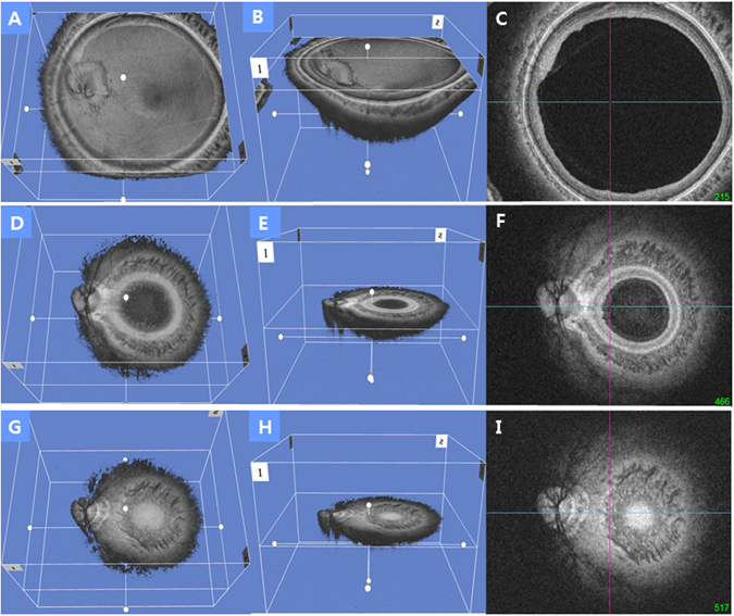
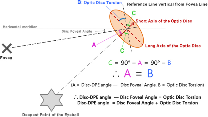
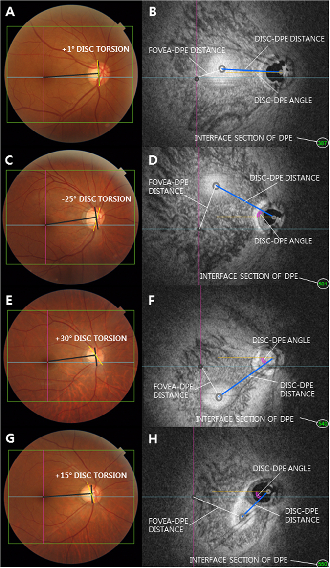
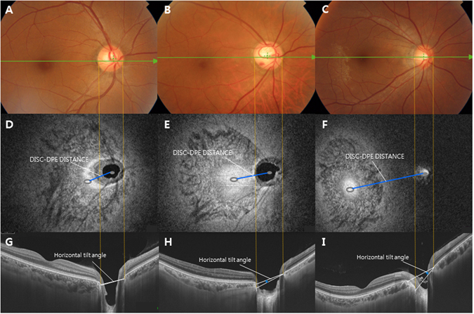
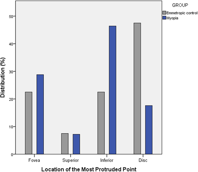
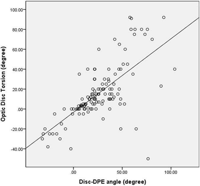
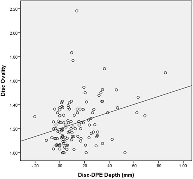
Tilted and rotated appearances are hallmarks of the myopic optic disc. As the eyeball grows axially, the posterior pole elongates not only globally but in a localized manner as well. In this process, the optic disc is pulled towards the deepest point of the elongated eyeball, which might result in a change in optic disc configuration. Thus, we hypothesized that analyzing the variation of posterior pole contour can play a major role in understanding optic disc configuration in myopic subjects. By analyzing consecutive images of swept source OCT coronal sections at the posterior pole, the deepest interface between Bruch's membrane and the choroid could be identified as the deepest point of the eyeball (DPE). The location and the properties of the DPE differed significantly between the 125 eyes of non-glaucomatous myopic group and the 40 eyes of non-glaucomatous emmetropic group classified based on 24 mm axial length. The results suggested that the larger disc to DPE angle and the larger disc to DPE depth strongly predicts the optic disc torsion degree and the optic disc tilt. Our findings suggest that identifying the posterior pole profile plays a major role in understanding the optic disc alterations found in myopic subjects.
倾斜和旋转的外观是近视性视盘的特征。随着眼球轴向生长,后极不仅在整体上而且在局部也会变长。在这个过程中,视盘被拉向伸长眼球的最深点,这可能导致视盘结构的改变。因此,我们假设分析后极轮廓的变化可以在理解近视患者视盘结构中发挥重要作用。通过分析扫频源 OCT 冠状截面的连续图像,可以将 Bruch 膜和脉络膜之间的最深界面确定为眼球的最深处 (DPE)。基于 24mm 眼轴长度,在非青光眼近视组的 125 只眼中和非青光眼正视组的 40 只眼中,DPE 的位置和特性存在显著差异。结果表明,较大的视盘至 DPE 角度和较大的视盘至 DPE 深度强烈预测视盘扭转程度和视盘倾斜。我们的发现表明,确定后极轮廓在理解近视患者中发现的视盘改变方面起着重要作用。