Department of Ophthalmology, College of medicine, Seoul St. Mary's Hospital, The Catholic University of Korea, Seoul, Korea.
Sci Rep. 2018 Jan 18;8(1):1121. doi: 10.1038/s41598-018-19242-z.
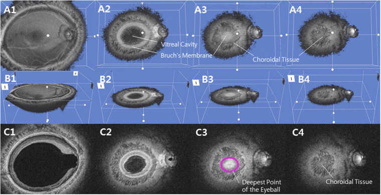
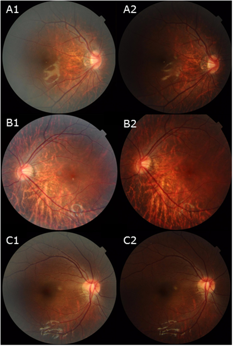
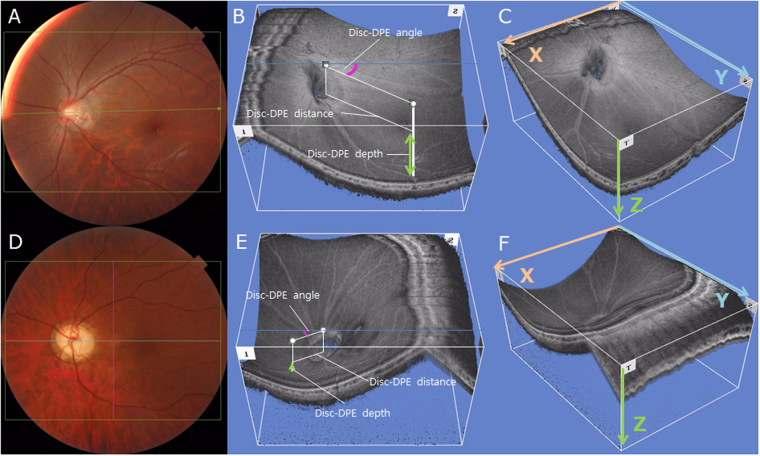
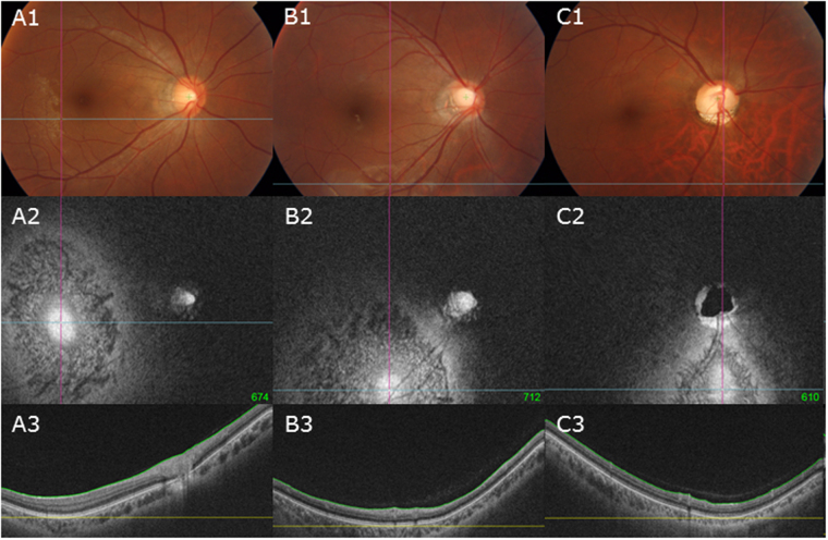
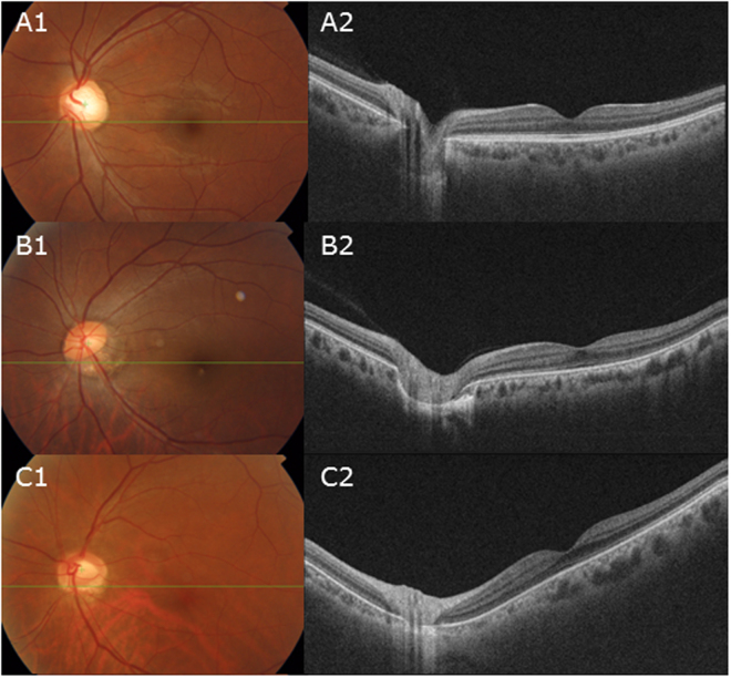
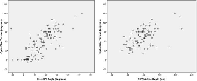
For over a century, tilted disc syndrome (TDS) has been defined vaguely. The lack of consensus of the terminology arises from the lack of understanding of the pathogenesis of this condition. Also, myopic discs with temporal crescents or peripapillary atrophy (PPA) are histologically indistinguishable from TDS. Therefore, we examined the morphological background of the extreme ONH appearances such as the myopic tilted disc and the TDS by analyzing the posterior segment of the eye from a three-dimensional (3D) perspective. 107 eyes of 107 subjects were classified into 3 groups with respect to the optic disc torsion degrees: (1) mild torsion (0-30 degrees; 35 eyes) and (2) moderate torsion (30-60 degrees; 35 eyes) and (3) severe torsion (60-90 degrees; 37 eyes). SSOCT images were analyzed in coronal view, which supplements anterior-posterior depth (z axis in Cartesian coordinates). The amount of optic disc torsion was significantly correlated with Disc-DPE angle and Fovea-Disc depth (r = 0.548, P < 0.001 and r = 0.544, P < 0.001). In conclusion, we describe specific types of posterior sclera configuration that corresponds to the increasing degree of optic disc torsion, even in the extreme ONH appearances such as the myopic tilted disc and the TDS. These findings suggest that the optic disc appearance is determined by the configuration of the posterior sclera.
一个多世纪以来,倾斜盘综合征(TDS)的定义一直很模糊。这种情况的发病机制尚未被充分理解,术语上缺乏共识。此外,具有颞侧新月形或视盘周围萎缩(PPA)的近视盘在组织学上与 TDS 无法区分。因此,我们通过从三维(3D)角度分析眼部后节,研究了近视倾斜盘和 TDS 等极端视盘外观的形态学背景。我们将 107 名受试者的 107 只眼分为 3 组,每组的视盘扭转程度分别为:(1)轻度扭转(0-30 度;35 只眼)、(2)中度扭转(30-60 度;35 只眼)和(3)重度扭转(60-90 度;37 只眼)。在冠状面分析 SSOCT 图像,这补充了前后深度(笛卡尔坐标系中的 z 轴)。视盘扭转量与 Disc-DPE 角和黄斑中心凹-视盘深度显著相关(r=0.548,P<0.001 和 r=0.544,P<0.001)。总之,我们描述了与视盘扭转程度增加相对应的特定类型的后部巩膜形态,即使在近视倾斜盘和 TDS 等极端视盘外观中也是如此。这些发现表明视盘外观由后部巩膜的形态决定。