Department of Diagnostic and Interventional Radiology, Lausanne University Hospital and University of Lausanne, Rue du Bugnon 46, CH-1011, Lausanne, Switzerland.
Advanced Clinical Imaging Technology, Siemens Healthcare AG, Lausanne, Switzerland.
Neuroradiology. 2020 Nov;62(11):1371-1380. doi: 10.1007/s00234-020-02477-x. Epub 2020 Jun 17.
We aimed at assessing the potential of automated MR morphometry to assess individual basal ganglia and thalamus volumetric changes at the chronic phase after cortical stroke.
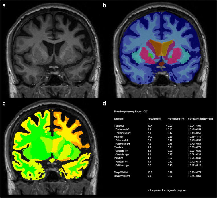
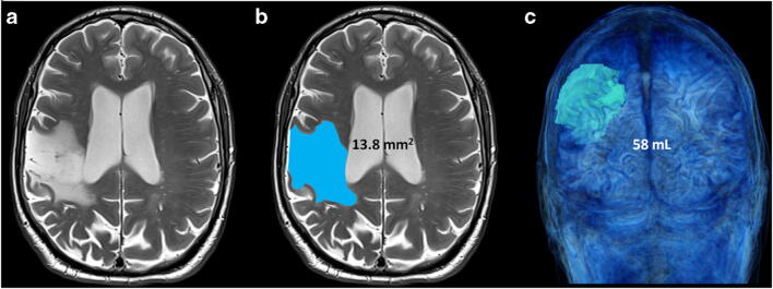
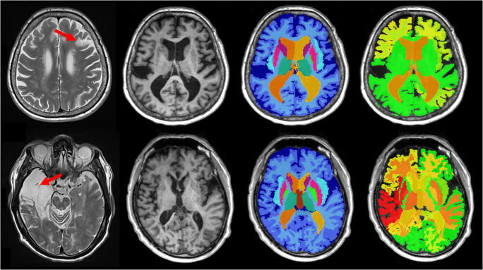
Ninety-six patients (mean age: 65 ± 18 years, male 55) with cortical stroke at the chronic phase were retrospectively included. Patients were scanned at 1.5 T or 3 T using a T1-MPRAGE sequence. Resulting 3D images were processed with the MorphoBox prototype software to automatically segment basal ganglia and thalamus structures, and to obtain Z scores considering the confounding effects of age and sex. Stroke volume was estimated by manual delineation on T2-SE imaging. Z scores were compared between ipsi- and contralateral stroke side and according to the vascular territory. Potential relationship between Z scores and stroke volume was assessed using the Spearman correlation coefficient.
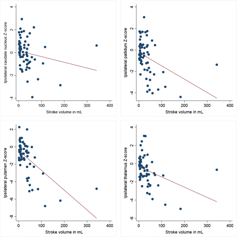
Basal ganglia and thalamus volume Z scores were lower ipsilaterally to MCA territory stroke (p values < 0.034) while they were not different between ipsi- and contralateral stroke sides in non-MCA territory stroke (p values > 0.37). In MCA territory stroke, ipsilateral caudate nucleus (rho = - 0.34, p = 0.007), putamen (rho = - 0.50, p < 0.001), pallidum (rho = - 0.44, p < 0.001), and thalamus (rho = - 0.48, p < 0.001) volume Z scores negatively correlated with the cortical stroke volume. This relation was not influenced by cardiovascular risk factors or time since stroke.
Automated MR morphometry demonstrated atrophy of ipsilateral basal ganglia and thalamus at the chronic phase after cortical stroke in the MCA territory. The atrophy was related to stroke volume. These results confirm the potential role for automated MRI morphometry to assess remote changes after stroke.
本研究旨在评估自动化磁共振形态测量学在皮质卒中慢性期评估个体基底节和丘脑容积变化的潜力。
回顾性纳入 96 例皮质卒中慢性期患者(平均年龄:65±18 岁,男性 55 例)。患者在 1.5T 或 3T 扫描仪上使用 T1-MPRAGE 序列进行扫描。使用 MorphoBox 原型软件对获得的 3D 图像进行处理,以自动分割基底节和丘脑结构,并获得考虑年龄和性别混杂因素影响的 Z 分数。通过手动勾画 T2-SE 图像来估计卒中体积。比较同侧和对侧卒中侧以及根据血管区域的 Z 分数。使用 Spearman 相关系数评估 Z 分数与卒中体积之间的潜在关系。
MCA 区域卒中时,基底节和丘脑体积 Z 分数较低(p 值<0.034),而非 MCA 区域卒中时,同侧和对侧卒中侧之间的 Z 分数无差异(p 值>0.37)。在 MCA 区域卒中时,同侧尾状核(rho=-0.34,p=0.007)、壳核(rho=-0.50,p<0.001)、苍白球(rho=-0.44,p<0.001)和丘脑(rho=-0.48,p<0.001)的体积 Z 分数与皮质卒中体积呈负相关。这种关系不受心血管危险因素或卒中后时间的影响。
自动化磁共振形态测量学在 MCA 区域皮质卒中慢性期显示出对侧基底节和丘脑的萎缩。这种萎缩与卒中体积有关。这些结果证实了自动化 MRI 形态测量学在评估卒中后远程变化方面的潜在作用。