Enam Syed Faaiz, Kader Sajidur Rahman, Bodkin Nicholas, Lyon Johnathan G, Calhoun Mark, Azrak Cesar, Tiwari Pooja Munnilal, Vanover Daryll, Wang Haichen, Santangelo Philip J, Bellamkonda Ravi Venkat
Department of Biomedical Engineering, Duke University, Durham, NC, USA.
Department of Biomedical Engineering, Georgia Institute of Technology, Atlanta, GA, USA.
J Neuroinflammation. 2020 Jun 20;17(1):197. doi: 10.1186/s12974-020-01860-y.
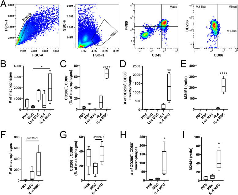
Appropriately modulating inflammation after traumatic brain injury (TBI) may prevent disabilities for the millions of those inflicted annually. In TBI, cellular mediators of inflammation, including macrophages and microglia, possess a range of phenotypes relevant for an immunomodulatory therapeutic approach. It is thought that early phenotypic modulation of these cells will have a cascading healing effect. In fact, an anti-inflammatory, "M2-like" macrophage phenotype after TBI has been associated with neurogenesis, axonal regeneration, and improved white matter integrity (WMI). There already exist clinical trials seeking an M2-like bias through mesenchymal stem/stromal cells (MSCs). However, MSCs do not endogenously synthesize key signals that induce robust M2-like phenotypes such as interleukin-4 (IL-4).
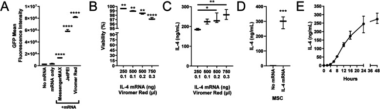
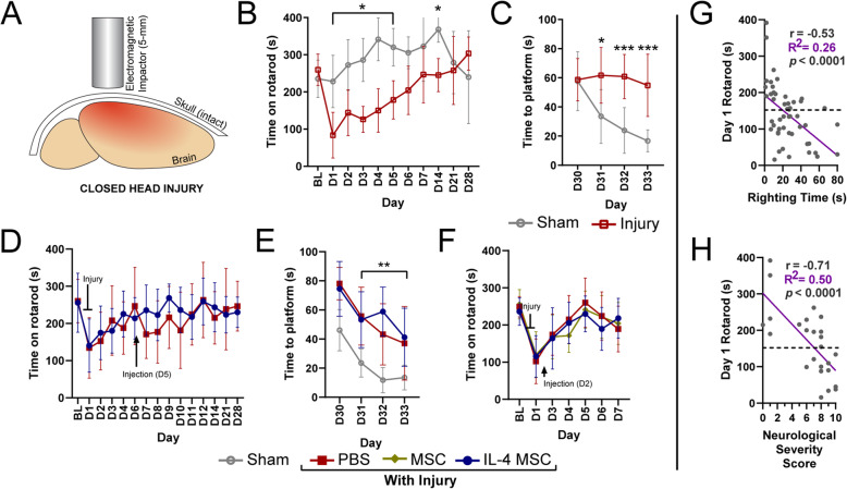
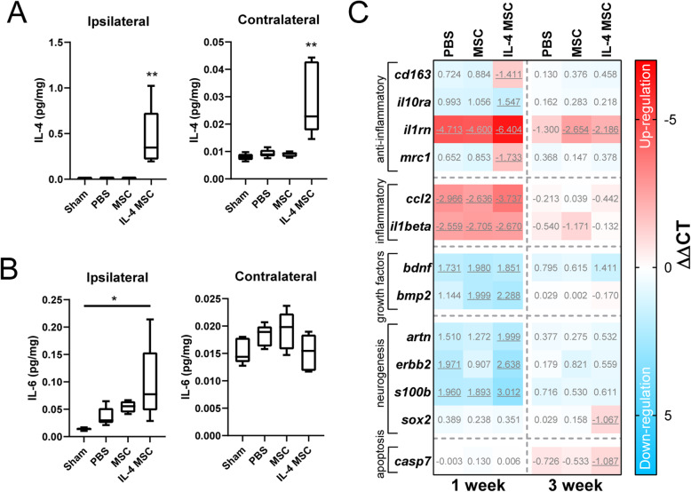
To enrich M2-like macrophages in a clinically relevant manner, we augmented MSCs with synthetic IL-4 mRNA to transiently express IL-4. These IL-4 expressing MSCs (IL-4 MSCs) were characterized for expression and functionality and then delivered in a modified mouse TBI model of closed head injury. Groups were assessed for functional deficits and MR imaging. Brain tissue was analyzed through flow cytometry, multi-plex ELISA, qPCR, histology, and RNA sequencing.
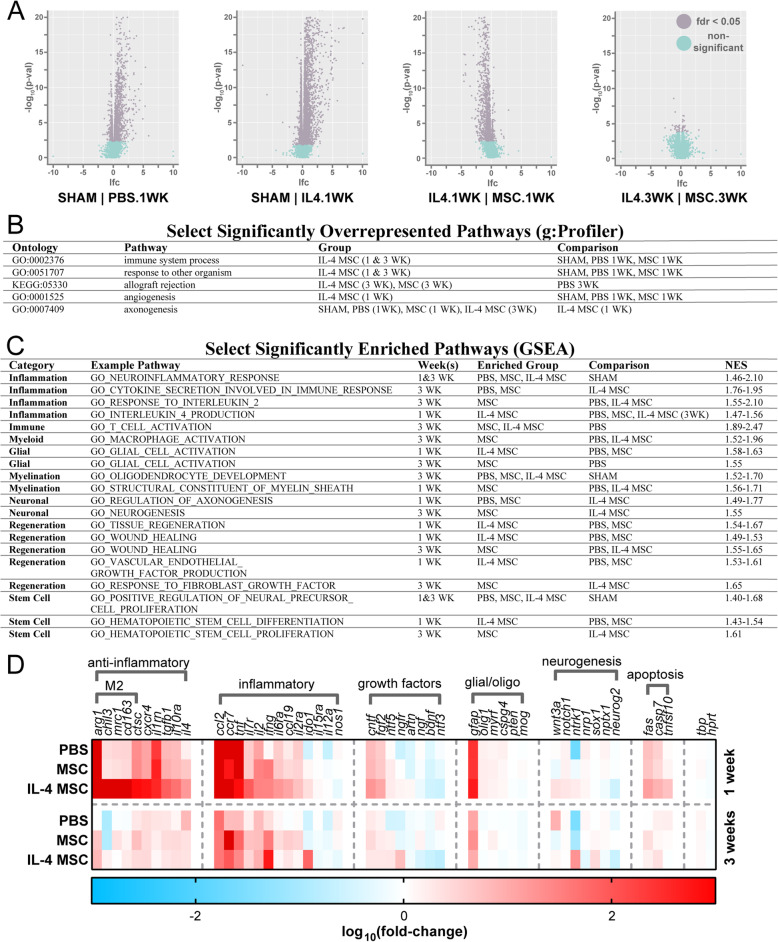
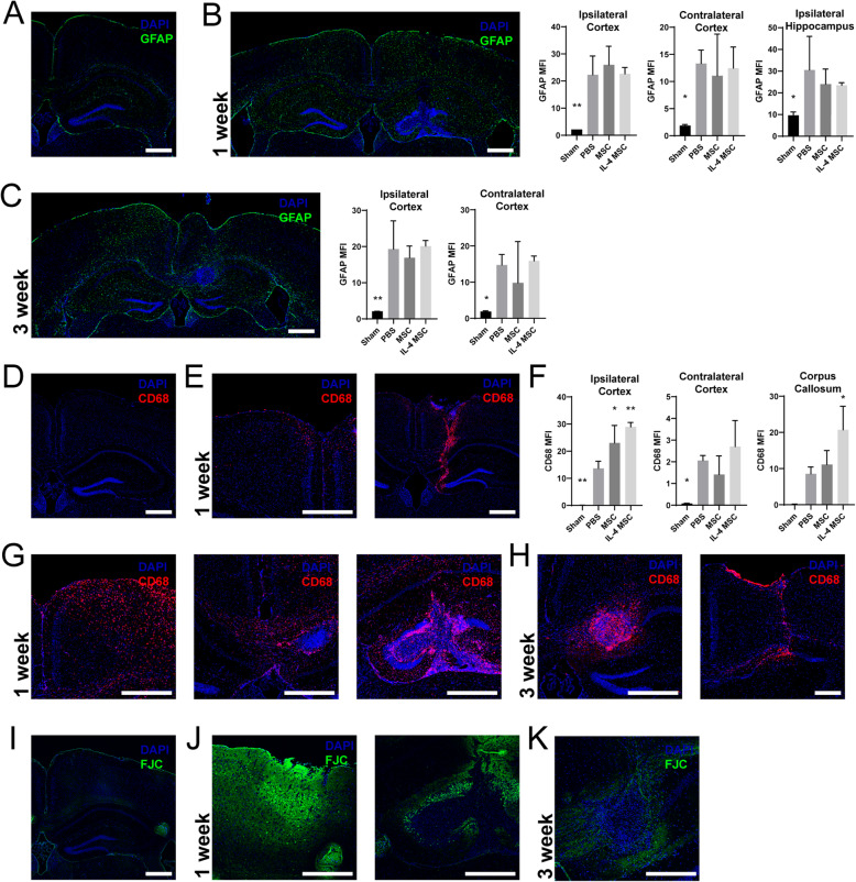
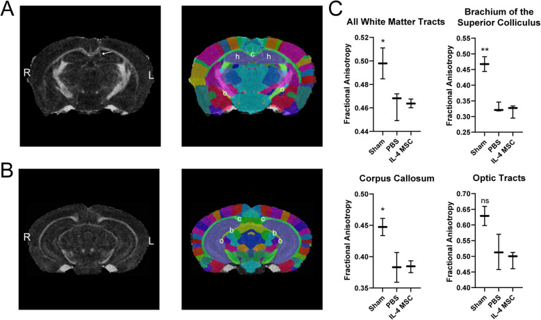
We observed that IL-4 MSCs indeed induce a robust M2-like macrophage phenotype and promote anti-inflammatory gene expression after TBI. However, here we demonstrate that acute enrichment of M2-like macrophages did not translate to improved functional or histological outcomes, or improvements in WMI on MR imaging. To further understand whether dysfunctional pathways underlie the lack of therapeutic effect, we report transcriptomic analysis of injured and treated brains. Through this, we discovered that inflammation persists despite acute enrichment of M2-like macrophages in the brain.
The results demonstrate that MSCs can be engineered to induce a stronger M2-like macrophage response in vivo. However, they also suggest that acute enrichment of only M2-like macrophages after diffuse TBI cannot orchestrate neurogenesis, axonal regeneration, or improve WMI. Here, we also discuss our modified TBI model and methods to assess severity, behavioral studies, and propose that IL-4 expressing MSCs may also have relevance in other cavitary diseases or in improving biomaterial integration into tissues.
对创伤性脑损伤(TBI)后的炎症进行适当调节,可能会预防每年数百万患者出现残疾。在TBI中,炎症的细胞介质,包括巨噬细胞和小胶质细胞,具有一系列与免疫调节治疗方法相关的表型。人们认为,对这些细胞进行早期表型调节将产生连锁的愈合效果。事实上,TBI后具有抗炎作用的“M2样”巨噬细胞表型与神经发生、轴突再生以及白质完整性(WMI)改善有关。目前已有通过间充质干/基质细胞(MSCs)寻求M2样偏向的临床试验。然而,MSCs不能内源性合成诱导强大M2样表型的关键信号,如白细胞介素-4(IL-4)。
为了以临床相关的方式富集M2样巨噬细胞,我们用合成的IL-4 mRNA增强MSCs,使其瞬时表达IL-4。对这些表达IL-4的MSCs(IL-4 MSCs)进行表达和功能表征,然后将其应用于改良的闭合性颅脑损伤小鼠TBI模型中。对各实验组进行功能缺陷评估和磁共振成像检查。通过流式细胞术、多重酶联免疫吸附测定、定量聚合酶链反应、组织学和RNA测序对脑组织进行分析。
我们观察到,IL-4 MSCs确实能诱导出强大的M2样巨噬细胞表型,并在TBI后促进抗炎基因表达。然而,我们在此证明,急性富集M2样巨噬细胞并不能转化为功能或组织学结果的改善,也不能改善磁共振成像上的WMI。为了进一步了解功能失调的通路是否是治疗效果不佳的原因,我们报告了对受伤和治疗后脑组织的转录组分析。通过这项分析,我们发现尽管大脑中急性富集了M2样巨噬细胞,但炎症仍然持续存在。
结果表明,可以对MSCs进行改造,使其在体内诱导更强的M2样巨噬细胞反应。然而,这些结果也表明,弥漫性TBI后仅急性富集M2样巨噬细胞并不能协调神经发生、轴突再生或改善WMI。在此,我们还讨论了我们改良的TBI模型以及评估严重程度的方法、行为学研究,并提出表达IL-4的MSCs可能在其他空洞性疾病或改善生物材料与组织的整合方面也具有相关性。